
How to read ecg easily in opd basis
Reading File
Now let me search the medical library and web in parallel for ECG reading content.
Finding Sources
Searching the Web
how to read ECG systematically OPD quick approach clinical
Finding Sources
Finding Sources
Found Harrison's 22E with a dedicated ECG interpretation section. Let me read it.
Reading File
Finding Sources
Reading File
Reading File
Reading File
Loading Image
Excellent - got the core ECG diagram and the normal intervals table. Let me get more key content - axis determination, ST changes, and common patterns.
Reading File
Finding Sources
Reading File
Reading File
Reading File
I now have all the information needed to write a comprehensive, practical ECG reading guide for OPD use. Let me compile the answer.
How to Read an ECG Easily in OPD
Here is a step-by-step systematic approach based on Harrison's Principles of Internal Medicine (22nd Ed.) and Goldman-Cecil Medicine. Following a fixed sequence prevents errors of omission - the most common ECG mistake.
First: Understand the ECG Paper
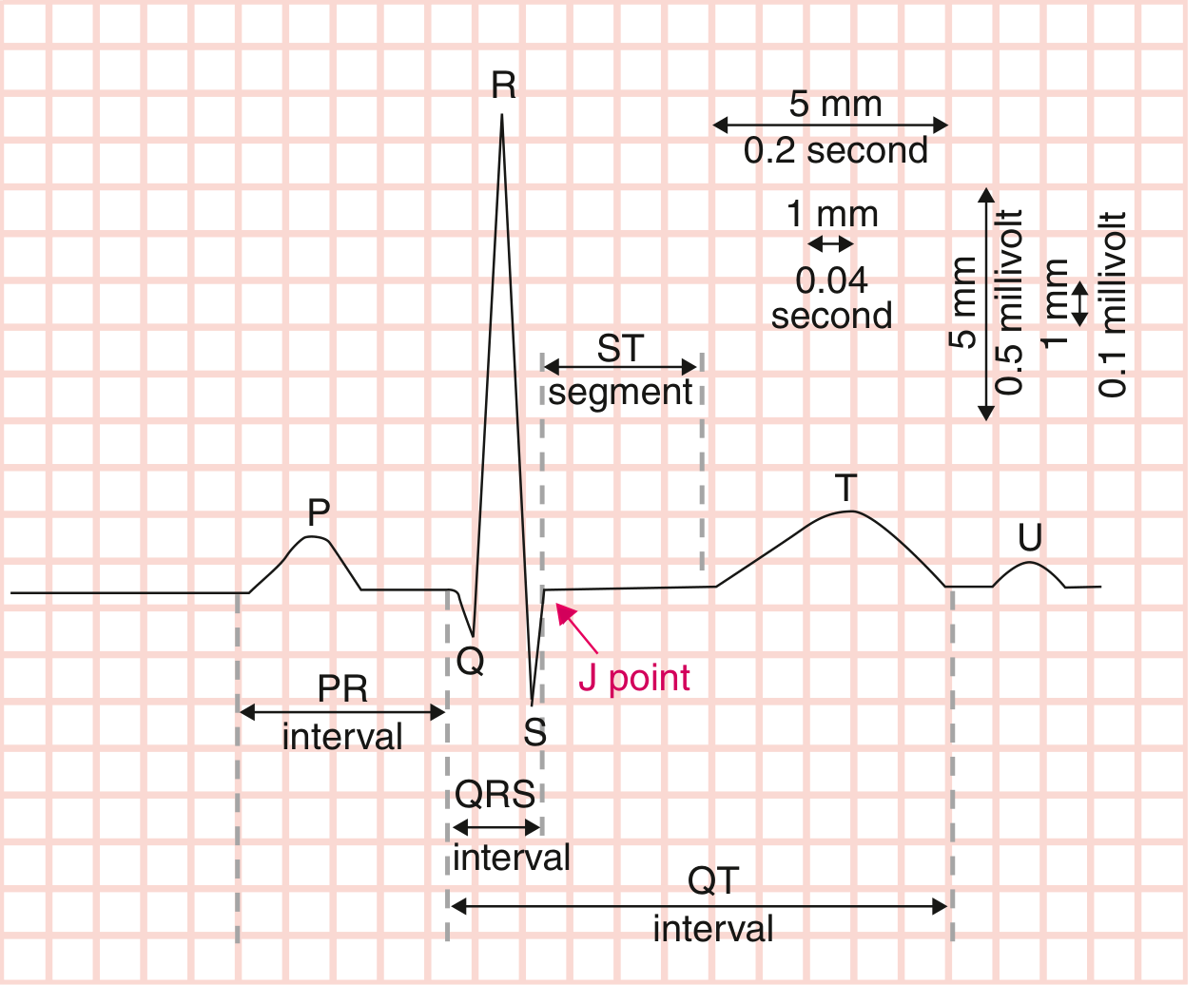
- Horizontal axis = Time: 1 small box = 0.04 sec; 1 large box = 0.2 sec
- Vertical axis = Voltage: 1 small box = 0.1 mV; 10 small boxes = 1 mV
- A standard ECG runs at 25 mm/sec for 10 seconds
The 10-Step OPD Reading Approach
Use this fixed mnemonic: "R-R-A-P-Q-Q-S-T-U-Compare"
Step 1 - Rate
Quick method (OPD-friendly): Count R waves in a 6-second strip (30 large boxes) × 10 = beats/min
Precise method: 300 ÷ (number of large boxes between two R waves)
| Rate | Interpretation |
|---|---|
| 60-100 bpm | Normal |
| > 100 bpm | Tachycardia |
| < 60 bpm | Bradycardia |
Normal range is 50-100 bpm; 60-100 is the classic teaching range.
Step 2 - Rhythm
Ask three questions:
- Is it regular? Are R-R intervals equal?
- Is there a P before every QRS, and a QRS after every P?
- Is the P wave upright in leads I and aVF? - if yes, it is sinus rhythm
Common abnormal patterns in OPD:
- Irregularly irregular + no clear P waves → Atrial fibrillation
- Regularly irregular with dropped beats → AV block
- Narrow complex tachycardia + P waves → SVT
Step 3 - Axis
Quick axis check (OPD method): Look at leads I and II (or I and aVF)
| Lead I | Lead aVF | Axis |
|---|---|---|
| Positive | Positive | Normal (-30° to +90°) |
| Positive | Negative | Left axis deviation |
| Negative | Positive | Right axis deviation |
| Negative | Negative | Extreme/indeterminate |
Causes to remember:
- Left axis deviation: LBBB, LAFB, inferior MI, LVH
- Right axis deviation: RBBB, RVH, pulmonary hypertension, tall thin individuals
Step 4 - P Wave
Normal P wave: upright in I, II, aVF; biphasic in V1; duration < 0.12 sec; amplitude < 2.5 mm
| P wave finding | Suggests |
|---|---|
| Broad, notched ("P mitrale") | Left atrial enlargement |
| Tall, peaked ("P pulmonale") | Right atrial enlargement |
| No P waves | Atrial fibrillation |
| P waves dissociated from QRS | Complete heart block |
Step 5 - PR Interval
Normal: 0.12 to 0.20 sec (3-5 small boxes)
| PR finding | Interpretation |
|---|---|
| > 0.20 sec (>1 large box) | 1st degree AV block |
| Progressively lengthening then dropped QRS | 2nd degree Mobitz I (Wenckebach) |
| Fixed PR with sudden dropped QRS | 2nd degree Mobitz II |
| No relationship between P and QRS | 3rd degree (complete) AV block |
| Short PR + delta wave | WPW syndrome |
Step 6 - QRS Complex
Normal duration: 0.075 to 0.11 sec (< 3 small boxes)
Width:
- Narrow QRS (< 0.12 sec) = supraventricular origin
- Wide QRS (≥ 0.12 sec) = bundle branch block, ventricular rhythm, or WPW
BBB pattern (quick recognition):
- RBBB: rSR' pattern in V1 ("rabbit ears"), wide S in V5-V6 and I
- LBBB: Wide, notched R in V5-V6, I, aVL; QS or rS in V1 (no normal R wave in V1)
Pathological Q waves:
- Q wave > 1 small box wide (0.04 sec) OR > 25% of R wave height = pathological
- Suggests old/previous myocardial infarction
Step 7 - QT Interval
Measure from start of QRS to end of T wave. Use corrected QTc (Bazett's formula: QTc = QT / √RR).
| QTc | Finding |
|---|---|
| Males < 450 msec | Normal |
| Females < 460 msec | Normal |
| > 500 msec | High risk of Torsades de Pointes |
OPD pearl: QTc prolongation is seen with hypokalemia, hypomagnesemia, certain drugs (haloperidol, azithromycin, certain antihistamines), and congenital syndromes.
Step 8 - ST Segment
This is the most clinically urgent part. Check all 12 leads.
ST Elevation (≥ 1 mm in limb leads, ≥ 2 mm in precordial):
| Leads with ST elevation | Territory |
|---|---|
| V1-V4 | Anterior (LAD) |
| II, III, aVF | Inferior (RCA) |
| I, aVL, V5-V6 | Lateral (LCx) |
| V1-V2 + right-sided leads (V4R) | Right ventricle |
Other causes of ST elevation (not STEMI):
- Saddle-shaped ST elevation in multiple leads → Pericarditis
- Concave ST elevation in young patients → Early repolarization (benign variant)
- LBBB (discordant ST changes)
ST Depression:
- Subendocardial ischemia, digoxin effect ("reverse tick"), posterior MI equivalent (leads V1-V3)
Step 9 - T Wave and U Wave
T wave:
- Normally upright in I, II, V3-V6; inverted in aVR (normal)
- Inverted T waves suggest ischemia, PE (V1-V4), RVH, or cardiomyopathy
- Tall, peaked/hyperacute T waves = earliest sign of STEMI or hyperkalemia
- Biphasic T waves in V2-V3 = Wellens' syndrome (critical LAD stenosis - do NOT stress test)
U wave:
- Small positive deflection after T wave - best seen in V2-V3
- Prominent U wave = hypokalemia or certain drugs
Step 10 - Compare with Previous ECG
Always compare. A "new" LBBB, new Q wave, or new ST change carries far more weight than a chronic finding. The Harrison's ECG chapter stresses that comparison with prior ECGs is "invaluable."
OPD Quick Reference Card
RATE → RHYTHM → AXIS → P wave → PR → QRS → QT → ST → T/U → COMPARE
Normal values at a glance:
| Parameter | Normal |
|---|---|
| Heart rate | 60-100 bpm |
| P wave duration | < 0.12 sec |
| PR interval | 0.12-0.20 sec |
| QRS duration | < 0.12 sec |
| QTc | < 450 ms (M), < 460 ms (F) |
| Axis | -30° to +90° |
Common OPD Diagnoses at a Glance
| Condition | ECG Pattern |
|---|---|
| Normal sinus rhythm | Regular, P before every QRS, rate 60-100 |
| Atrial fibrillation | Irregularly irregular, no P waves, narrow QRS |
| STEMI | ST elevation in contiguous leads + reciprocal depression |
| NSTEMI/UA | ST depression ± T inversion, no ST elevation |
| First-degree AV block | PR > 0.20 sec, all beats conducted |
| Complete heart block | P and QRS completely dissociated |
| RBBB | rSR' in V1, wide S in I and V6 |
| LBBB | Wide R in I, aVL, V5-V6; QS in V1 |
| LVH | Sokolow-Lyon: S in V1 + R in V5 or V6 > 35 mm |
| WPW | Short PR + delta wave + wide QRS |
| Pericarditis | Saddle-shaped ST elevation, PR depression |
| Hyperkalemia | Tall peaked T → wide QRS → sine wave |
| Hypokalemia | Flat T, prominent U wave, prolonged QT |
A great free resource to practice is the Harvard ECG Wave-Maven self-assessment program at ecg.bidmc.harvard.edu.
For a comprehensive video walkthrough, this Ninja Nerd lecture is excellent:
Sources: Harrison's Principles of Internal Medicine 22E (2025); Goldman-Cecil Medicine, International Edition
This is a shared conversation. Sign in to Orris to start your own chat.