
Role of ultrasound in airway management
Reading File
Now let me search the medical library for content on ultrasound in airway management, running parallel searches:
Finding Sources
Finding Sources
Searching PubMed
"ultrasound" AND "airway management"
Good. Let me now read the most relevant sections in parallel:
Reading File
Reading File
Reading Article
PMID: 40891437
Now let me get more detailed content from Morgan & Mikhail and search for additional specific sections:
Reading File
Finding Sources
Finding Sources
Excellent content. Now let me get a few more sections:
Reading File
Reading File
Finding Sources
Reading File
Finding Sources
Finding Sources
Finding Sources
Now let me fetch the key ultrasound images and get the Barash section on airway anatomy/ultrasound:
Loading Image
Loading Image
Loading Image
Loading Image
Excellent - I have comprehensive content from multiple authoritative sources. Now I can compose a thorough, well-illustrated answer.
Role of Ultrasound in Airway Management
Point-of-care ultrasound (POCUS) has become an important adjunct across all phases of airway management - from pre-intubation assessment through post-intubation confirmation and procedural guidance. The applications span the entire sequence of events: predict, prepare, perform, and confirm.
1. Pre-intubation Airway Assessment
Prediction of Difficult Airway
Conventional bedside tests (Mallampati, thyromental distance, mouth opening) have limited sensitivity and specificity for predicting a difficult airway. Ultrasound provides a quantitative, operator-independent adjunct.
A 2025 systematic review and meta-analysis in Anaesthesia (Soni et al., PMID 40891437) - the most comprehensive evidence synthesis to date, covering 60 studies and 10,580 patients evaluating 58 POCUS parameters - found the following:
| Parameter | Sensitivity | Specificity | AUROC | Evidence Quality |
|---|---|---|---|---|
| Skin-to-vocal-cords distance (difficult laryngoscopy) | 0.84 | 0.81 | 0.87 | High |
| Skin-to-epiglottis distance (difficult intubation) | 0.80 | 0.86 | - | High |
| Skin-to-hyoid distance (difficult intubation) | 0.78 | 0.81 | 0.86 | Moderate |
| Tongue thickness (difficult mask ventilation) | Qualitative association | - | - | Narrative |
The key insight is that deeper tissue distances from skin to these structures (vocal cords, epiglottis, hyoid) predict difficulty because they reflect increased soft tissue bulk - seen in obesity, edema, or redundant pharyngeal tissue.
Assessment in Obesity
Morbidly obese patients (BMI ≥30 kg/m²) may have increased neck circumference and redundant pharyngeal tissue, making both intubation and bag-mask ventilation difficult. Ultrasound allows direct measurement of pre-tracheal soft tissue depth and tongue thickness, which correlates with intubation difficulty better than external anthropometrics alone. - Morgan and Mikhail's Clinical Anesthesiology, 7e
2. Identifying the Cricothyroid Membrane (CTM)
The CTM is the primary "front-of-neck access" (FONA) landmark for emergency surgical airways. It is typically 8-9 mm in height, located between the inferior edge of the thyroid cartilage superiorly and the superior surface of the cricoid cartilage inferiorly.
The problem with palpation: Palpation fails to correctly identify the CTM in up to 67% of cases, particularly in females, obese patients, and those with neck pathology (prior surgery, tumor, hematoma, radiation).
SMART mnemonic for anticipated difficult cricothyrotomy:
S - Surgery (prior)
M - Mass (abscess, hematoma)
A - Access/anatomy problems (obesity, edema)
R - Radiation
T - Tumor
(Rosen's Emergency Medicine)
In any patient with these features, ultrasound should be used proactively to mark the CTM on the skin surface before airway manipulation begins. A high-frequency linear probe placed transversely on the anterior neck identifies the thyroid cartilage, cricothyroid membrane (as a hypoechoic gap), and cricoid cartilage in sequence.
The 2025 meta-analysis found POCUS was significantly better than palpation for CTM identification: OR 3.61 (95% CI 2.20-5.92), moderate-high certainty evidence. - Soni et al., PMID 40891437
3. Confirming Endotracheal Tube Placement
This is arguably the most immediately impactful role of POCUS - rapid confirmation that the tube is in the trachea, not the esophagus.
Tracheal Anatomy on Ultrasound
With a linear probe placed transversely on the anterior neck, the trachea appears as a curved hyperechoic structure with posterior shadowing from the air-cartilage interface. The thyroid gland sits lateral to it.
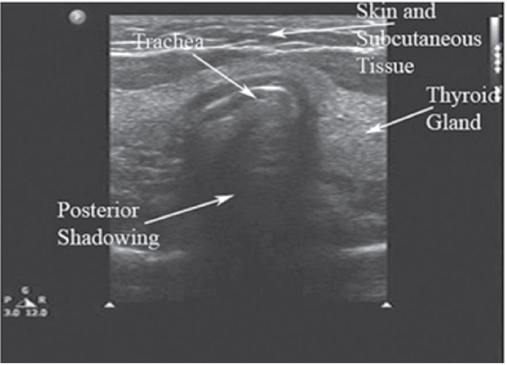
Dynamic Confirmation During Intubation
The probe is placed transversely at the suprasternal notch during laryngoscopy. As the ETT passes through the vocal cords into the trachea, increased echogenicity (the "snowstorm" artifact) appears within the tracheal lumen in real time.
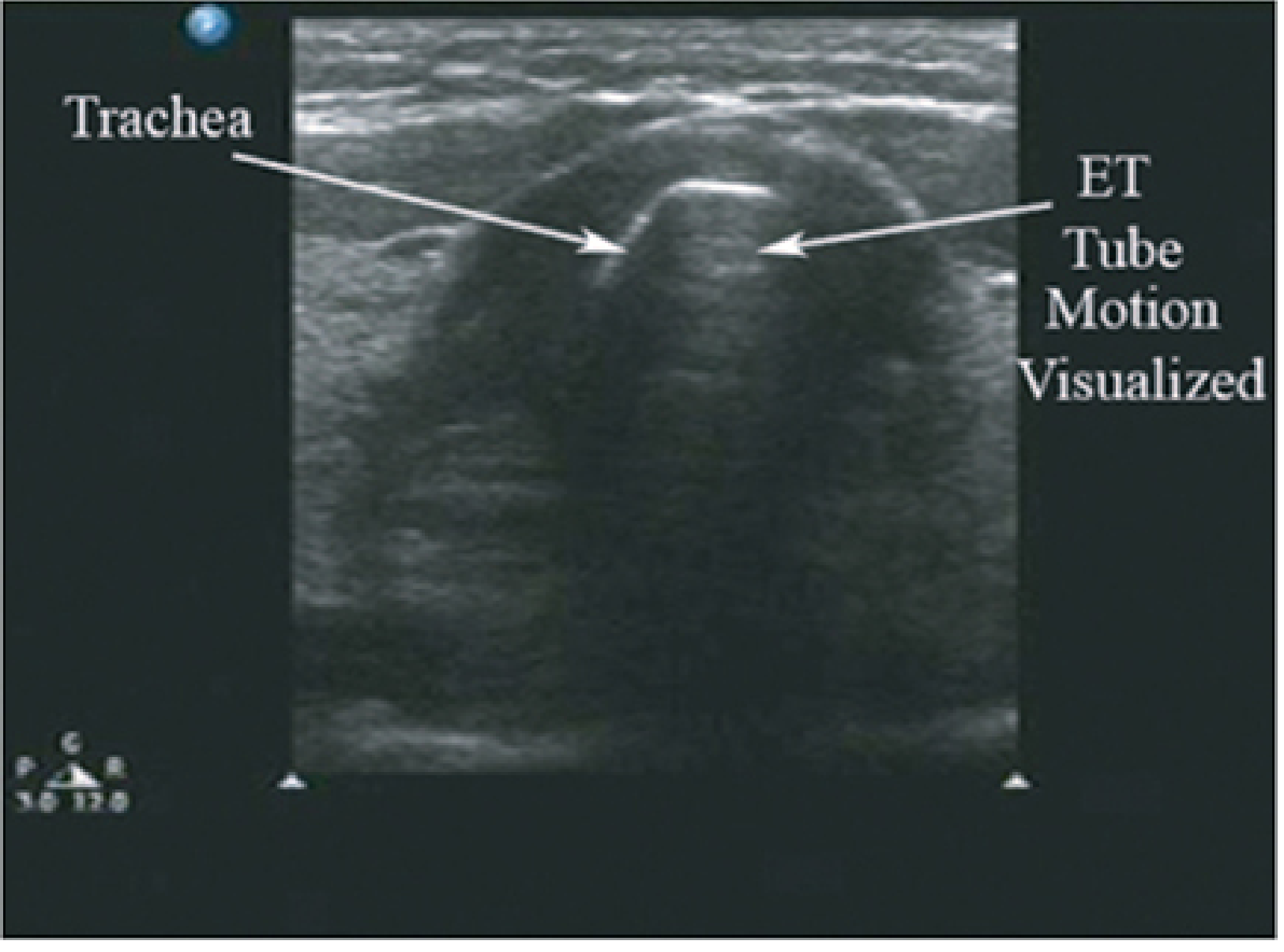
Detecting Esophageal Intubation
When the ETT is placed in the esophagus, the esophagus (posterior and lateral to the trachea) expands and two parallel echogenic lines are seen within it - the inner and outer walls of the tube.
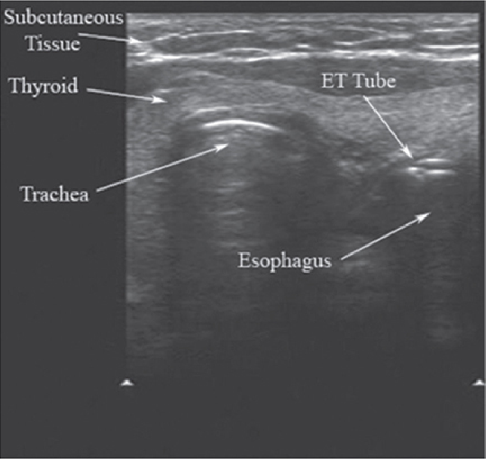
Side-by-side Tintinalli comparison:
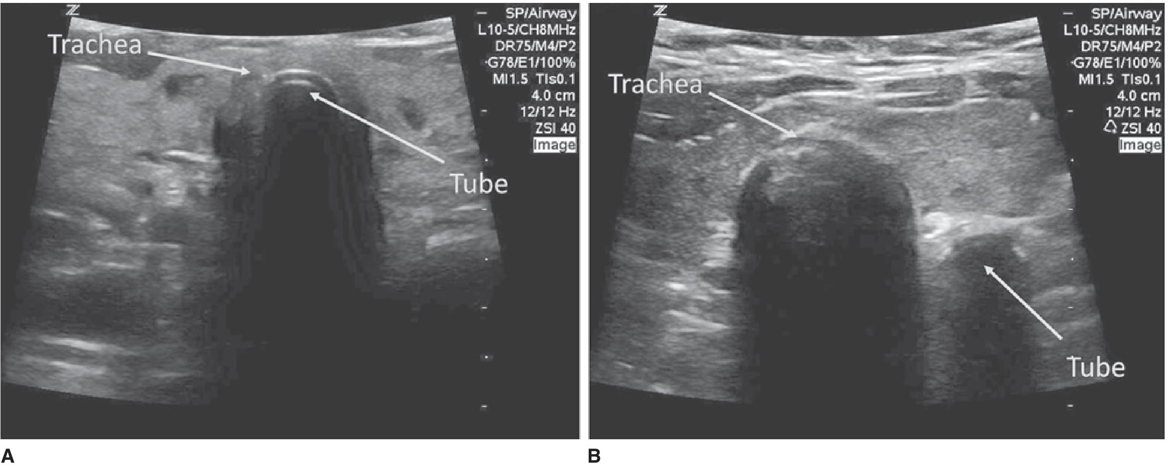
Key advantage over capnography: POCUS is immediately available even during cardiac arrest (where ETCO2 may be unreliable due to low cardiac output) and does not require equipment at the head of the bed. - Miller's Anesthesia, 10e
Confirming Bilateral Lung Ventilation
Lung sliding (the "sliding sign" of visceral-parietal pleura moving together) can be assessed bilaterally to confirm bilateral ETT placement and rule out inadvertent mainstem intubation or tension pneumothorax. Absence of lung sliding on one side after intubation should prompt tube adjustment.
4. Percutaneous Tracheostomy Guidance
POCUS significantly improves the safety of bedside percutaneous dilatational tracheostomy (PDT):
- Pre-procedure mapping: Identify tracheal rings, midline, and ensure no midline vessels (thyroid arteries, aberrant veins) overlie the insertion site
- ETT size selection: Measure internal tracheal diameter to guide cuffed tube size
- Real-time guidance: Visualize needle entry between tracheal rings (usually between rings 2-3) during the Seldinger technique
- Avoid thyroid isthmus: Locate and avoid the isthmus to prevent hemorrhage
The 2025 meta-analysis found POCUS use was associated with higher first-pass success in PDT: OR 3.9 (2.1-7.1), low-moderate certainty evidence. - Soni et al., PMID 40891437
Miller's Anesthesia further notes: "POCUS can help determine size of trachea and endotracheal tube, accurately locate cricothyroid membrane, and improves success of procedures like cricothyrotomy, tracheal rings for percutaneous tracheostomy." - Miller's Anesthesia, 10e
5. Subglottic ETT Cuff Position
The ETT cuff can be inflated with saline and visualized ultrasonographically as it fills in the subglottic/suprastomal area. This allows confirmation that:
- The cuff lies below the vocal cords (not in the subglottis where it would cause mucosal ischemia)
- The tube tip is not in a mainstem bronchus
6. Lung Ultrasound After Intubation
Post-intubation lung assessment is a critical extension of airway POCUS:
| Finding | Significance |
|---|---|
| Bilateral lung sliding | Correct ETT position, both lungs ventilating |
| Absent lung sliding (unilateral) | Mainstem intubation or pneumothorax |
| B-lines | Pulmonary edema, aspiration, ARDS |
| Consolidation with dynamic air bronchograms | Pneumonia (guides antibiotic initiation) |
| Pleural effusion | Identify before repositioning/procedures |
7. Probe Selection and Technique Summary
| Application | Probe | Orientation |
|---|---|---|
| CTM identification, tracheal anatomy | High-frequency linear (10-15 MHz) | Transverse, then longitudinal on anterior neck |
| ETT confirmation | High-frequency linear | Transverse at suprasternal notch |
| Lung sliding / pleural assessment | Phased array or linear | Intercostal spaces, bilateral |
| Percutaneous tracheostomy | Linear | Transverse anterior neck |
| Gastric content / aspiration risk | Curvilinear | Sagittal/coronal, subxiphoid for antrum |
Key Takeaways
- Pre-intubation: Skin-to-epiglottis and skin-to-vocal-cord distances are the best POCUS predictors of difficult intubation and laryngoscopy, respectively (high-certainty evidence).
- CTM marking: POCUS outperforms palpation (OR 3.6) - should be used proactively whenever FONA is anticipated to be difficult.
- ETT confirmation: Real-time tracheal tube visualization distinguishes tracheal from esophageal intubation quickly, even when capnography is unreliable.
- Percutaneous tracheostomy: POCUS guidance improves first-pass success (OR ~4) and reduces vascular injury.
- Post-intubation: Bilateral lung sliding assessment detects mainstem intubation and pneumothorax at the bedside without X-ray delay.
Recent evidence note: The 2025 meta-analysis by Soni et al. (Anaesthesia, PMID 40891437) is now the largest pooled analysis on upper airway POCUS, reinforcing high-level evidence for several parameters. A 2025 pediatric-specific meta-analysis (PMID 41430150) also confirmed high diagnostic accuracy of POCUS for ETT confirmation in pediatric acute care.
This is a shared conversation. Sign in to Orris to start your own chat.