
Essay from General Medicine Topic: Nephrotic Syndrome
Reading File
Now let me search the medical library for comprehensive content on Nephrotic Syndrome.
Finding Sources
Finding Sources
Excellent. Let me now read the most comprehensive sections - the Frameworks for Internal Medicine dedicated section and the NKF Primer sections, plus the Kidney textbook's dedicated section.
Reading File
Reading File
Reading File
Excellent data coming in. Let me continue reading the Frameworks section (for causes and management) and NKF Primer for the specific diseases (MCD, FSGS, MN, etc.), plus search for complications and treatment.
Reading File
Reading File
Finding Sources
Reading File
Reading File
Reading File
Reading File
Reading File
I now have rich, comprehensive content. Let me fetch a couple of key images and get Goldman-Cecil's FSGS/MN sections before writing the essay.
Reading File
Loading Image
I now have comprehensive data from multiple authoritative textbooks. Let me write the full essay.
Nephrotic Syndrome
Definition
Nephrotic syndrome is a clinical syndrome defined by the tetrad of:
- Proteinuria >3.5 g/24 hours (nephrotic-range)
- Hypoalbuminemia - serum albumin <3.5 g/dL (some sources use <2.5 g/dL as the threshold for clinically significant disease)
- Hyperlipidemia - total cholesterol usually >180 mg/dL
- Edema - often anasarca
While edema and hyperlipidemia are the most common clinical characteristics, they are not strictly required for the biochemical diagnosis. - National Kidneys Foundation Primer on Kidney Diseases, 8e, p.189
Pathophysiology
Glomerular Filtration Barrier
The glomerular filtration barrier normally maintains size and charge selectivity through a combination of:
- The glomerular basement membrane (GBM)
- Endothelial cells (fenestrated, covered by a glycocalyx)
- Epithelial cells (podocytes) - the foot processes with their slit diaphragms are the final barrier
Nephrotic syndrome occurs when there is impairment of this charge and size selectivity. The resultant increase in glomerular permeability allows large molecules such as albumin (MW ~69,000 Da) to escape into the urine. - Frameworks for Internal Medicine, p.464
Why Edema Develops - The Underfill vs. Overfill Debate
Two mechanisms explain sodium and water retention in nephrotic syndrome:
1. Underfill hypothesis:
Urinary albumin loss + reduced hepatic synthesis → hypoalbuminemia → fall in plasma oncotic pressure → increased flux of fluid into interstitial spaces → reduced effective circulating volume → activation of RAAS → renal sodium retention.
2. Overfill hypothesis:
A primary intrinsic renal defect causes sodium retention independent of hypoalbuminemia, leading to volume expansion.
In practice, both mechanisms coexist to varying degrees. Patients with minimal change disease tend to have contracted plasma volume and stimulated RAAS (underfill), while those with other causes often have expanded plasma volume and suppressed RAAS (overfill). - Brenner and Rector's The Kidney, p.2277
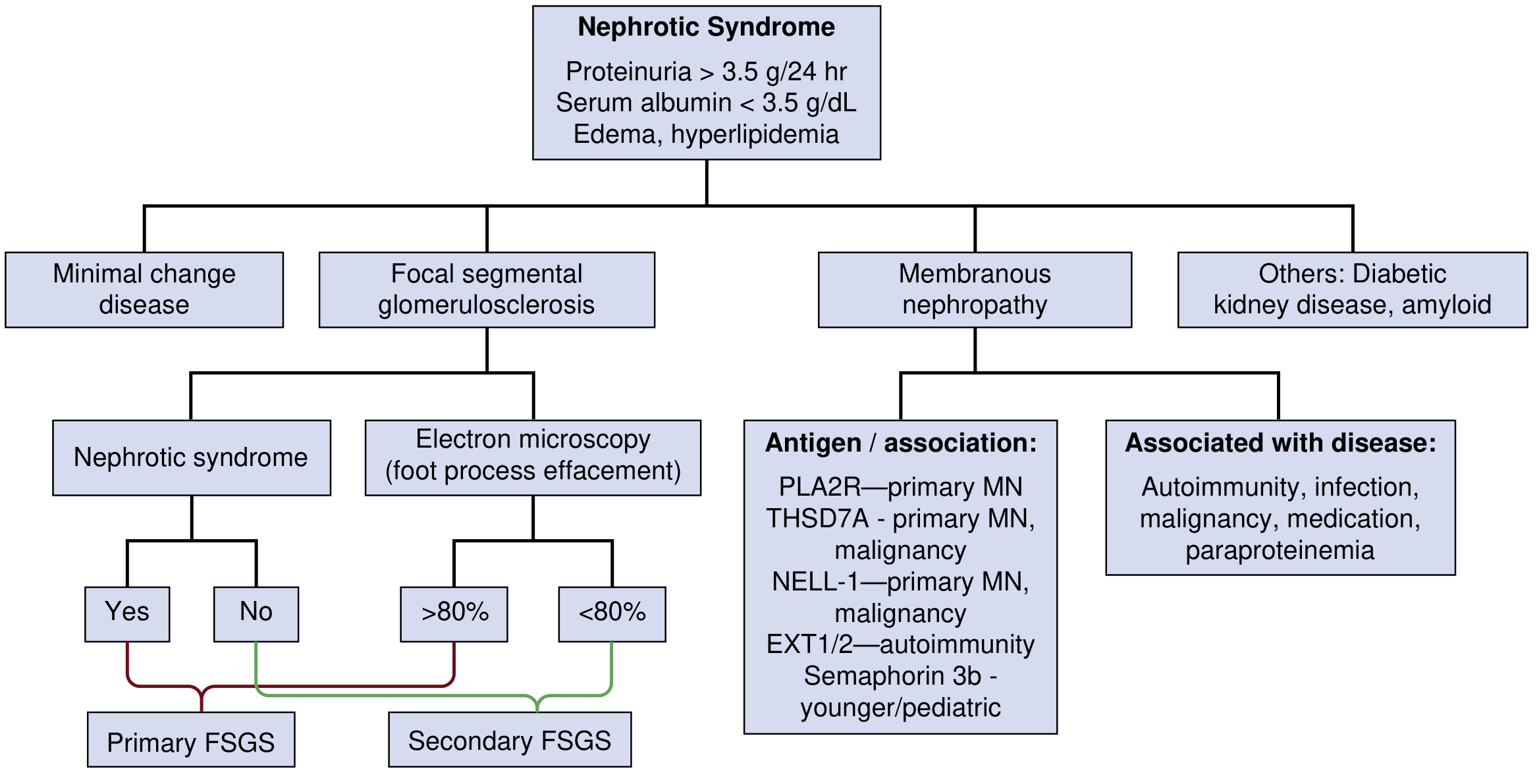
Etiology and Classification
Causes are divided into primary (idiopathic) - arising from the kidney without identifiable systemic disease - and secondary - associated with systemic conditions or exposures.
Primary (Idiopathic) Causes
1. Minimal Change Disease (MCD)
- Epidemiology: Most common cause in children (70-90%); accounts for only ~15% of adult nephrotic syndrome
- Pathogenesis: Unknown, but T-cell-mediated dysfunction is suspected. Recent discovery of autoantibodies targeting the podocyte protein nephrin points to an autoimmune etiology in at least some cases. Associated with NSAIDs, lithium, thymoma, and Hodgkin lymphoma.
- Histology:
- Light microscopy: No glomerular abnormalities ("minimal change"); tubules may show lipid droplet accumulation (hence the older term "lipoid nephrosis")
- Immunofluorescence: No immune deposits
- Electron microscopy: Diffuse, extensive foot process effacement across all capillary loops; no electron-dense deposits
- Treatment: Systemic corticosteroids (prednisone 1 mg/kg/day, max 80 mg, or alternate-day 2 mg/kg/day) with remission in >80% of adults. Relapses occur in ~40% by 1 year. Steroid-resistant or frequently relapsing cases: rituximab (1000 mg IV × 2), cyclosporine, tacrolimus, mycophenolate mofetil, or cyclophosphamide.
- Prognosis: Excellent. Most patients have relapses but maintain long-term kidney function. Progressive decline in kidney function should prompt re-biopsy to rule out FSGS. - Goldman-Cecil Medicine, p.1252
2. Focal Segmental Glomerulosclerosis (FSGS)
- Epidemiology: ~25% of adult nephrotic syndrome; most common cause of idiopathic nephrotic syndrome in Black Americans (APOL1 risk alleles increase susceptibility 10-20-fold)
- Classification: Primary (circulating podocyte permeability factor), Secondary (adaptive, drug/viral, maladaptive), Genetic (mutations in podocyte proteins: NPHS1/nephrin, NPHS2/podocin, ACTN4/α-actinin-4, TRPC6, INF2, collagen 4, APOL1)
- Secondary causes: HIV-associated nephropathy, parvovirus B19, COVID-19 (collapsing variant), heroin, pamidronate, sirolimus, obesity-related glomerulopathy, reduced nephron mass
- Histology:
- Light microscopy: Focal (not all glomeruli) and segmental (not entire glomerulus) sclerosis. Five variants: cellular, tip, perihilar, collapsing, NOS
- Electron microscopy: Varying degrees of foot process effacement - >80% diffuse effacement favors primary FSGS; <80% segmental effacement favors secondary FSGS
- Collapsing FSGS: Strongly associated with viral etiologies (HIV, SARS-CoV-2); presents with abrupt, severe nephrotic syndrome and rapid progression to ESKD
- Treatment: Primary FSGS - corticosteroids, calcineurin inhibitors, mycophenolate mofetil. Secondary FSGS - treat underlying cause; avoid unnecessary immunosuppression.
- Prognosis: Non-nephrotic-range proteinuria confers best outcomes. Persistent nephrotic syndrome or steroid resistance predicts progression to end-stage kidney disease (ESKD). - NKF Primer on Kidney Diseases, 8e, p.190-191
3. Membranous Nephropathy (MN)
- Epidemiology: Most common cause of adult nephrotic syndrome in whites; peak incidence in the 4th-5th decade; male:female ratio 2:1
- Pathogenesis: Autoantibody-mediated. Anti-phospholipase A2 receptor (PLA2R) antibodies are present in ~70% of primary MN. Other antigens include THSD7A (1-3%), NELL-1, EXT1/2, and Semaphorin 3B (Sema3B)
- Secondary causes: Autoimmunity (SLE), infections (hepatitis B), drugs (NSAIDs, penicillamine, gold), malignancies (typically solid tumors - colon, lung, breast cancer), paraproteinemias. EXT1/2-associated MN is linked to autoimmune disease.
- Histology:
- Light microscopy: Thickened GBM with subepithelial "spikes" on methenamine silver stain (due to GBM growing around deposits)
- Immunofluorescence: Granular deposition of IgG and C3 along capillary walls
- Electron microscopy: Subepithelial and intramembranous electron-dense immune deposits; foot process effacement
- Treatment: One-third of patients undergo spontaneous remission, one-third partial remission. Immunosuppression (cyclophosphamide + steroids, rituximab, or calcineurin inhibitors) is indicated for persistent nephrotic syndrome or declining kidney function. PLA2R antibody titers guide response to therapy.
- Prognosis: The "rule of thirds" - 1/3 spontaneous remission, 1/3 persistent proteinuria, 1/3 progressive renal failure. Membranous nephropathy carries the highest thromboembolism risk among nephrotic causes. - NKF Primer on Kidney Diseases, 8e, p.192
Secondary Causes
| Etiology | Features / Associations |
|---|---|
| Diabetic Kidney Disease (DKD) | Most common cause of ESKD in the US; Kimmelstiel-Wilson nodular glomerulosclerosis on biopsy; associated with retinopathy |
| Amyloidosis | Congo red staining with apple-green birefringence under polarized light |
| Systemic Lupus Erythematosus | Can cause both nephrotic (membranous class V) and nephritic patterns; "full house" immunofluorescence |
| Preeclampsia | Nephrotic-range proteinuria in pregnancy after 20 weeks with hypertension |
| Infections | Hepatitis B/C (MN or MPGN pattern), HIV (collapsing FSGS), endocarditis |
| Drugs | Gold, penicillamine (in RA) → MN pattern; NSAIDs → MCD pattern |
| Malignancy | Hodgkin lymphoma → MCD; solid tumors → MN |
| Amyloidosis | AL (plasma cell dyscrasia) or AA (chronic inflammation) |
Clinical Features
Symptoms
- Peripheral edema (often anasarca - generalized pitting edema), particularly periorbital on waking
- Fatigue
- Dyspnea (from pleural effusions or ascites)
- Foamy urine (due to proteinuria)
Physical Findings
- Hypertension
- Generalized dependent pitting edema
- Ascites
- Pleural effusions
- Muehrcke's lines: Paired white bands on fingernails due to hypoalbuminemia (transverse leukonychia)
- Eruptive xanthomata and xanthelasma (from hyperlipidemia)
-
- Frameworks for Internal Medicine, p.464
Laboratory Investigations
Urine
- Proteinuria >3.5 g/24 hours (gold standard)
- Spot urine protein:creatinine ratio: A single untimed sample correlates with 24-hour protein excretion (value in g/g approximates g/day); practical and validated alternative
- Lipiduria: Oval fat bodies (tubular cells laden with lipid), free fat droplets, fatty casts with "Maltese cross" pattern under polarized light
- Microscopic hematuria may be present (especially in FSGS, MCD in adults)
Blood
- Hypoalbuminemia (<3.5 g/dL)
- Hyperlipidemia - elevated total cholesterol, LDL, VLDL, triglycerides (due to increased hepatic lipoprotein synthesis in response to low oncotic pressure)
- Serum creatinine: variable; may reflect intravascular volume depletion or true GFR reduction
- Complement levels (C3, C4): normal in primary nephrotic syndrome; reduced in SLE, MPGN, infection-related GN
Additional Work-Up
- ANA, anti-dsDNA (SLE)
- Anti-PLA2R antibody (membranous nephropathy - positive in ~70% of primary MN)
- Hepatitis B, C serologies
- HIV test
- Serum and urine protein electrophoresis (paraproteinemia/amyloidosis)
- Blood glucose/HbA1c (diabetic nephropathy)
- Renal biopsy - required for definitive diagnosis in adults (with exception of diabetic nephropathy with typical features and in children where MCD can be assumed and empirically treated)
Complications
1. Thromboembolism
The mechanism is incompletely understood but relates to an imbalance of procoagulant and anticoagulant factors:
- Loss in urine: Antithrombin III, proteins C and S, plasminogen (anticoagulant proteins)
- Increased hepatic production: Fibrinogen, factor V, factor VIII (procoagulant proteins)
The most common thrombotic sites are the renal veins and veins of the lower extremities. Renal vein thrombosis is particularly associated with membranous nephropathy. Risk is highest when proteinuria >10 g/24h and albumin <2 g/dL. Membranous nephropathy and amyloidosis carry the highest thrombotic risk. - NKF Primer on Kidney Diseases, 8e, p.189
2. Infection
- Loss of immunoglobulins (IgG) and opsonins in the urine → hypogammaglobulinemia → predisposition to bacterial infections (e.g., cellulitis, spontaneous bacterial peritonitis, pneumococcal infections)
- Loss of complement factor B may impair the alternate pathway
- Patients are particularly susceptible to encapsulated organisms (Streptococcus pneumoniae)
3. Acute Kidney Injury (AKI)
- Reduced effective circulating volume from hypoalbuminemia → prerenal AKI
- Interstitial edema compressing tubules
- Drug toxicity (nephrotoxic medications, diuretic-induced hypovolemia)
4. Hyperlipidemia
- Driven by increased hepatic synthesis of lipoproteins as a compensatory response to oncotic pressure loss
- Increased cardiovascular risk with prolonged nephrotic syndrome
- Urinary loss of HDL components accelerates atherogenesis
5. Nutritional Deficiencies
- Iron deficiency anemia: loss of transferrin in the urine
- Vitamin D deficiency: loss of vitamin D-binding protein → reduced 25-OH vitamin D → secondary hyperparathyroidism and bone disease
- Thyroid hormone loss: loss of thyroxine-binding globulin (may result in low total T4, but free T4 and TSH are usually normal)
Management
Non-Specific / Supportive Measures
1. Diet
- Low-salt diet to manage edema
- Protein-restricted diet (0.8-1 g/kg/day) - reduces proteinuria load but must balance against ongoing protein losses
2. Edema management
- Loop diuretics (furosemide) are first-line; resistance is common in nephrotic syndrome because:
- Furosemide is heavily protein-bound and its tubular secretion depends on albumin
- Hypoalbuminemia reduces delivery of furosemide to its tubular site of action
- A rational approach is to combine loop diuretics with ACE inhibitor/ARB to limit albuminuria
- Combination of IV albumin with furosemide may help in severe diuretic-resistant cases (though evidence is limited)
3. Proteinuria reduction
- ACE inhibitors or ARBs: Cornerstone of non-disease-specific management; reduce intraglomerular pressure, decrease proteinuria, and have additional benefits on coagulability, dyslipidemia, and progression of renal disease
4. Hyperlipidemia
- Statin therapy is recommended; reduces cardiovascular risk
5. Thromboembolism prophylaxis
- Anticoagulation (warfarin) for established thrombosis
- Prophylactic anticoagulation is recommended in membranous nephropathy with albumin <2.8 g/dL and low bleeding risk
- Data on direct oral anticoagulants (DOACs) in this setting are still insufficient - Goldman-Cecil Medicine, p.1252
6. Infection prevention
- Pneumococcal vaccination
- Consider prophylactic penicillin in children with severe nephrotic syndrome
Disease-Specific Therapy
| Disease | First-Line | Alternatives |
|---|---|---|
| Minimal Change Disease | Prednisone 1 mg/kg/day (max 80 mg) × 5-6 months | Rituximab, cyclosporine, tacrolimus, mycophenolate, cyclophosphamide |
| Primary FSGS | Corticosteroids | Calcineurin inhibitors (CNI), mycophenolate, rituximab |
| Membranous Nephropathy | Watchful waiting (30% spontaneous remission) | Rituximab, cyclophosphamide + steroids, CNIs |
| Diabetic Nephropathy | Glycemic control, ACE inhibitor/ARB, SGLT2 inhibitors | - |
| MCD (steroid-resistant/dependent) | Rituximab | Cyclosporine, tacrolimus, mycophenolate |
Nephrotic vs. Nephritic Syndrome (Key Distinction)
| Feature | Nephrotic | Nephritic |
|---|---|---|
| Proteinuria | >3.5 g/day (massive) | <3.5 g/day (mild-moderate) |
| Hematuria | Absent (or mild) | Present (RBC casts - hallmark) |
| Edema | Prominent | Mild |
| Hypertension | Variable | Common |
| Oliguria | Absent | Common |
| Serum albumin | Markedly reduced | Normal/mildly reduced |
| Complement | Normal (primary) | Often reduced (PSGN, SLE) |
Note: Some entities can overlap - IgA nephropathy and MPGN can present with features of both syndromes. SLE can cause both patterns. - Frameworks for Internal Medicine, p.465
Prognosis
Prognosis depends heavily on the underlying etiology:
- MCD: Excellent; steroid-responsive in most cases; kidney function preserved long-term even with relapses
- FSGS: Variable; primary FSGS recurs post-transplant (supporting circulating factor hypothesis); secondary FSGS progresses slowly if underlying cause is addressed; collapsing FSGS has poor prognosis
- Membranous nephropathy: Rule of thirds (spontaneous remission, partial remission, progressive disease)
- Diabetic nephropathy: Progressive in absence of metabolic control; leading cause of ESKD
Summary
Nephrotic syndrome represents a spectrum of glomerular diseases unified by the clinical signature of massive proteinuria, hypoalbuminemia, edema, and hyperlipidemia. The underlying pathophysiology involves disruption of the glomerular filtration barrier - primarily at the level of the podocyte - allowing unrestricted albumin passage into the urine. Its causes range from primary glomerular diseases (MCD, FSGS, membranous nephropathy) to systemic conditions (diabetes, SLE, amyloidosis). Management requires both addressing the specific underlying etiology with immunosuppression or disease-targeted therapy and mitigating life-threatening complications including thromboembolism, infection, and progressive CKD. The prognosis varies widely: MCD carries an excellent outlook, while FSGS and membranous nephropathy may progress to ESKD without appropriate treatment.
References:
- National Kidneys Foundation Primer on Kidney Diseases, 8e - Chapters 16, 18, 19, 20
- Goldman-Cecil Medicine International Edition - Chapter 107
- Frameworks for Internal Medicine - Chapter 34
- Brenner and Rector's The Kidney, 2-Volume Set - Chapter 50
This is a shared conversation. Sign in to Orris to start your own chat.