
Best ppi drugs
Reading File
Proton pump inhibitors (PPIs) are among the most widely used drugs in gastroenterology. They block the H+/K+-ATPase enzyme in gastric parietal cells, reducing acid secretion. Common PPIs include omeprazole, pantoprazole, esomeprazole, lansoprazole, and rabeprazole. Esomeprazole and rabeprazole are often considered the most potent. Now let me pull detailed textbook information.
Searching sources...
Finding Sources
Searching PubMed
"proton pump inhibitors" comparison efficacy
Reading File
Finding Sources
Reading File
Reading File
Reading File
Reading File
Reading File
I now have comprehensive information to give a detailed, authoritative answer.
Loading Image
Here is a complete, textbook-based overview of PPI drugs.
Proton Pump Inhibitors (PPIs)
What They Are
PPIs are substituted benzimidazole prodrugs that irreversibly block the H+/K+-ATPase (proton pump) in gastric parietal cells. Because they block the final common pathway of acid secretion, they suppress both fasting and meal-stimulated acid, regardless of the stimulus (histamine, gastrin, acetylcholine). In standard doses, PPIs inhibit 90-98% of 24-hour acid secretion - far more than H2-blockers.
Molecular structures of the four main agents:
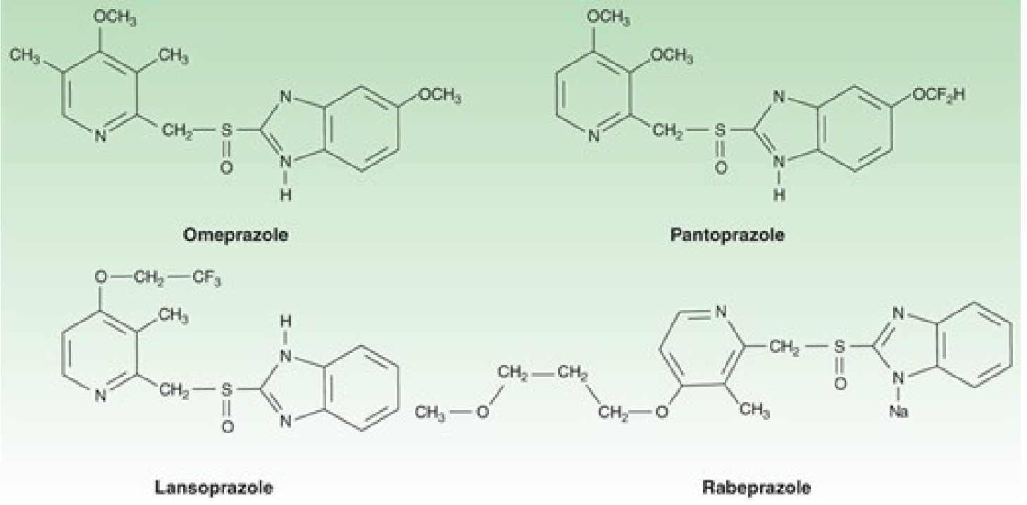
The 6 Available PPIs
| Drug | Brand Name | Bioavailability | Half-life (h) | Standard Dose |
|---|---|---|---|---|
| Omeprazole | Prilosec | 40-65% | 0.5-1.0 | 20-40 mg once daily |
| Esomeprazole | Nexium | >80% | 1.5 | 20-40 mg once daily |
| Lansoprazole | Prevacid | >80% | 1.0-2.0 | 30 mg once daily |
| Dexlansoprazole | Dexilant | NA | 1.0-2.0 | 30-60 mg once daily |
| Pantoprazole | Protonix | 77% | 1.0-1.9 | 40 mg once daily |
| Rabeprazole | Aciphex | 52% | 1.0-2.0 | 20 mg once daily |
- Esomeprazole is the S-isomer of omeprazole (same molecule, improved bioavailability)
- Dexlansoprazole is the R-enantiomer of lansoprazole
- IV formulations exist for esomeprazole, pantoprazole, and omeprazole sodium
(Katzung's Basic & Clinical Pharmacology, 16e, p. 1700)
Mechanism in Detail
- PPIs are prodrugs - inactive at neutral pH
- After oral absorption, they diffuse into the parietal cell's acidic secretory canaliculi
- Acid-catalyzed conversion produces an active sulfenamide that covalently binds to cysteine residues on H+/K+-ATPase
- Because binding is irreversible, acid suppression lasts 24-48 hours despite the drug's plasma half-life of only 0.5-3 hours
- Acid secretion only resumes after new pump synthesis (18+ hours)
Because of irreversible binding, it takes 3-4 days of daily dosing to reach maximal acid suppression, and 3-4 days after stopping for full secretion to return.
(Goodman & Gilman's Pharmacological Basis of Therapeutics, p. 1093-1094)
How to Take Them Correctly
- Take 30-60 minutes before the first meal of the day (so peak drug levels coincide with maximal pump activity after meal stimulation)
- Food reduces bioavailability by ~50%, so take on an empty stomach
- In fasting state, only 10% of pumps are active - this is why taking them before eating matters
- Dose reduction is generally not needed for renal insufficiency or mild-moderate liver disease, but should be considered in severe hepatic impairment
Clinical Uses
| Indication | Notes |
|---|---|
| GERD / Erosive esophagitis | First-line therapy; heal >90% of erosive esophagitis cases |
| Peptic ulcer disease | Heal >90% of duodenal ulcers in 4 weeks; gastric ulcers in 6-8 weeks |
| H. pylori eradication | Used as part of triple or quadruple therapy with antibiotics |
| NSAID-associated ulcers | Prevention and healing, even with continued NSAID use |
| Zollinger-Ellison syndrome | Mainstay therapy for this hypersecretory condition |
| GI bleed from peptic ulcer | High-dose IV PPI reduces rebleeding risk for high-risk ulcers |
| Extraesophageal reflux (asthma, chronic cough, laryngitis) | Twice-daily PPIs for ≥3 months |
OTC versions (omeprazole, esomeprazole, lansoprazole) are approved for self-treatment of acid reflux.
Which PPI is "Best"?
Per Goodman & Gilman: "All PPIs have equivalent efficacy at comparable doses."
However, subtle differences exist:
- Esomeprazole 40 mg showed the highest mean 24-hour intragastric pH (4.0) and longest time pH >4 (14 hours) in crossover studies vs. pantoprazole 40 mg (pH 3.3, 10.1 hours) - making it slightly more potent by lab measures, though the clinical significance is debated
- Pantoprazole has fewest drug interactions and is preferred with clopidogrel (least CYP2C19 inhibition - does not reduce clopidogrel's antiplatelet effect as much)
- Omeprazole has the most drug interactions - it inhibits CYP2C19 and induces CYP1A2, affecting warfarin, phenytoin, diazepam, and others
- Rabeprazole has the highest pKa (5.0), which means it activates at less acidic conditions and may have a faster onset, and it is less dependent on CYP2C19 metabolism (more predictable in poor metabolizers)
- Dexlansoprazole has a dual delayed-release formulation with a longer Tmax, useful for patients with nocturnal symptoms, though overall acid suppression is comparable
Adverse Effects
- Generally very well tolerated; strong safety record
- Common: headache, nausea, abdominal pain, flatulence, diarrhea, constipation
- Long-term concerns: hypomagnesemia, increased risk of C. difficile infection, possible increased fracture risk, acute interstitial nephritis, vitamin B12 deficiency
- Rebound acid hypersecretion can occur after stopping - taper gradually over a few weeks
Drug Interactions Summary
| PPI | Key Interactions |
|---|---|
| Omeprazole | Inhibits CYP2C19 (warfarin, phenytoin, diazepam); induces CYP1A2 (theophylline, antipsychotics) |
| Esomeprazole | Warfarin, diazepam, atazanavir |
| Lansoprazole | Warfarin, atazanavir |
| Pantoprazole | Safest with clopidogrel; atazanavir interaction |
| Rabeprazole | Warfarin, cyclosporine; least CYP2C19-dependent |
| All PPIs | Reduce absorption of drugs needing acidic environment (atazanavir, ketoconazole); can raise methotrexate levels |
(Goodman & Gilman's, p. 1095)
Quick Practical Summary
- For most patients: any PPI at standard dose works equally well
- Need IV PPI: esomeprazole or pantoprazole
- On clopidogrel: prefer pantoprazole
- CYP2C19 poor metabolizers or need predictable levels: rabeprazole
- Nocturnal/breakthrough symptoms: dexlansoprazole (dual-release formulation)
- Most potent by pH data: esomeprazole 40 mg
Ppi means
PPI stands for Proton Pump Inhibitor.
It refers to a class of drugs that reduce stomach acid production by blocking the proton pump (H⁺/K⁺-ATPase enzyme) in the stomach's parietal cells — the final step in acid secretion.
Common examples:
- Omeprazole (Prilosec)
- Pantoprazole (Protonix)
- Esomeprazole (Nexium)
- Lansoprazole (Prevacid)
- Rabeprazole (Aciphex)
Used for: Acidity, GERD, peptic ulcers, H. pylori treatment, and heartburn.
Deibetic foot treatment
Diabetic foot treatment involves wound care, infection control (antibiotics), offloading pressure, blood sugar control, and surgery if needed. Severe cases may require amputation. Early intervention is key to preventing limb loss.
Searching sources...
Reading File
Finding Sources
Searching PubMed
"diabetic foot" treatment management
Reading File
Reading File
Reading File
Reading File
Reading File
Reading File
Reading File
Now I have thorough, authoritative content from multiple surgical and medical textbooks. Here is the complete answer:
Diabetic Foot Treatment
Background & Pathophysiology
Diabetic foot ulcers (DFUs) arise from a triad of neuropathy, ischemia, and infection. Approximately 1 in 4 diabetic patients will develop a foot complication during their lifetime, and 15-20% of DFUs will result in lower extremity amputation (LEA). Nearly 85% of all amputations are preceded by a diabetic foot ulcer.
The key contributing factors are:
- Sensorimotor neuropathy - loss of protective sensation, muscle atrophy, structural deformity (claw/hammertoe)
- Autonomic neuropathy - dry skin, fissures
- Peripheral arterial disease (PAD) - ischemia impairs healing
- Immune dysfunction - impaired bacterial clearance
- Structural abnormalities - calluses, bunions, Charcot deformity create pressure points
Classification
The WIfI (Wound, Ischemia, foot Infection) classification is the preferred modern system, grading each domain from 0-3 to predict:
- Risk of major amputation at 1 year
- Likely benefit of revascularization
Wound grades:
- Grade 0 - no wound / pre-ulceration
- Grade 1 - shallow ulcer, no exposed bone (except distal phalanx), no gangrene
- Grade 2 - exposed bone, gangrene limited to digits, or shallow heel ulcer
- Grade 3 - extensive ulcer/gangrene, full-thickness heel ulcer
(Current Surgical Therapy, 14e, p. 1186-1187)
Treatment: The 6 Pillars
1. Glycemic Control
- Integration of endocrinology into the care team is essential
- HbA1c target ≤8% (not necessarily ≤7%) has been shown to predict avoidance of major amputation - a more liberalized approach may be appropriate
- Poor glycemic control directly impairs wound healing and immune function
2. Wound Care & Debridement
- Start with callus debridement - overlying callus hides the wound and blocks wound care products
- Wounds are probed to assess depth and bone penetration ("probe-to-bone" test)
- 3-view X-ray of foot to evaluate for osteomyelitis and structural deformities
- MRI is the most sensitive modality for bone involvement
- Dirty/infected wounds are left open initially for serial debridement, then closed in staged fashion
- For wounds with exposed tendon or bone, Integra dermal regeneration template can be used, followed by split-thickness skin grafting (12-month healing rate ~79%)
3. Offloading Pressure
- Removing mechanical pressure is fundamental - without offloading, ulcers will not heal
- Options: total contact casting (gold standard), removable cast walkers, therapeutic footwear, custom orthotics
- Structural deformities causing recurring ulcers may require surgical correction (excision + foot rebalancing) for long-term resolution
4. Infection Management
Microbiology:
- Causative organisms: S. aureus, beta-haemolytic streptococci, aerobic Gram-negative bacilli
- Pseudomonas is over-represented - empirical therapy for severe infections must cover it
- Anaerobes (add metronidazole for abscesses/devitalized tissue)
Antibiotic approach:
- Mild/superficial infection: oral antibiotics guided by culture
- Grade 2-3 infection (deep tissue): IV parenteral antibiotics + hospitalization
- Antibiotics should be guided by deep tissue cultures (not surface swab cultures - these are unreliable)
- Simple toe amputation: 2-week oral antibiotics post-op
- Metatarsal resection (ray/proximal amputation): 4-6 weeks antibiotics regardless of margins
Surgical management of infection:
- Drainage of abscesses
- Debridement of necrotic soft tissue and bone
- Digit or ray amputation when bone is infected
(Bailey and Love's Short Practice of Surgery, 28e, p. 628)
5. Revascularization (for Ischemia)
- Full vascular assessment is mandatory in all patients with poor peripheral pulses
- Angiography + endovascular intervention for wounds with toe pressure 60-80 mm Hg that fail to heal in 4-6 weeks
- Near all grade 1+ ischemia are referred for angiography
- Options:
- Endovascular (angioplasty/stenting) - preferred for tibial disease; lower perioperative risk
- Open bypass (single-segment saphenous vein graft) - for long-segment or heavily calcified lesions not crossable endovascularly
- Hyperbaric oxygen therapy - last resort for severe forefoot ischemia not amenable to surgical/endovascular treatment; indications: osteomyelitis, gangrene, or open wound with inadequate perfusion
6. Amputation (when necessary)
- Required for extensive infection, gangrene, or tissue not salvageable
- Minor amputations: toe, ray, or transmetatarsal amputation (limb-preserving)
- Major amputations: below-knee (BKA) or above-knee (AKA) when more distal options are exhausted
- Excision must be adequate to remove all infected material; excess bone resected for tension-free closure
- In extensive peripheral neuropathy, BKA in a better-sensate area may be more appropriate than foot-level amputation
Advanced/Adjunctive Therapies
| Therapy | Indication |
|---|---|
| Hyperbaric oxygen | Ischemic wounds, osteomyelitis not amenable to revascularization |
| Negative pressure wound therapy (VAC) | Large open wounds after debridement |
| Dermal substitutes (Integra) | Exposed bone/tendon, large defects |
| Split-thickness skin graft | After wound bed preparation |
| Growth factors / biologics | Adjuncts in non-healing wounds |
Multidisciplinary Team Approach
Standard of care involves a limb preservation service including:
- Vascular surgery
- Surgical podiatry
- Wound care nursing
- Endocrinology
- Infectious disease (complex infections)
- Plastic surgery / orthopaedic surgery (as needed)
Multidisciplinary management is associated with improved wound healing, lower major amputation rates, and decreased readmissions. Follow-up every 3 months for foot inspection, noninvasive vascular testing, and glucose management.
(Current Surgical Therapy, 14e, p. 1190)
Prevention (Long-Term)
- Regular foot inspection (daily self-exam)
- Custom therapeutic footwear
- Strict glycemic control
- Smoking cessation
- Patient and family education on foot care
The 2023 IWGDF guidelines (PMID 37302121) provide updated evidence-based recommendations on foot ulcer prevention in persons with diabetes.
Key point: Early recognition and multidisciplinary management are the most effective tools for preventing limb loss. Approximately 85% of amputations are preceded by an ulcer - most are preventable with proper care.
Find recent clinical guidelines on diabetic foot care
The most recent major guidelines on diabetic foot care come from the IWGDF (2023 update), ADA Standards of Care (2024/2025), and IDSA. Let me search for the latest evidence now.
Searching sources...
Reading File
Searching PubMed
"diabetic foot" guidelines
Searching the Web
diabetic foot care clinical guidelines 2024 2025 IWGDF ADA IDSA
Reading Article
PMID: 37302121
Reading Article
PMID: 37226568
Reading Article
PMID: 37232034
Reading Article
PMID: 37779457
Searching PubMed
"diabetic foot" infection peripheral artery disease IWGDF
Reading Article
PMID: 37218537
Reading Article
PMID: 37179483
Searching PubMed
"diabetic foot" peripheral artery disease revascularization guideline
Searching PubMed
"diabetic foot" ADA standards care 2025 2026
Reading Web Page
https://pmc.ncbi.nlm.nih.gov/articles/PMC12690177
I now have everything needed for a comprehensive, well-cited answer on the latest clinical guidelines.
Recent Clinical Guidelines on Diabetic Foot Care
Here is a complete rundown of the most current, authoritative guidelines, organized by issuing body and topic.
1. IWGDF 2023 Guidelines (Published 2024) - The Gold Standard
The International Working Group on the Diabetic Foot (IWGDF) released a comprehensive suite of updated evidence-based guidelines in 2023/2024, each covering a distinct clinical area. These are the most widely cited international standards and use the GRADE methodology.
A. Prevention of Foot Ulcers
Bus SA et al. (2024). IWGDF 2023 Prevention Guideline [PMID: 37302121]
Key recommendations:
- Screen all diabetic patients annually for loss of protective sensation (LOPS) and peripheral artery disease (PAD); screen higher-risk patients more frequently
- Educate patients on appropriate foot self-care and not walking barefoot
- Treat any pre-ulcerative lesions (callus, blister, fissure) promptly
- Prescribe therapeutic footwear with demonstrated plantar pressure-relieving effect to prevent plantar ulcer recurrence
- Consider supervised foot-ankle exercise programme to reduce ulcer risk factors in low-to-moderate risk patients
- Consider flexor tendon tenotomy in non-rigid hammertoe with pre-ulcerative lesion
- Provide integrated multidisciplinary foot care for moderate-to-high risk patients
B. Offloading Foot Ulcers
Bus SA, Armstrong DG et al. (2024). IWGDF 2023 Offloading Guideline [PMID: 37226568]
Key recommendations (in order of preference):
- Non-removable knee-high offloading device (total contact cast) = first choice for neuropathic plantar forefoot/midfoot ulcer
- Removable knee-high or ankle-high device = second choice if contraindication/intolerance to non-removable device
- Therapeutic footwear + felted foam = third choice if devices unavailable
- If non-surgical offloading fails: consider Achilles tendon lengthening, metatarsal head resection, joint arthroplasty, or metatarsal osteotomy
- For neuropathic digit ulcers from flexible toe deformity: digital flexor tendon tenotomy
C. Wound Healing Interventions
Chen P, Vilorio NC, Dhatariya K et al. (2024). IWGDF 2023 Wound Healing Guideline [PMID: 37232034]
29 separate recommendations. Conditional support for:
- Sucrose octasulfate dressings (TLC-NOSF)
- Negative pressure wound therapy (NPWT/VAC) for post-operative wounds
- Placental-derived products
- Autologous leucocyte/platelet/fibrin patch
- Topical oxygen therapy
- Hyperbaric oxygen therapy
Note: All adjuncts should only be used when best standard care alone has failed, and resources are available. Overall certainty of evidence remains low - the guideline calls for higher-quality RCTs.
D. Classification of Foot Ulcers
Monteiro-Soares M, Hamilton EJ et al. (2024). IWGDF 2023 Classification Guideline [PMID: 37179483]
Recommended classification systems:
- SINBAD (Site, Ischaemia, Neuropathy, Bacterial infection, Area, Depth) - first choice for communication between clinicians and for auditing population outcomes
- WIfI (Wound, Ischaemia, foot Infection) - alternative for stratifying healing likelihood and amputation risk when equipment/expertise available
- IDSA/IWGDF - for characterizing infected ulcers
E. Diabetic Foot Infections (IWGDF/IDSA Joint Guideline)
Senneville E et al. (2023). IWGDF/IDSA 2023 Infection Guideline [PMID: 37779457]
(Joint guideline with Infectious Diseases Society of America)
Key areas covered:
- Diagnosis and classification of soft tissue vs. bone infections
- Severity grading (mild/moderate/severe) using IDSA/IWGDF criteria
- How to collect microbiological samples correctly (deep tissue, not surface swabs)
- Empiric vs. culture-directed antimicrobial therapy
- Surgical indications and approach
- Adjunctive treatments - which work and which don't
- Emphasis: individualize treatment based on infection severity and comorbidities including PAD
⚠️ Note: An erratum was published for this guideline (PMID: 38809257, July 2024) - check the correction before clinical use.
F. Charcot Neuro-osteoarthropathy
Wukich DK, Schaper NC et al. (2024). IWGDF 2023 Charcot Guideline [PMID: 37218537]
- First-ever IWGDF guideline dedicated to Charcot foot
- Covers diagnosis and treatment of active Charcot neuro-osteoarthropathy in diabetes
- GRADE-based systematic review methodology
2. ADA Standards of Care in Diabetes - 2026
American Diabetes Association. Section 12: Retinopathy, Neuropathy, and Foot Care. Diabetes Care 2026;49(Suppl. 1):S261-S276. PMC12690177
Key ADA 2026 foot care recommendations:
- Annual comprehensive foot exam for all diabetic patients (inspection, assessment of protective sensation, vascular assessment)
- No-risk/low-risk: education and self-care
- Moderate-to-high risk (LOPS, PAD, structural deformity, Charcot): refer to foot care specialist for further evaluation and regular surveillance
- Urgent referral for any open ulceration, unexplained swelling, erythema, or increased skin temperature
- Treatment of foot infections should be individualized based on severity and comorbidities including PAD (references the IWGDF/IDSA 2023 joint guideline)
- Explicitly endorses the IWGDF 2023 practical guidelines (Schaper NC et al., PMID: 37243927)
3. Wound Healing Society (WHS) 2024 Update
Lavery LA et al. WHS Guidelines Update: Diabetic Foot Ulcer Treatment Guidelines. Wound Repair Regen. 2024;32:34-46.
- Updated USA-based guidelines from the Wound Healing Society
- Focuses specifically on DFU treatment algorithms
- Free access guideline
4. IWGDF/ESVS/SVS - Peripheral Artery Disease Guideline (2023)
Fitridge R, Chuter V, Mills J et al. The intersocietal IWGDF, ESVS, SVS guidelines on peripheral artery disease in people with diabetes and a foot ulcer. Diabetes Metab Res Rev. 2024;40:e3686.
- Joint guideline from IWGDF + European/American vascular surgery societies
- Covers vascular assessment, revascularization thresholds, and techniques specific to the diabetic foot
Summary Table
| Guideline | Year | Focus | Organization |
|---|---|---|---|
| IWGDF Prevention | 2023/2024 | Ulcer prevention, footwear, screening | IWGDF |
| IWGDF Offloading | 2023/2024 | Total contact cast, offloading hierarchy | IWGDF |
| IWGDF Wound Healing | 2023/2024 | Dressings, NPWT, HBO, biologics | IWGDF |
| IWGDF Classification | 2023/2024 | SINBAD, WIfI, IDSA grading | IWGDF |
| IWGDF/IDSA Infection | 2023 | Diagnosis, antibiotics, surgery | IWGDF + IDSA |
| IWGDF Charcot | 2023/2024 | Charcot neuro-osteoarthropathy | IWGDF |
| ADA Standards 2026 | 2026 | Risk stratification, referral, exam | ADA |
| IWGDF/ESVS/SVS PAD | 2023/2024 | Revascularization | IWGDF + Vascular societies |
| WHS Update | 2024 | DFU treatment algorithms | Wound Healing Society |
Bottom line: The IWGDF 2023 suite (published in Diabetes Metab Res Rev, March 2024) is the most comprehensive and current international standard. The ADA 2026 standards formally endorse and cross-reference these guidelines. For infection specifically, the joint IWGDF/IDSA 2023 document is the definitive reference.
This is a shared conversation. Sign in to Orris to start your own chat.