
Help me make a seminar presentation on coagulation of blood
Who is the target audience for this seminar? · What format do you want? · How detailed should the content be?
coagulation cascade clotting factors intrinsic extrinsic pathway diagram
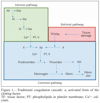
Summary : This diagram illustrates the traditional coagulation cascade, detailing the sequence of events and factors involved in blood clotting via the intrinsic, extrinsic, and common pathways. flowchart: # Pathways : • Intrinsic pathway (green box): Initiated by factor XII, proceeds through XI, IX, and VIII, with activation steps indicated by "a" (e.g., XIIa, XIa, IXa). • Extrinsic pathway (red box): Initiated by tissue damage, which activates tissue factor (TF) and factor VIIa. • Common pathway (blue box): Both intrinsic and extrinsic pathways converge at factor X, leading to the activation of prothrombin to thrombin, which then converts fibrinogen to fibrin, forming a fibrin clot. # Nodes : • XII → XIIa (Intrinsic) • XI → XIa (Intrinsic) • IX → IXa (Intrinsic) • Ca²⁺, PT, V (Intrinsic, Common) • TF/VIIa (Extrinsic) • Tissue damage (Extrinsic) • X → Xa (Common) • Prothrombin → Thrombin (Common) • Fibrinogen → Fibrin (Common) • XIII (Common) • Fibrin clot (Common) # Connectors : • Arrows indicate the direction of activation or conversion between factors. • Intrinsic and extrinsic pathways both feed into the activation of factor X in the common pathway. • Ca²⁺ and PT, V are required cofactors at specific steps. # Layout : • Three main colored sections: green (intrinsic, top left), red (extrinsic, top right), blue (common, bottom). • Flow proceeds from top (initiation) to bottom (clot formation). • Arrows show sequential activation and convergence of pathways. # Analysis : • The diagram clearly separates the intrinsic and extrinsic initiation mechanisms, both converging on the common pathway at factor X. • The cascade is highly sequential, with each activated factor catalyzing the next step. • Calcium ions (Ca²⁺) and phospholipids (PT) are essential cofactors at multiple steps. • The final outcome is the formation of a stable fibrin clot, highlighting the coordinated nature of the coagulation process.
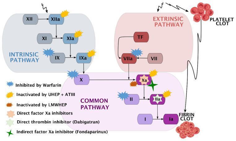
A comprehensive pathophysiology diagram illustrating the human coagulation cascade and the specific pharmacodynamic sites of action for various anticoagulants. The diagram is divided into three functional zones: the Intrinsic Pathway (blue), starting with Factor XII; the Extrinsic Pathway (red), initiated by Tissue Factor (TF) and Factor VII; and the Common Pathway (purple), where the pathways converge at Factor X. The cascade terminates in the conversion of Fibrinogen (I) to Fibrin (Ia), forming a fibrin clot. A secondary visual element depicts a platelet clot associated with the extrinsic pathway. Key clinical pharmacotherapeutic targets are marked with color-coded symbols: Warfarin inhibits Factors IX, VII, X, and II; Unfractionated Heparin (UHEP) + Antithrombin III (ATIII) inactivates Factors XIIa, XIa, IXa, Xa, and IIa; Low-Molecular-Weight Heparin (LMWHEP) and Fondaparinux target Factor Xa; Direct Factor Xa inhibitors act on Factor Xa; and Dabigatran acts as a direct thrombin inhibitor (Factor IIa). This schematic is designed for medical education regarding hemostasis and thromboprophylaxis.
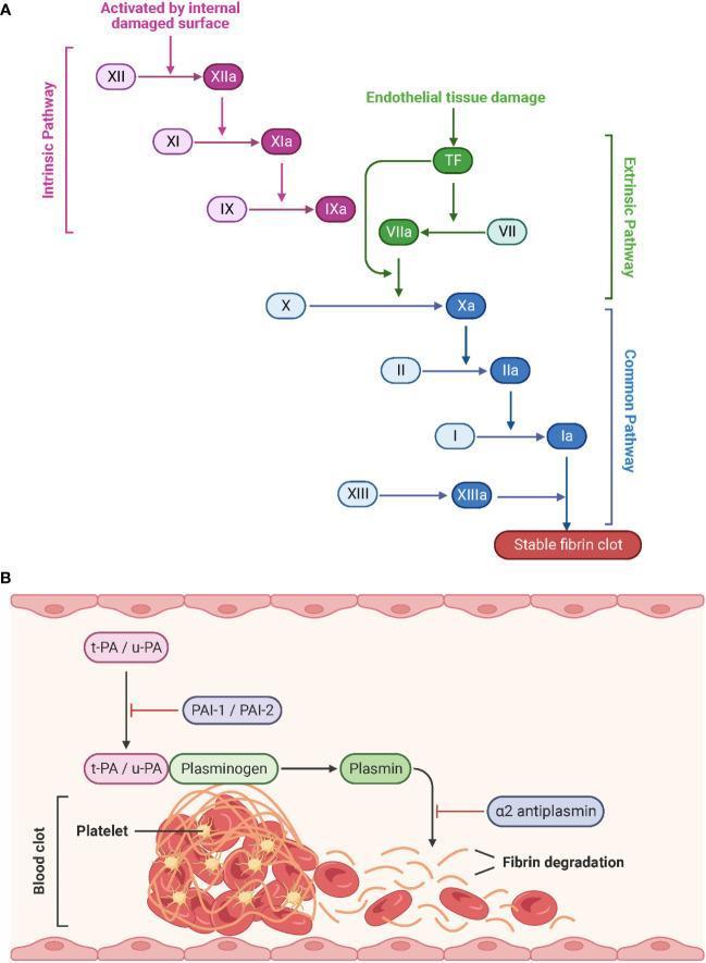
This medical schematic illustrates the physiological processes of hemostasis and fibrinolysis. Part A depicts the Coagulation Cascade, divided into the Intrinsic, Extrinsic, and Common pathways. The Intrinsic pathway (purple) is triggered by internal damaged surfaces, activating Factor XII to XIIa, followed by XI and IX. The Extrinsic pathway (green) begins with endothelial tissue damage and Tissue Factor (TF), activating Factor VII to VIIa. Both pathways converge at the Common pathway (blue) starting with the activation of Factor X to Xa. This leads to the conversion of prothrombin (II) to thrombin (IIa), and fibrinogen (I) to fibrin (Ia), culminating in a stable fibrin clot stabilized by Factor XIIIa. Activated factors are denoted by an 'a' suffix. Part B illustrates Fibrinolysis, where tissue-type (t-PA) and urokinase-type (u-PA) plasminogen activators convert plasminogen to plasmin, which then degrades the fibrin network of the blood clot. Inhibitory regulators including PAI-1, PAI-2, and ̡2-antiplasmin are also shown. The diagram represents the structural breakdown of a clot containing platelets and red blood cells into fibrin degradation products.
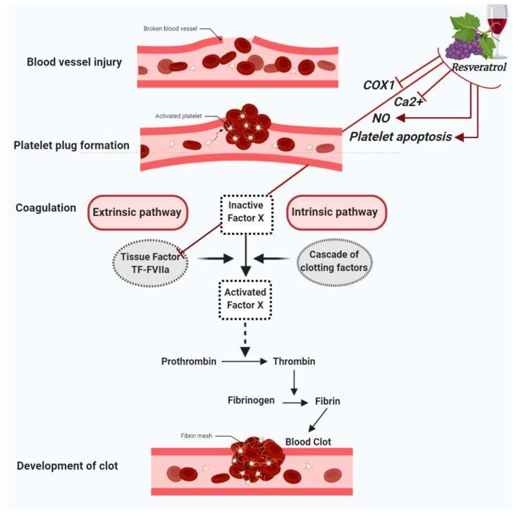
This pathophysiology diagram illustrates the process of hemostasis and the inhibitory mechanisms of resveratrol on platelet function. The central vertical flow depicts the sequence of events following a blood vessel injury: (1) Blood vessel injury, (2) Platelet plug formation involving activated platelets, (3) Coagulation, and (4) Development of a blood clot with a fibrin mesh. The coagulation cascade highlights the convergence of the extrinsic pathway (Tissue Factor: TF-FVIIa) and the intrinsic pathway (cascade of clotting factors) onto Inactive Factor X. Activated Factor X then facilitates the conversion of Prothrombin to Thrombin, and Fibrinogen to Fibrin. Interacting with these pathways, the diagram shows Resveratrol's antiplatelet effects through the inhibition of COX1, reduction of calcium (Ca2+) and nitric oxide (NO) concentrations, and the induction of platelet apoptosis. Additionally, Resveratrol is shown to inhibit the TF-FVIIa complex within the extrinsic pathway. The illustration uses a combination of anatomical cross-sections and biochemical flowcharts to detail secondary hemostasis and pharmacodynamic interactions relevant to cardiovascular health.
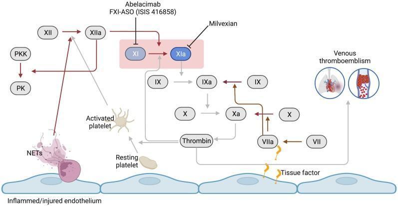
This pathophysiology diagram illustrates the coagulation cascade focusing on the central role of Factor XI (FXI) in venous thromboembolism. The visual depicts an inflamed/injured endothelial layer at the base, leading to the activation of the contact pathway (via FXII and NETs) and the extrinsic pathway (via Tissue Factor and FVII). Key clotting factors, including FXII, FXI, FIX, FX, and Thrombin, are shown as oval nodes interconnected by arrows representing biochemical activation. A highlighted pink box focuses on the conversion of FXI to FXIa, noting pharmacologic inhibitors like Abelacimab, FXI-ASO, and Milvexian. The diagram differentiates between resting and activated platelets and includes an icon for venous thromboembolism featuring pulmonary embolism and deep vein thrombosis. Dark brown arrows indicate pathways unaffected by FXI inhibition, while lighter grey arrows signify the attenuated thrombin formation pathway. This educational material is designed for medical students and clinicians studying hematology, pharmacology, and cardiovascular medicine.
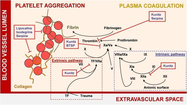
A pathophysiology diagram illustrating the mechanisms of hemostasis and the inhibitory effects of tick salivary proteins on platelet aggregation and plasma coagulation. The diagram is divided into the 'Blood Vessel Lumen' and the 'Extravascular Space.' On the left, 'Platelet Aggregation' is shown as a cluster of red spheres representing platelets cross-linked by fibrin strands; this process is inhibited by Lipocalins, Ixodegrins, and Serpins. On the right, the 'Plasma Coagulation' cascade is detailed, featuring both the Extrinsic pathway (initiated by Tissue Factor/TF due to trauma) and the Intrinsic pathway (activated via anionic surfaces). The diagram highlights key enzymatic conversions, such as Factor X to Xa/Va, Prothrombin to Thrombin, and Fibrinogen to Fibrin. Red annotation boxes identify specific tick protein families—including Kunitz-type inhibitors, BTSPs, and Serpins—that target and inhibit various coagulation factors like Thrombin, Factor Xa, and the XIIa/XIa complex. This visual resource effectively demonstrates the interference of exogenous biological agents with human blood clotting mechanisms.
platelet activation primary hemostasis plug formation
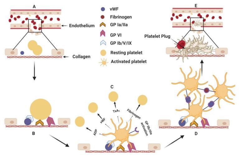
This hematology diagram illustrates the multistep pathophysiology of primary hemostasis following endothelial injury. The process is divided into five sequential stages (A–E). (A) Endothelial disruption exposes subendothelial collagen. (B) Platelet adhesion occurs as resting platelets (smooth, discoid) bind to collagen via surface glycoprotein receptors GP Ia/IIa, GP VI, and GP Ib/V/IX mediated by von Willebrand factor (vWF). (C) Platelet activation results in a dramatic morphological transformation, with platelets developing multiple cytoplasmic pseudopodia (starburst shape). This stage shows the release of granular contents including ADP, TxA2, vWF, and fibrinogen, which recruit additional platelets. (D) Platelet aggregation is shown, where activated GP IIb/IIIa receptors form cross-links between adjacent platelets using vWF and fibrinogen as molecular bridges. (E) The sequence concludes with the formation of a stable platelet plug at the injury site, effectively sealing the vascular breach. This educational visual explains the cellular signaling and mechanical interactions required for initial thrombus formation.
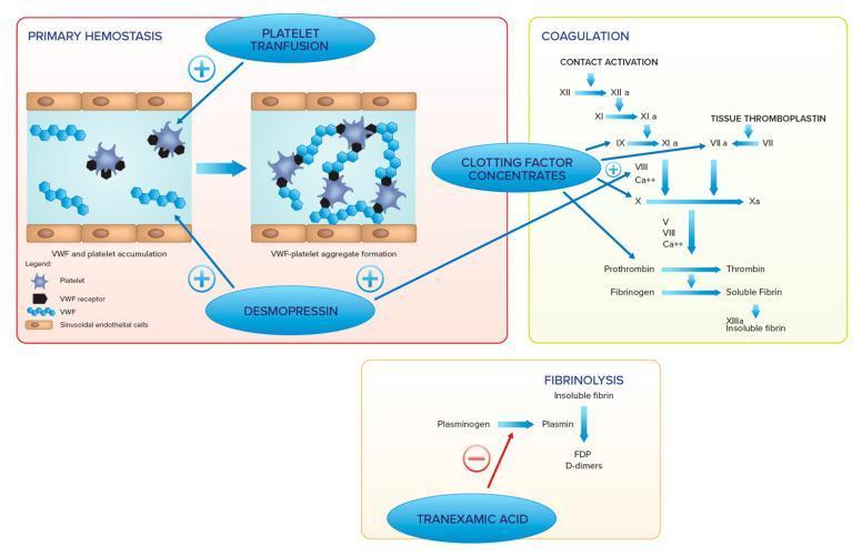
A medical infographic and pathway diagram detailing the mechanisms of hemostasis and associated therapeutic interventions. The content is divided into three main sections: Primary Hemostasis, Coagulation (Secondary Hemostasis), and Fibrinolysis. The 'Primary Hemostasis' section illustrates the transition from von Willebrand Factor (VWF) and platelet accumulation to VWF-platelet aggregate formation within a vessel lined by sinusoidal endothelial cells; it highlights Platelet Transfusion and Desmopressin as therapeutic options to enhance this stage. The 'Coagulation' section depicts the classical cascade, including the intrinsic (contact activation) pathway through Factor XII, XI, and IX, and the extrinsic pathway via Tissue Thromboplastin and Factor VII. These pathways converge on the common pathway (Factor Xa, Prothrombin to Thrombin, and Fibrinogen to Fibrin), with Clotting Factor Concentrates and Desmopressin indicated as interventions. The 'Fibrinolysis' section shows the conversion of Plasminogen to Plasmin leading to the breakdown of insoluble fibrin into D-dimers, where Tranexamic Acid is shown as an inhibitory agent. The diagram serves as an educational summary of hematologic pathways and pharmacological targets for managing coagulopathies.
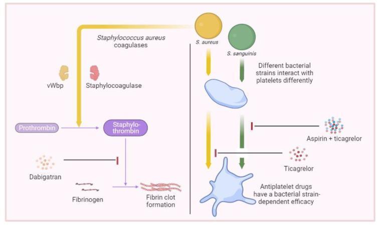
This medical illustration depicts the mechanisms of bacteria-induced hemostasis and the site of action for various antithrombotic agents. The diagram is split into two primary sections: Coagulation (left) and Platelet Interaction (right). The left panel illustrates the Staphylococcus aureus-mediated coagulation pathway, where vWbp (von Willebrand factor-binding protein) and staphylocoagulase facilitate the non-proteolytic activation of prothrombin into staphylo-thrombin. This complex subsequently catalyzes the conversion of fibrinogen to fibrin, leading to clot formation. Dabigatran is shown as a pharmacological inhibitor targeting the conversion step from prothrombin to staphylo-thrombin. The right panel demonstrates bacterial-platelet interactions involving S. aureus and S. sanguinis. It illustrates how these different bacterial strains interact with quiescent and activated platelets. The graphic highlights that antiplatelet drug efficacy is strain-dependent: Ticagrelor is indicated as an inhibitor of S. aureus-induced platelet activation, while the combination of aspirin and ticagrelor is shown targeting S. sanguinis-induced pathways. This educational figure is intended to explain the pathophysiology of infective endocarditis and the potential roles of anticoagulants and antiplatelet therapy in managing bacterial-induced thrombosis.
fibrinolysis plasmin tPA fibrin degradation products D-dimer
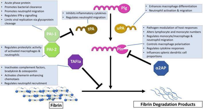
A pathophysiology diagram illustrating the fibrinolytic system's role in immune regulation. The central pathway shows the conversion of Plasminogen (Plg) to the active enzyme Plasmin (Pln) via tissue plasminogen activator (tPA) and urokinase (uPA), subsequently leading to the degradation of fibrin into fibrin degradation products. Regulatory mechanisms include inhibition of tPA/uPA by Plasminogen Activator Inhibitors 1 and 2 (PAI-1, PAI-2), direct inhibition of Plasmin by alpha2-antiplasmin (a2AP), and inhibition of fibrinolysis by activated Thrombin Activatable Fibrinolysis Inhibitor (TAFIa). Annotated text boxes describe non-fibrinolytic immune functions: PAI-1 regulates IFN-gamma signaling and viral replication; PAI-2 modulates macrophage proteolytic activity; a2AP influences macrophage polarization and splenic dendritic cell proportions; TAFIa inactivates complement factors and bradykinin; and tPA/uPA/Pln influence neutrophil migration and macrophage differentiation. This educational schematic demonstrates the crosstalk between coagulation, fibrinolysis, and innate immune responses.
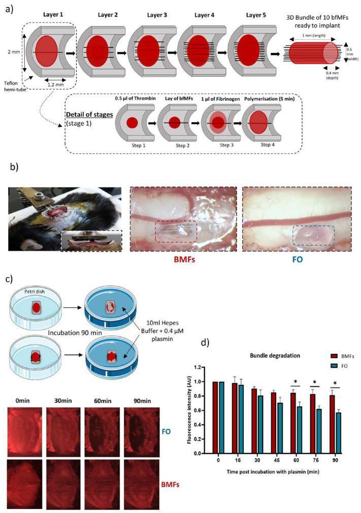
Educational diagram and experimental results depicting the fabrication and enzymatic degradation of a neuroprosthetic fibrin-based implant. Panel (a) illustrates a layer-by-layer construction process within a Teflon hemi-tube, alternating thrombin (0.5 µL) and fibrinogen (1 µL) with biofunctionalized microfibers (BMFs) to create a 3D bundle measuring approximately 1 x 0.5 x 0.4 mm. Panel (b) shows bright-field clinical photographs of the implant being positioned within a spinal cord lesion cavity in an animal model, comparing BMFs to Fibrin Only (FO) controls. Panel (c) details the in vitro fibrinolysis assay where implants are immersed in plasmin/HEPES buffer; fluorescence microscopy images track the degradation of sulforhodamine B-labeled fibrin over 90 minutes. Panel (d) provides a line graph showing normalized fluorescence intensity, demonstrating that the inclusion of microfibers (BMFs) significantly slows fibrin degradation compared to FO implants (p < 0.05), indicating enhanced structural stability in the presence of enzymatic plasmin.
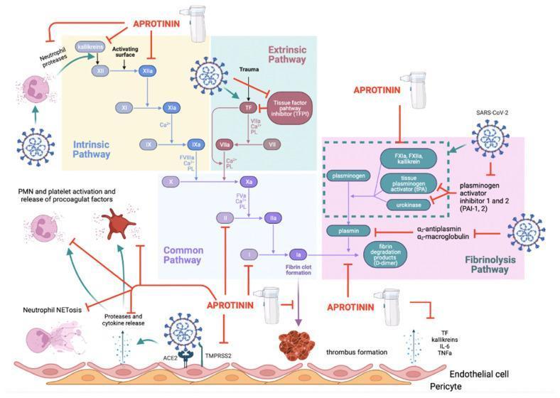
This pathophysiology diagram illustrates the complex relationship between SARS-CoV-2 infection, thromboinflammation, and the therapeutic inhibitory effects of aprotinin. The visual represents three integrated systems: the Intrinsic Pathway, Extrinsic Pathway, and Fibrinolysis Pathway. At the cellular level, the diagram shows SARS-CoV-2 interacting with endothelial ACE2 and TMPRSS2 receptors, triggering protease and cytokine release, neutrophil NETosis, and PMN/platelet activation. In the intrinsic pathway, kallikreins and factors XII through IX are shown sequentially activating. The extrinsic pathway is triggered by tissue factor (TF) release, which SARS-CoV-2 further enhances by inhibiting tissue factor pathway inhibitor (TFPI). Both pathways converge at the common pathway (Factor X to IIa/thrombin and I/fibrin), leading to thrombus formation. The fibrinolysis pathway depicts the conversion of plasminogen to plasmin via tPA and urokinase, resulting in D-dimer production. Aprotinin is depicted as a multi-modal inhibitor (indicated by red T-bars) acting on viral entry, neutrophil proteases, kallikreins, Factor XIIa, thrombin, and fibrinogen. The illustration emphasizes aprotinin’s role in restoring the balance between procoagulant and fibrinolytic forces in the context of COVID-19 coagulopathy.
anticoagulant drugs warfarin heparin factor Xa inhibitors mechanism of action
A comprehensive pathophysiology diagram illustrating the human coagulation cascade and the specific pharmacodynamic sites of action for various anticoagulants. The diagram is divided into three functional zones: the Intrinsic Pathway (blue), starting with Factor XII; the Extrinsic Pathway (red), initiated by Tissue Factor (TF) and Factor VII; and the Common Pathway (purple), where the pathways converge at Factor X. The cascade terminates in the conversion of Fibrinogen (I) to Fibrin (Ia), forming a fibrin clot. A secondary visual element depicts a platelet clot associated with the extrinsic pathway. Key clinical pharmacotherapeutic targets are marked with color-coded symbols: Warfarin inhibits Factors IX, VII, X, and II; Unfractionated Heparin (UHEP) + Antithrombin III (ATIII) inactivates Factors XIIa, XIa, IXa, Xa, and IIa; Low-Molecular-Weight Heparin (LMWHEP) and Fondaparinux target Factor Xa; Direct Factor Xa inhibitors act on Factor Xa; and Dabigatran acts as a direct thrombin inhibitor (Factor IIa). This schematic is designed for medical education regarding hemostasis and thromboprophylaxis.
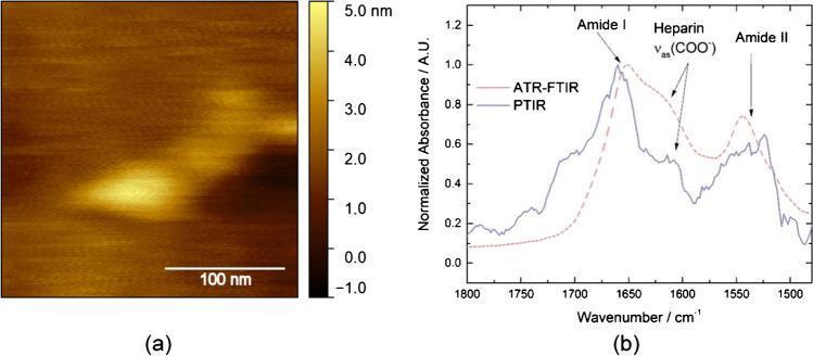
This composite educational image showcases the characterization of an individual APAC (antiplatelet and anticoagulant) molecule, a dual-action agent targeting von Willebrand factor (VWF). Panel (a) is a high-resolution Atomic Force Microscopy (AFM) topography image of a single APAC molecule adsorbed on an atomically flat gold substrate. A 100 nm scale bar and a vertical color-gradient scale (ranging from -1.0 to 5.0 nm) facilitate quantitative analysis, showing the molecule's morphology and a height of approximately 3-4 nm. Panel (b) presents a comparison of molecular vibration spectra: Photothermal Induced Resonance (PTIR) in solid blue and Attenuated Total Reflection Fourier-Transform Infrared (ATR-FTIR) in dashed red. The graph plots Normalized Absorbance against Wavenumber (1800-1500 cm⁻¹). Key chemical signatures are identified: Amide I (approx. 1650 cm⁻¹), Heparin asymmetric carboxylate stretching νas(COO-) (approx. 1600 cm⁻¹), and Amide II (approx. 1550 cm⁻¹). These measurements confirm the structural integrity and chemical composition of the APAC molecule at the nanoscale, essential for understanding its mechanism in preventing arterial thrombosis.
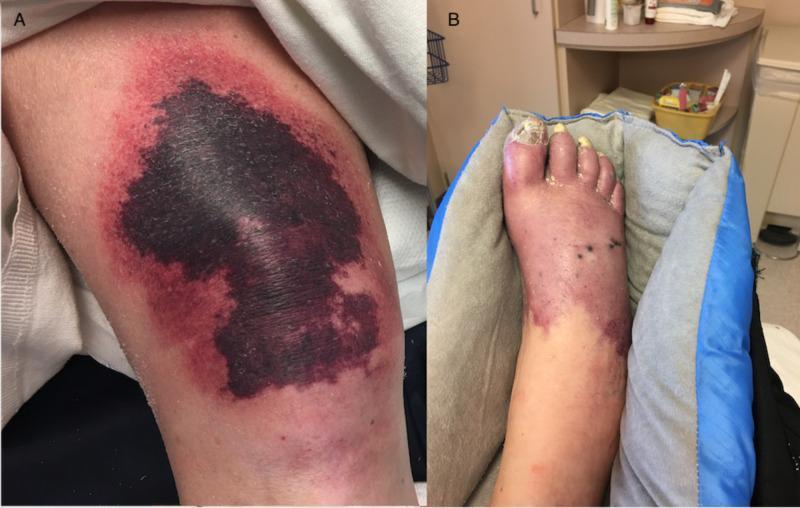
This clinical photograph consists of two panels illustrating severe cutaneous manifestations of warfarin-induced skin necrosis and heparin-induced thrombocytopenia (HIT) with secondary protein C deficiency. Panel A displays the right knee, featuring a large, irregularly shaped, well-demarcated purpuric plaque. The lesion exhibits central black eschar formation indicating tissue necrosis, surrounded by a violaceous, erythematous inflammatory border. Panel B shows the distal right foot and ankle, demonstrating diffuse, deep purplish-blue discoloration consistent with retiform purpura and impending gangrene. The foot exhibits significant edema, thickened and yellowed toenails (onychomycosis), and scattered petechiae or small necrotic foci. These visual findings are classic for a hypercoagulable state leading to microvascular thrombosis and subsequent skin infarction. The images serve as educational examples of critical dermatologic emergencies associated with anticoagulant complications and severe thrombotic disorders.
mkdir -p /tmp/workspace/coagulation-seminar && ls /tmp/workspace/
~/coagulation-seminar/build_pptx.py
import json, subprocess, base64
from io import BytesIO
from pptx import Presentation
from pptx.util import Inches, Pt, Emu
from pptx.dml.color import RGBColor
from pptx.enum.text import PP_ALIGN, MSO_ANCHOR
from pptx.oxml.ns import qn
from lxml import etree

# ── Palette ──────────────────────────────────────────────────────────────────
DARK_NAVY   = RGBColor(0x0D, 0x1B, 0x3E)   # slide backgrounds / header bars
MID_BLUE    = RGBColor(0x1A, 0x4A, 0x8A)   # accent boxes
LIGHT_BLUE  = RGBColor(0xE8, 0xF1, 0xFB)   # content area background
ACCENT_RED  = RGBColor(0xC0, 0x39, 0x2B)   # highlight / bullet accent
WHITE       = RGBColor(0xFF, 0xFF, 0xFF)
GOLD        = RGBColor(0xF0, 0xB4, 0x29)
LIGHT_GREY  = RGBColor(0xF4, 0xF6, 0xF8)
DARK_TEXT   = RGBColor(0x1C, 0x1C, 0x2E)

# ── Helpers ───────────────────────────────────────────────────────────────────
def filled_rect(slide, left, top, width, height, fill_color):
    shape = slide.shapes.add_shape(
        1,  # MSO_SHAPE_TYPE.RECTANGLE = 1
        Inches(left), Inches(top), Inches(width), Inches(height)
    )
    shape.fill.solid()
    shape.fill.fore_color.rgb = fill_color
    shape.line.fill.background()
    return shape

def add_text(slide, text, left, top, width, height,
             font_name="Calibri", font_size=18, bold=False, italic=False,
             color=WHITE, align=PP_ALIGN.LEFT, wrap=True, v_anchor=MSO_ANCHOR.TOP):
    tb = slide.shapes.add_textbox(Inches(left), Inches(top), Inches(width), Inches(height))
    tf = tb.text_frame
    tf.word_wrap = wrap
    tf.vertical_anchor = v_anchor
    tf.margin_left  = Inches(0.05)
    tf.margin_right = Inches(0.05)
    tf.margin_top   = Inches(0.03)
    tf.margin_bottom = Inches(0.03)
    p = tf.paragraphs[0]
    p.alignment = align
    run = p.add_run()
    run.text = text
    run.font.name = font_name
    run.font.size = Pt(font_size)
    run.font.bold = bold
    run.font.italic = italic
    run.font.color.rgb = color
    return tb

def add_bullet_slide(prs, title_text, bullets, image_url=None, image_left=7.3, image_top=1.3, image_w=5.7, image_h=5.5):
    """Standard content slide: dark header + light body + optional right image"""
    blank = prs.slide_layouts[6]
    slide = prs.slides.add_slide(blank)

    # Background
    filled_rect(slide, 0, 0, 13.333, 7.5, LIGHT_GREY)

    # Header bar
    filled_rect(slide, 0, 0, 13.333, 1.1, DARK_NAVY)

    # Title
    add_text(slide, title_text, 0.25, 0.1, 12.5, 0.9,
             font_size=28, bold=True, color=WHITE, v_anchor=MSO_ANCHOR.MIDDLE)

    # Gold accent line
    line_shape = slide.shapes.add_shape(1, Inches(0.25), Inches(1.1), Inches(12.8), Inches(0.04))
    line_shape.fill.solid()
    line_shape.fill.fore_color.rgb = GOLD
    line_shape.line.fill.background()

    content_right = 12.9 if image_url is None else (image_left - 0.2)

    # Bullet box
    filled_rect(slide, 0.25, 1.25, content_right - 0.25, 5.9, WHITE)

    # Bullets
    tb = slide.shapes.add_textbox(Inches(0.45), Inches(1.35), Inches(content_right - 0.5), Inches(5.7))
    tf = tb.text_frame
    tf.word_wrap = True
    tf.margin_left = Inches(0.05)
    tf.margin_right = Inches(0.05)
    tf.margin_top = Inches(0.05)

    first = True
    for bullet in bullets:
        if first:
            p = tf.paragraphs[0]
            first = False
        else:
            p = tf.add_paragraph()

        # Section header style (starts with **)
        if bullet.startswith("**") and bullet.endswith("**"):
            p.space_before = Pt(8)
            run = p.add_run()
            run.text = bullet[2:-2]
            run.font.name = "Calibri"
            run.font.size = Pt(15)
            run.font.bold = True
            run.font.color.rgb = MID_BLUE
        elif bullet.startswith("  "):   # sub-bullet
            p.level = 1
            p.space_before = Pt(2)
            run = p.add_run()
            run.text = bullet.strip()
            run.font.name = "Calibri"
            run.font.size = Pt(13)
            run.font.bold = False
            run.font.color.rgb = DARK_TEXT
        else:
            p.space_before = Pt(4)
            # Bullet char
            run0 = p.add_run()
            run0.text = "• "
            run0.font.name = "Calibri"
            run0.font.size = Pt(14)
            run0.font.bold = True
            run0.font.color.rgb = ACCENT_RED
            run = p.add_run()
            run.text = bullet
            run.font.name = "Calibri"
            run.font.size = Pt(14)
            run.font.bold = False
            run.font.color.rgb = DARK_TEXT

    # Embed image if provided
    if image_url:
        result = json.loads(subprocess.check_output(
            ["python", "/tmp/skills/shared/scripts/fetch_images.py", image_url]
        ))
        if result and result[0].get("base64"):
            raw = base64.b64decode(result[0]["base64"].split(",", 1)[-1])
            slide.shapes.add_picture(BytesIO(raw),
                Inches(image_left), Inches(image_top),
                Inches(image_w), Inches(image_h))
    return slide

def add_two_col_slide(prs, title_text, left_bullets, right_bullets,
                      left_title="", right_title="",
                      left_color=MID_BLUE, right_color=ACCENT_RED):
    blank = prs.slide_layouts[6]
    slide = prs.slides.add_slide(blank)
    filled_rect(slide, 0, 0, 13.333, 7.5, LIGHT_GREY)
    filled_rect(slide, 0, 0, 13.333, 1.1, DARK_NAVY)
    add_text(slide, title_text, 0.25, 0.1, 12.5, 0.9,
             font_size=28, bold=True, color=WHITE, v_anchor=MSO_ANCHOR.MIDDLE)
    line_shape = slide.shapes.add_shape(1, Inches(0.25), Inches(1.1), Inches(12.8), Inches(0.04))
    line_shape.fill.solid()
    line_shape.fill.fore_color.rgb = GOLD
    line_shape.line.fill.background()

    for i, (col_title, bullets, xcol, col_color) in enumerate([
        (left_title, left_bullets, 0.25, left_color),
        (right_title, right_bullets, 6.8, right_color)
    ]):
        # Header strip
        filled_rect(slide, xcol, 1.2, 6.2, 0.45, col_color)
        add_text(slide, col_title, xcol + 0.1, 1.2, 6.0, 0.45,
                 font_size=14, bold=True, color=WHITE, v_anchor=MSO_ANCHOR.MIDDLE)
        # Content box
        filled_rect(slide, xcol, 1.65, 6.2, 5.5, WHITE)
        tb = slide.shapes.add_textbox(Inches(xcol + 0.15), Inches(1.75),
                                       Inches(5.9), Inches(5.3))
        tf = tb.text_frame
        tf.word_wrap = True
        first = True
        for bullet in bullets:
            if first:
                p = tf.paragraphs[0]
                first = False
            else:
                p = tf.add_paragraph()
            p.space_before = Pt(4)
            run0 = p.add_run()
            run0.text = "• "
            run0.font.name = "Calibri"
            run0.font.size = Pt(13)
            run0.font.bold = True
            run0.font.color.rgb = col_color
            run = p.add_run()
            run.text = bullet
            run.font.name = "Calibri"
            run.font.size = Pt(13)
            run.font.color.rgb = DARK_TEXT
    return slide

# ═══════════════════════════════════════════════════════════════════════════
# BUILD PRESENTATION
# ═══════════════════════════════════════════════════════════════════════════
prs = Presentation()
prs.slide_width  = Inches(13.333)
prs.slide_height = Inches(7.5)

# ── SLIDE 1: Title Slide ────────────────────────────────────────────────────
blank = prs.slide_layouts[6]
s1 = prs.slides.add_slide(blank)
filled_rect(s1, 0, 0, 13.333, 7.5, DARK_NAVY)

# Decorative top bar
filled_rect(s1, 0, 0, 13.333, 0.08, GOLD)

# Central white content panel
filled_rect(s1, 1.5, 1.0, 10.333, 5.2, WHITE)

# Red accent left strip
filled_rect(s1, 1.5, 1.0, 0.15, 5.2, ACCENT_RED)

# Title
add_text(s1, "COAGULATION OF BLOOD", 1.9, 1.4, 9.7, 1.4,
         font_size=40, bold=True, color=DARK_NAVY,
         align=PP_ALIGN.LEFT, v_anchor=MSO_ANCHOR.MIDDLE)

# Subtitle bar
filled_rect(s1, 1.65, 2.9, 10.1, 0.05, MID_BLUE)

# Subtitle
add_text(s1, "Mechanisms, Regulation & Clinical Correlations", 1.9, 3.0, 9.7, 0.7,
         font_size=22, bold=False, italic=True, color=MID_BLUE,
         align=PP_ALIGN.LEFT)

# Details
add_text(s1, "Seminar Presentation  |  Postgraduate Residents", 1.9, 3.85, 9.7, 0.5,
         font_size=16, bold=False, color=DARK_TEXT, align=PP_ALIGN.LEFT)
add_text(s1, "Department of Medicine", 1.9, 4.3, 9.7, 0.5,
         font_size=14, bold=False, italic=True, color=RGBColor(0x55, 0x55, 0x66),
         align=PP_ALIGN.LEFT)

# Bottom info
add_text(s1, "Sources: Schwartz's Surgery | Tietz Lab Medicine | Rosen's Emergency Medicine | Harper's Biochemistry | Goodman & Gilman",
         0.3, 6.85, 12.8, 0.5, font_size=10, color=RGBColor(0x99, 0xAA, 0xBB),
         align=PP_ALIGN.CENTER)

# ── SLIDE 2: Overview / Learning Objectives ─────────────────────────────────
add_bullet_slide(prs, "Learning Objectives", [
    "Describe the three phases of hemostasis: vascular, platelet, and coagulation",
    "Understand the intrinsic, extrinsic, and common coagulation pathways",
    "Explain the cell-based model of coagulation (initiation, amplification, propagation)",
    "Identify the roles of individual clotting factors (I–XIII)",
    "Describe the natural anticoagulant mechanisms (Protein C/S, ATIII, TFPI)",
    "Understand fibrinolysis and its regulation",
    "Correlate laboratory tests (PT, aPTT, TT, fibrinogen, D-dimer) with pathology",
    "Apply these concepts to clinical disorders: Hemophilia, vWD, DIC, thrombophilia",
    "Identify pharmacologic targets: warfarin, heparins, DOACs, thrombolytics",
])

# ── SLIDE 3: Overview of Hemostasis ─────────────────────────────────────────
add_bullet_slide(prs, "Overview of Hemostasis", [
    "**Definition**",
    "Physiologic process that stops bleeding after vascular injury while maintaining blood fluidity",
    "**Four Interrelated Processes**",
    "1. Vascular spasm - immediate vasoconstriction reduces blood flow",
    "2. Primary hemostasis - platelet adhesion, activation, and plug formation",
    "3. Secondary hemostasis (coagulation cascade) - fibrin clot reinforcement",
    "4. Fibrinolysis - clot dissolution after healing",
    "**Key Players**",
    "Endothelium (anti-thrombotic at rest; pro-thrombotic when injured)",
    "Platelets (circulate inactively; activated by sub-endothelial exposure)",
    "Clotting factors (mostly zymogens synthesized in the liver)",
    "Inhibitors (Protein C, Protein S, ATIII, TFPI) - prevent propagation",
],
image_url="https://cdn.orris.care/cdss_images/pmc_clinical_VQA_1dc53d345851d43758418822c64f18e401f284d7723b40eb1b9d198b43d0ede3.jpg",
image_left=7.5, image_top=1.25, image_w=5.5, image_h=5.9)

# ── SLIDE 4: Primary Hemostasis ──────────────────────────────────────────────
add_bullet_slide(prs, "Primary Hemostasis: Platelet Plug Formation", [
    "**Step 1 - Adhesion**",
    "Endothelial injury exposes subendothelial collagen and von Willebrand Factor (vWF)",
    "Platelets bind via GP Ib/V/IX (to vWF) and GP Ia/IIa (to collagen directly)",
    "**Step 2 - Activation**",
    "Shape change: disc to spiny sphere with pseudopodia",
    "Granule release: ADP, TXA2, serotonin, fibrinogen recruit more platelets",
    "TXA2 (thromboxane A2) is a potent vasoconstrictor and platelet activator",
    "**Step 3 - Aggregation**",
    "GP IIb/IIIa receptor activation - binds fibrinogen as molecular bridge",
    "Cross-links platelets into primary plug (unstable, temporary)",
    "**Clinical Pearls**",
    "Aspirin: irreversibly inhibits COX, blocks TXA2 synthesis",
    "Clopidogrel/Ticagrelor: block ADP receptor P2Y12",
    "GP IIb/IIIa inhibitors: abciximab, eptifibatide, tirofiban",
],
image_url="https://cdn.orris.care/cdss_images/pmc_clinical_VQA_3d192d54b5b0e2251f9af9b9a5e3ba2c29ba45780c60c394edd05306a203cc6c.jpg",
image_left=7.4, image_top=1.25, image_w=5.6, image_h=5.9)

# ── SLIDE 5: Clotting Factors Table Slide ────────────────────────────────────
blank = prs.slide_layouts[6]
s5 = prs.slides.add_slide(blank)
filled_rect(s5, 0, 0, 13.333, 7.5, LIGHT_GREY)
filled_rect(s5, 0, 0, 13.333, 1.1, DARK_NAVY)
add_text(s5, "Coagulation Factors: A Quick Reference", 0.25, 0.1, 12.5, 0.9,
         font_size=28, bold=True, color=WHITE, v_anchor=MSO_ANCHOR.MIDDLE)
line_shape = s5.shapes.add_shape(1, Inches(0.25), Inches(1.1), Inches(12.8), Inches(0.04))
line_shape.fill.solid()
line_shape.fill.fore_color.rgb = GOLD
line_shape.line.fill.background()

table_data = [
    ["Factor", "Name", "Pathway", "Vitamin K?", "Key Note"],
    ["I", "Fibrinogen", "Common", "No", "Converted to fibrin by thrombin"],
    ["II", "Prothrombin", "Common", "YES", "Converted to thrombin (IIa)"],
    ["III", "Tissue Factor", "Extrinsic", "No", "Initiates extrinsic pathway"],
    ["IV", "Calcium (Ca²⁺)", "All", "No", "Cofactor for multiple reactions"],
    ["V", "Labile Factor", "Common", "No", "Cofactor for Xa; consumed in DIC"],
    ["VII", "Stable Factor", "Extrinsic", "YES", "Shortest half-life (~4-6 h)"],
    ["VIII", "Anti-hemophilic A", "Intrinsic", "No", "Deficient in Hemophilia A"],
    ["IX", "Anti-hemophilic B", "Intrinsic", "YES", "Deficient in Hemophilia B"],
    ["X", "Stuart-Prower", "Common", "YES", "Convergence point of both paths"],
    ["XI", "Plasma thromboplastin", "Intrinsic", "No", "Hemophilia C (mild-moderate)"],
    ["XII", "Hageman Factor", "Intrinsic", "No", "Contact activation; NOT needed in vivo"],
    ["XIII", "Fibrin stabilizing", "Common", "No", "Cross-links fibrin monomers"],
]

rows = len(table_data)
cols = len(table_data[0])
table = s5.shapes.add_table(rows, cols,
    Inches(0.25), Inches(1.2), Inches(12.8), Inches(5.95)).table
col_widths = [0.6, 2.0, 1.6, 1.3, 7.3]
for ci, w in enumerate(col_widths):
    table.columns[ci].width = Inches(w)

header_colors = [DARK_NAVY, DARK_NAVY, DARK_NAVY, DARK_NAVY, DARK_NAVY]
vk_yes_color = RGBColor(0xE8, 0xF5, 0xE9)

for ri, row_data in enumerate(table_data):
    for ci, cell_text in enumerate(row_data):
        cell = table.cell(ri, ci)
        cell.text = cell_text
        tf = cell.text_frame
        tf.word_wrap = True
        p = tf.paragraphs[0]
        run = p.add_run() if not p.runs else p.runs[0]
        if ri == 0:
            run.font.color.rgb = WHITE
            run.font.bold = True
            run.font.size = Pt(12)
            p.alignment = PP_ALIGN.CENTER
            tc = cell._tc
            tcPr = tc.get_or_add_tcPr()
            solidFill = etree.SubElement(tcPr, qn('a:solidFill'))
            srgbClr = etree.SubElement(solidFill, qn('a:srgbClr'))
            srgbClr.set('val', '0D1B3E')
        else:
            run.font.color.rgb = DARK_TEXT
            run.font.bold = (ci == 0)
            run.font.size = Pt(11)
            if ci == 3 and cell_text == "YES":
                run.font.color.rgb = RGBColor(0x27, 0x7E, 0x3C)
                run.font.bold = True
            bg = LIGHT_GREY if ri % 2 == 0 else WHITE
            tc = cell._tc
            tcPr = tc.get_or_add_tcPr()
            solidFill = etree.SubElement(tcPr, qn('a:solidFill'))
            srgbClr = etree.SubElement(solidFill, qn('a:srgbClr'))
            hex_bg = '{:02X}{:02X}{:02X}'.format(bg.r, bg.g, bg.b)
            srgbClr.set('val', hex_bg)

# ── SLIDE 6: Coagulation Cascade (Classical) ─────────────────────────────────
add_bullet_slide(prs, "The Coagulation Cascade: Classical Model", [
    "**Extrinsic Pathway (tissue factor pathway)**",
    "Triggered by tissue factor (TF/Factor III) released from damaged endothelium",
    "TF + Factor VII → TF-VIIa complex (extrinsic tenase)",
    "Activates Factor X → Xa; also Factor IX → IXa (crossover)",
    "Measured by Prothrombin Time (PT) / INR",
    "**Intrinsic Pathway (contact activation)**",
    "Triggered by negatively charged surfaces (glass, collagen, kallikrein)",
    "Factor XII → XIIa → XI → XIa → IX → IXa + VIIIa (intrinsic tenase)",
    "VIIIa-IXa complex is 50x more potent than TF-VIIa for factor X activation",
    "Measured by aPTT (activated partial thromboplastin time)",
    "**Common Pathway**",
    "Factor Xa + Va (prothrombinase complex) → converts Prothrombin → Thrombin (IIa)",
    "Thrombin cleaves fibrinogen → fibrin monomers",
    "Factor XIII (activated by thrombin) cross-links fibrin → stable clot",
],
image_url="https://cdn.orris.care/cdss_images/GLGCA_4589275_1766503740499_cf028afd-3032-40dd-adf5-6e85434c207d_8249caac-80d2-4940-9015-2d7611df005c.png",
image_left=7.35, image_top=1.25, image_w=5.7, image_h=5.9)

# ── SLIDE 7: Cell-Based Model ─────────────────────────────────────────────────
add_bullet_slide(prs, "Cell-Based Model of Coagulation (In Vivo)", [
    "**Phase 1: Initiation (on adventitial fibroblasts)**",
    "TF exposed on subendothelial cells after vessel injury",
    "TF-VIIa → activates Factor X → Xa and IX → IXa",
    "Small amounts of thrombin generated (insufficient for clotting)",
    "Rapidly inhibited by Tissue Factor Pathway Inhibitor (TFPI)",
    "**Phase 2: Amplification (on platelet surface)**",
    "Small thrombin burst activates platelets, Factors V, VIII, XI",
    "Platelets adhere to matrix; GP receptors recruit more platelets",
    "Factor VIIIa and Va assemble on platelet phospholipid surface",
    "**Phase 3: Propagation (massive thrombin generation)**",
    "Intrinsic tenase (VIIIa-IXa) and prothrombinase (Va-Xa) assembled on platelets",
    "Explosive 'thrombin burst' - converts fibrinogen to fibrin en masse",
    "Fibrin monomers polymerize; Factor XIIIa cross-links for stability",
    "Thrombin-activatable fibrinolysis inhibitor (TAFI) stabilizes clot",
])

# ── SLIDE 8: Natural Anticoagulant Mechanisms ─────────────────────────────────
add_two_col_slide(prs, "Natural Anticoagulant Mechanisms",
    left_title="Protein-Based Inhibitors",
    right_title="Enzymatic & Cell-Based Mechanisms",
    left_bullets=[
        "Antithrombin III (ATIII): neutralizes thrombin, Xa, IXa, XIa, XIIa. Heparin accelerates ATIII 1000-fold",
        "Protein C: activated by Thrombin-Thrombomodulin complex; degrades Factors Va and VIIIa",
        "Protein S: cofactor for Protein C; deficiency = hypercoagulable state",
        "TFPI: rapidly inhibits TF-VIIa and Xa after initiation phase",
        "Alpha-2 macroglobulin: secondary thrombin inhibitor",
        "Heparin cofactor II: specifically inhibits thrombin",
    ],
    right_bullets=[
        "Thrombomodulin (TM): endothelial surface protein; binds thrombin, redirects it to activate Protein C",
        "Prostacyclin (PGI2): released by intact endothelium; potent platelet inhibitor and vasodilator",
        "Nitric Oxide (NO): endothelium-derived; inhibits platelet activation",
        "tPA / uPA: released post-injury to initiate fibrinolysis",
        "Factor V Leiden: mutation makes Factor V resistant to APC cleavage → thrombophilia",
        "Prothrombin G20210A mutation: elevated prothrombin levels → VTE risk",
    ]
)

# ── SLIDE 9: Fibrinolysis ────────────────────────────────────────────────────
add_bullet_slide(prs, "Fibrinolysis: Clot Dissolution", [
    "**Purpose**: Limit clot size; restore vascular patency after healing",
    "**Key Enzyme**: Plasmin - serine protease that digests fibrin",
    "**Activation Pathway**",
    "Tissue plasminogen activator (tPA) released from endothelium (triggered by thrombin, stasis)",
    "Urokinase plasminogen activator (uPA) - extravascular fibrinolysis",
    "Both convert plasminogen → plasmin (fibrin-bound, localized)",
    "**Products of Fibrinolysis**",
    "Fibrin degradation products (FDPs) and D-dimers (cross-linked fibrin fragments)",
    "D-dimer elevated in DVT, PE, DIC, post-surgery, pregnancy",
    "**Regulation (Anti-fibrinolytic System)**",
    "PAI-1 (Plasminogen Activator Inhibitor-1): inhibits tPA and uPA",
    "Alpha-2 antiplasmin: directly inhibits free plasmin",
    "TAFI (thrombin-activatable fibrinolysis inhibitor): slows fibrinolysis",
    "**Pharmacology**: tPA (alteplase), streptokinase, urokinase for thrombolysis",
    "Tranexamic acid / aminocaproic acid: antifibrinolytics (trauma, surgery)",
],
image_url="https://cdn.orris.care/cdss_images/pmc_clinical_VQA_94efa0724af446f5371e4a01af8c6ece4588617fbd6ff472c6935dab2a64e17b.jpg",
image_left=7.4, image_top=1.25, image_w=5.6, image_h=5.9)

# ── SLIDE 10: Laboratory Assessment ─────────────────────────────────────────
blank = prs.slide_layouts[6]
s10 = prs.slides.add_slide(blank)
filled_rect(s10, 0, 0, 13.333, 7.5, LIGHT_GREY)
filled_rect(s10, 0, 0, 13.333, 1.1, DARK_NAVY)
add_text(s10, "Laboratory Assessment of Coagulation", 0.25, 0.1, 12.5, 0.9,
         font_size=28, bold=True, color=WHITE, v_anchor=MSO_ANCHOR.MIDDLE)
line_shape = s10.shapes.add_shape(1, Inches(0.25), Inches(1.1), Inches(12.8), Inches(0.04))
line_shape.fill.solid()
line_shape.fill.fore_color.rgb = GOLD
line_shape.line.fill.background()

lab_data = [
    ["Test", "Pathway Assessed", "Normal Range", "Elevated In", "Clinical Use"],
    ["PT / INR", "Extrinsic + Common\n(Factors I, II, V, VII, X)", "11-13 s / INR 0.8-1.2", "Warfarin, Liver disease,\nVit K deficiency, DIC", "Monitor warfarin;\npre-op screening"],
    ["aPTT", "Intrinsic + Common\n(Factors I, II, V, VIII, IX, X, XI, XII)", "25-35 seconds", "Hemophilia A/B, vWD,\nheparin therapy, DIC", "Monitor unfractionated\nheparin; screen for lupus anticoagulant"],
    ["Thrombin Time (TT)", "Fibrinogen → Fibrin conversion", "14-19 seconds", "Hypofibrinogenemia,\nheparin, dabigatran", "Assess fibrinogen\nfunction"],
    ["Fibrinogen", "Clot substrate", "200-400 mg/dL", "Consumed in DIC;\nlow in liver failure", "DIC monitoring;\ntransfusion guidance"],
    ["D-dimer", "Fibrinolysis marker", "< 0.5 mg/L FEU", "DVT, PE, DIC, sepsis,\npregnancy, malignancy", "High sensitivity for VTE\n(low specificity)"],
    ["Platelet Count", "Primary hemostasis", "150-400 × 10⁹/L", "ITP, HIT, DIC, sepsis,\nbone marrow suppression", "Bleeding risk;\nthrombocytopenia workup"],
    ["TEG/ROTEM", "Whole blood viscoelastic", "Amplitude, time parameters", "Identifies clot initiation,\nstrength, and lysis defects", "Intraoperative; trauma;\nliver transplant"],
]

rows = len(lab_data)
cols = len(lab_data[0])
table = s10.shapes.add_table(rows, cols,
    Inches(0.2), Inches(1.18), Inches(12.93), Inches(6.1)).table
col_widths2 = [1.5, 2.3, 1.7, 2.6, 2.6]
for ci, w in enumerate(col_widths2):
    table.columns[ci].width = Inches(w)
for ri, row_data in enumerate(lab_data):
    for ci, cell_text in enumerate(row_data):
        cell = table.cell(ri, ci)
        cell.text = cell_text
        tf = cell.text_frame
        tf.word_wrap = True
        p = tf.paragraphs[0]
        run = p.add_run() if not p.runs else p.runs[0]
        if ri == 0:
            run.font.color.rgb = WHITE
            run.font.bold = True
            run.font.size = Pt(11)
            p.alignment = PP_ALIGN.CENTER
            tc = cell._tc
            tcPr = tc.get_or_add_tcPr()
            solidFill = etree.SubElement(tcPr, qn('a:solidFill'))
            srgbClr = etree.SubElement(solidFill, qn('a:srgbClr'))
            srgbClr.set('val', '0D1B3E')
        else:
            run.font.size = Pt(10)
            run.font.color.rgb = DARK_TEXT
            if ci == 0:
                run.font.bold = True
                run.font.color.rgb = MID_BLUE
            bg = LIGHT_GREY if ri % 2 == 0 else WHITE
            tc = cell._tc
            tcPr = tc.get_or_add_tcPr()
            solidFill = etree.SubElement(tcPr, qn('a:solidFill'))
            srgbClr = etree.SubElement(solidFill, qn('a:srgbClr'))
            hex_bg = '{:02X}{:02X}{:02X}'.format(bg.r, bg.g, bg.b)
            srgbClr.set('val', hex_bg)

# ── SLIDE 11: Bleeding Disorders ────────────────────────────────────────────
add_two_col_slide(prs, "Bleeding Disorders: Clinical Correlations",
    left_title="Hemophilia A & B",
    right_title="von Willebrand Disease (vWD)",
    left_bullets=[
        "Hemophilia A: X-linked recessive, Factor VIII deficiency",
        "Hemophilia B (Christmas disease): X-linked, Factor IX deficiency",
        "Presents: hemarthroses, deep muscle hematomas, post-procedural bleeding",
        "Laboratory: prolonged aPTT, normal PT, normal platelet count",
        "Treatment A: Factor VIII concentrates, desmopressin (DDAVP) for mild",
        "Treatment B: Factor IX concentrates, recombinant FIX",
        "Inhibitor development: 30% of severe Hemophilia A; use bypassing agents (aPCC, rFVIIa)",
        "Gene therapy: valoctocogene roxaparvovec (Hemophilia A) now approved",
    ],
    right_bullets=[
        "Most common inherited bleeding disorder (1:100–1000)",
        "Deficiency or dysfunction of von Willebrand Factor (vWF)",
        "vWF roles: platelet adhesion (GP Ib) + carrier protein for FVIII",
        "Types: Type 1 (quantitative decrease, 70%), Type 2 (qualitative), Type 3 (absent)",
        "Presents: mucocutaneous bleeding, menorrhagia, post-surgical bleeding",
        "Laboratory: prolonged aPTT (low FVIII), abnormal PFA-100, low vWF:Ag",
        "Ristocetin Cofactor assay tests vWF function",
        "Treatment: DDAVP (Type 1); vWF/FVIII concentrate; tranexamic acid",
    ]
)

# ── SLIDE 12: Disseminated Intravascular Coagulation (DIC) ───────────────────
add_bullet_slide(prs, "Disseminated Intravascular Coagulation (DIC)", [
    "**Pathophysiology**",
    "Uncontrolled systemic activation of coagulation → consumption of factors and platelets",
    "Simultaneous thrombosis (microvascular fibrin deposition) AND hemorrhage",
    "**Common Triggers (STOP Making New Thrombi)**",
    "Sepsis, Trauma/burns, Obstetric complications (abruption, amniotic fluid embolism, HELLP)",
    "Pancreatitis, Malignancy (esp. AML-M3), Transfusion reactions",
    "**Laboratory Profile**",
    "Thrombocytopenia + prolonged PT + prolonged aPTT + low fibrinogen",
    "Elevated D-dimer + elevated FDPs + schistocytes on blood film",
    "Depleted: Factors V, VIII, XIII, Antithrombin III, Protein C",
    "**Management Principles**",
    "Treat the underlying cause - paramount",
    "Active bleeding: platelets (target >50K), FFP (INR <1.5 for procedures), cryoprecipitate (fibrinogen >150)",
    "Thrombosis dominant: heparin (cautious use)",
    "Antifibrinolytics (TXA) generally CONTRAINDICATED in DIC",
])

# ── SLIDE 13: Thrombophilia (Hypercoagulable States) ─────────────────────────
add_two_col_slide(prs, "Thrombophilia: Hypercoagulable States",
    left_title="Inherited Thrombophilias",
    right_title="Acquired Thrombophilias",
    left_bullets=[
        "Factor V Leiden (FVL): Most common inherited thrombophilia in Caucasians (~5%); Factor V resistant to APC cleavage; 3-7x VTE risk (heterozygous)",
        "Prothrombin G20210A: 2nd most common; elevated prothrombin levels; 2-3x VTE risk",
        "Protein C deficiency: 3-5% VTE patients; neonatal purpura fulminans in homozygous",
        "Protein S deficiency: cofactor for Protein C; AD inheritance; similar risk to Protein C",
        "Antithrombin deficiency: highest risk; 25-50x baseline VTE risk",
        "MTHFR mutation: hyperhomocysteinemia; arterial and venous thrombosis",
    ],
    right_bullets=[
        "Antiphospholipid Syndrome (APS): antibodies against phospholipid-binding proteins; recurrent VTE + arterial thrombosis + obstetric loss; treat with anticoagulation",
        "Heparin-Induced Thrombocytopenia (HIT): IgG against PF4-heparin; paradoxical thrombosis; 4Ts score; stop heparin; use argatroban or bivalirudin",
        "Malignancy: Trousseau syndrome; tissue factor release; migratory thrombophlebitis",
        "Pregnancy: increased FVIII, fibrinogen, vWF; reduced Protein S; stasis",
        "Oral contraceptives: increase VTE risk 3-4x; higher with FVL",
        "Nephrotic syndrome: antithrombin III loss in urine",
    ]
)

# ── SLIDE 14: Anticoagulant Drug Targets ─────────────────────────────────────
add_bullet_slide(prs, "Pharmacology: Anticoagulant Drug Targets", [
    "**Vitamin K Antagonists**",
    "Warfarin: inhibits VKORC1 → depletes Vit K-dependent factors (II, VII, IX, X, Protein C/S)",
    "Monitor with INR; narrow therapeutic window; multiple drug interactions",
    "**Heparins (via Antithrombin III)**",
    "Unfractionated heparin (UFH): inhibits IIa + Xa; monitor with aPTT; reversible with protamine",
    "LMWH (enoxaparin): predominantly anti-Xa; predictable PK; once daily dosing",
    "Fondaparinux: selective anti-Xa only; no HIT risk",
    "**Direct Oral Anticoagulants (DOACs)**",
    "Direct Factor Xa inhibitors: rivaroxaban, apixaban, edoxaban",
    "Direct Thrombin inhibitors: dabigatran (oral); argatroban, bivalirudin (IV)",
    "No routine monitoring needed; specific reversal agents: idarucizumab (dabigatran), andexanet alfa (Xa inhibitors)",
    "**Thrombolytics (Fibrinolytics)**",
    "Alteplase (tPA): activates plasminogen; used in STEMI, PE, ischemic stroke",
    "Streptokinase, tenecteplase, reteplase (longer acting)",
],
image_url="https://cdn.orris.care/cdss_images/pmc_clinical_VQA_0572dc7609856271b0f6d9ff739016942f62064a55eb89161138beeca5a9637f.jpg",
image_left=7.4, image_top=1.25, image_w=5.6, image_h=5.9)

# ── SLIDE 15: Coagulation & Liver Disease ────────────────────────────────────
add_bullet_slide(prs, "Coagulation in Liver Disease", [
    "**Pathophysiology**",
    "Liver synthesizes ALL clotting factors EXCEPT Factor VIII (endothelial origin) and vWF",
    "Cirrhosis → reduced procoagulants (I, II, V, VII, IX, X, XI) AND anticoagulants (Protein C/S, ATIII)",
    "This 'rebalanced' coagulopathy: PT/INR unreliable predictor of bleeding risk",
    "**Pro-thrombotic Mechanisms in Liver Disease**",
    "Elevated Factor VIII (acute phase reactant)",
    "Elevated vWF (endothelial activation)",
    "Reduced ADAMTS-13 (cleaves vWF multimers) → prothrombotic large vWF multimers",
    "Portal vein thrombosis common in cirrhosis",
    "**Bleeding Mechanisms**",
    "Thrombocytopenia (splenomegaly + reduced thrombopoietin)",
    "Hypofibrinogenemia + dysfibrinogenemia",
    "**Monitoring**: TEG/ROTEM preferred over standard PT/aPTT in surgery",
    "**ACLF (Acute-on-Chronic Liver Failure)**: both hemorrhage and thrombosis can occur simultaneously",
])

# ── SLIDE 16: Vitamin K and Coagulation ──────────────────────────────────────
add_bullet_slide(prs, "Vitamin K and Coagulation", [
    "**Vitamin K-Dependent Clotting Factors**",
    "Factors II, VII, IX, X (procoagulant) + Protein C, Protein S (anticoagulant)",
    "Mnemonic: '1972' = Factors II, VII, IX, X are Vit K-dependent",
    "**Mechanism of Action**",
    "Vitamin K is cofactor for gamma-carboxylase enzyme",
    "Adds gamma-carboxyl group to glutamic acid residues on clotting factor precursors",
    "Carboxylation enables Ca²⁺-dependent membrane binding (essential for activity)",
    "Without Vit K: factors synthesized but non-functional (PIVKAs)",
    "**Clinical Causes of Vitamin K Deficiency**",
    "Newborns: VKDB - hemorrhagic disease of the newborn (no gut flora); give IM Vit K at birth",
    "Malabsorption: fat-soluble vitamin; requires bile salts for absorption",
    "Broad-spectrum antibiotics: eliminate gut flora (Vit K2 production)",
    "Warfarin toxicity: blocks Vitamin K epoxide reductase",
    "**Treatment**: Vitamin K1 (phytonadione) oral/IV; or FFP for immediate reversal",
])

# ── SLIDE 17: Summary Diagram Slide ─────────────────────────────────────────
add_bullet_slide(prs, "Coagulation Cascade: Anticoagulant Drug Targets", [
    "This diagram illustrates the cascade with pharmacologic inhibition points",
    "Note the convergence of intrinsic and extrinsic pathways at Factor X",
    "Warfarin acts at Factors II, VII, IX, X synthesis (Vitamin K antagonism)",
    "UFH + ATIII inhibits Factors XIIa, XIa, IXa, Xa, and IIa",
    "LMWH and fondaparinux selectively inhibit Factor Xa",
    "Direct Xa inhibitors (rivaroxaban, apixaban): block the Factor X convergence point",
    "Dabigatran: direct thrombin (IIa) inhibitor - final common pathway",
],
image_url="https://cdn.orris.care/cdss_images/pmc_clinical_VQA_0572dc7609856271b0f6d9ff739016942f62064a55eb89161138beeca5a9637f.jpg",
image_left=6.5, image_top=1.25, image_w=6.5, image_h=5.9)

# ── SLIDE 18: Key Take-Home Points ───────────────────────────────────────────
blank = prs.slide_layouts[6]
s18 = prs.slides.add_slide(blank)
filled_rect(s18, 0, 0, 13.333, 7.5, DARK_NAVY)
filled_rect(s18, 0, 0, 13.333, 0.08, GOLD)
filled_rect(s18, 0, 7.42, 13.333, 0.08, GOLD)

add_text(s18, "Key Take-Home Points", 0.3, 0.15, 12.7, 0.85,
         font_size=30, bold=True, color=GOLD,
         align=PP_ALIGN.CENTER, v_anchor=MSO_ANCHOR.MIDDLE)

points = [
    ("1", "Hemostasis = vascular spasm + platelet plug + coagulation cascade + fibrinolysis"),
    ("2", "In vivo coagulation: cell-based model (TF initiation → amplification → thrombin burst propagation)"),
    ("3", "aPTT = intrinsic pathway (VIII, IX, XI, XII); PT/INR = extrinsic pathway (VII) + common"),
    ("4", "Natural anticoagulants (Protein C/S, ATIII, TFPI) prevent runaway clotting"),
    ("5", "Hemophilia A: FVIII deficiency; B: FIX deficiency - both prolong aPTT, normal PT"),
    ("6", "DIC: consumption coagulopathy; treat the cause first; replace (platelets, FFP, cryo) for bleeding"),
    ("7", "Liver disease creates a 'rebalanced' but fragile coagulation state - TEG/ROTEM guides therapy"),
    ("8", "Warfarin blocks II, VII, IX, X; heparin amplifies ATIII; DOACs target specific factors"),
]

for i, (num, text) in enumerate(points):
    row = i // 2
    col = i % 2
    x = 0.3 + col * 6.5
    y = 1.15 + row * 1.5
    filled_rect(s18, x, y, 6.1, 1.35, MID_BLUE)
    # Number circle-like box
    filled_rect(s18, x + 0.05, y + 0.05, 0.45, 1.2, GOLD)
    add_text(s18, num, x + 0.05, y + 0.05, 0.45, 1.2,
             font_size=20, bold=True, color=DARK_NAVY,
             align=PP_ALIGN.CENTER, v_anchor=MSO_ANCHOR.MIDDLE)
    add_text(s18, text, x + 0.55, y + 0.07, 5.5, 1.2,
             font_size=12.5, bold=False, color=WHITE,
             align=PP_ALIGN.LEFT, v_anchor=MSO_ANCHOR.MIDDLE)

# ── SLIDE 19: References ─────────────────────────────────────────────────────
blank = prs.slide_layouts[6]
s19 = prs.slides.add_slide(blank)
filled_rect(s19, 0, 0, 13.333, 7.5, LIGHT_GREY)
filled_rect(s19, 0, 0, 13.333, 1.1, DARK_NAVY)
add_text(s19, "References & Further Reading", 0.25, 0.1, 12.5, 0.9,
         font_size=28, bold=True, color=WHITE, v_anchor=MSO_ANCHOR.MIDDLE)
line_shape = s19.shapes.add_shape(1, Inches(0.25), Inches(1.1), Inches(12.8), Inches(0.04))
line_shape.fill.solid()
line_shape.fill.fore_color.rgb = GOLD
line_shape.line.fill.background()

filled_rect(s19, 0.25, 1.2, 12.8, 5.9, WHITE)

refs = [
    "1.  Brunicardi FC et al. Schwartz's Principles of Surgery, 11th Edition. McGraw-Hill, 2019. Chapter 4: Hemostasis, Surgical Bleeding and Transfusion.",
    "2.  Rifai N et al. Tietz Textbook of Laboratory Medicine, 7th Edition. Elsevier, 2023. Chapter 79: Coagulation.",
    "3.  Walls R et al. Rosen's Emergency Medicine: Concepts and Clinical Practice, 10th Edition. Elsevier, 2023. Chapter 111: Disseminated Intravascular Coagulation.",
    "4.  Harper HA et al. Harper's Illustrated Biochemistry, 32nd Edition. McGraw-Hill, 2023. Chapter: Hemostasis and Platelet Disorders.",
    "5.  Brunton LL et al. Goodman & Gilman's Pharmacological Basis of Therapeutics, 14th Edition. McGraw-Hill, 2023. Chapter: Hemostasis and Anticoagulants.",
    "6.  Miller RD et al. Miller's Anesthesia, 10th Edition. Elsevier, 2023. Chapters on Coagulation and Liver Transplantation.",
    "7.  Hoffman M, Monroe DM. A cell-based model of hemostasis. Thromb Haemost. 2001;85(6):958-965.",
    "8.  Connors JM. Thrombophilia Testing and Venous Thrombosis. N Engl J Med. 2017;377:1177-1187.",
    "9.  Levi M, van der Poll T. Disseminated Intravascular Coagulation. N Engl J Med. 2022 (Review).",
    "10. Witt DM et al. Direct oral anticoagulants. Am J Health Syst Pharm. 2020.",
]

tb = s19.shapes.add_textbox(Inches(0.5), Inches(1.3), Inches(12.3), Inches(5.7))
tf = tb.text_frame
tf.word_wrap = True
first = True
for ref in refs:
    if first:
        p = tf.paragraphs[0]
        first = False
    else:
        p = tf.add_paragraph()
    p.space_before = Pt(4)
    run = p.add_run()
    run.text = ref
    run.font.name = "Calibri"
    run.font.size = Pt(11)
    run.font.color.rgb = DARK_TEXT

# ── SAVE ─────────────────────────────────────────────────────────────────────
output_path = "/tmp/workspace/coagulation-seminar/Coagulation_of_Blood_Seminar.pptx"
buf = BytesIO()
prs.save(buf)
with open(output_path, "wb") as f:
    f.write(buf.getvalue())

print(f"Saved: {output_path}")
print(f"Slides: {len(prs.slides)}")
cd /tmp/workspace/coagulation-seminar && python build_pptx.py 2>&1
fetch_images.py in /tmp/skills
python /tmp/skills/shared/scripts/fetch_images.py "https://cdn.orris.care/cdss_images/pmc_clinical_VQA_1dc53d345851d43758418822c64f18e401f284d7723b40eb1b9d198b43d0ede3.jpg" 2>&1 | head -5
~/coagulation-seminar/build_pptx.py
Successfully replaced 1 instance(s) of the string in '/tmp/workspace/coagulation-seminar/build_pptx.py'
cd /tmp/workspace/coagulation-seminar && python build_pptx.py 2>&1