Hi

Hi! How can I help you today?

Myesthenia gravis?

Reading File
Finding Sources
Searching PubMed

"myasthenia gravis"[MeSH Terms] AND treatment

Reading File
Reading File
Reading File
Reading File
Reading File
Reading File
Loading Image
I now have comprehensive, authoritative content. Let me compile the full answer.

Myasthenia Gravis (MG)

Definition

MG is the most common primary disorder of neuromuscular transmission (NMT). Autoantibodies bind to proteins at the neuromuscular junction (NMJ) - most commonly the acetylcholine receptor (AChR) - disrupting normal NMT and producing fatigable muscle weakness that worsens with activity and improves with rest.
  • Bradley and Daroff's Neurology in Clinical Practice

Epidemiology

  • US prevalence ~20/100,000 (~60,000 patients total)
  • Women affected ~3x more than men before age 40; incidence higher in males after age 50
  • Bimodal distribution: peaks in the 20s (young women) and 60s (older men)
  • Prevalence has been increasing over the past 50 years, partly due to improved diagnosis and increased longevity

Pathophysiology

The NMJ lacks a blood-nerve barrier, making it vulnerable to circulating immune factors.
Mechanism of antibody-mediated injury:
  1. Anti-AChR antibodies (IgG) target the α-subunit of the nicotinic AChR - specifically the main immunogenic region (MIR)
  2. Antibodies cause disease by:
    • Activating complement → lysis of the postsynaptic muscle membrane
    • Cross-linking AChRs → accelerated receptor degradation
    • Blocking ACh binding directly
  3. Net result: reduced AChR density at junctional folds → decreased amplitude of miniature endplate potentials (MEPPs) → failure of neuromuscular transmission
Antibody subtypes:
AntibodyFrequencyFeatures
Anti-AChR80-90% of generalized MG; 50% of ocular MGGold standard
Anti-MuSK (muscle-specific tyrosine kinase)~5-8% of AChR-seronegative casesMore prominent facial/bulbar weakness, more severe
Anti-LRP4 (lipoprotein receptor-related protein 4)1-3%Mild-moderate symptoms
Thymus role: Thymic hyperplasia or thymoma is found in ~10% of MG patients. Thymic cells express nicotinic AChRs; thymoma-associated MG almost invariably has anti-AChR antibodies (not anti-MuSK or LRP4). Patients with thymoma may also have antibodies against titin and ryanodine receptor.
  • Medical Physiology and Bradley and Daroff's Neurology in Clinical Practice

Clinical Features

Hallmark

Fatigable weakness - worst at end of day or after sustained effort, better with rest

Initial Symptoms

  • Ptosis and diplopia in ~2/3 of patients at onset (virtually all develop ocular symptoms within 2 years)
  • Difficulty chewing, swallowing, or talking (bulbar) in ~1/6
  • Limb weakness in ~10%

Ocular MG

  • Ptosis (unilateral or bilateral, often asymmetric)
  • Diplopia from ophthalmoplegia - any pattern of extraocular muscle weakness
  • Remains ocular-only in ~10-15% (up to 58% in Asian populations, especially children)
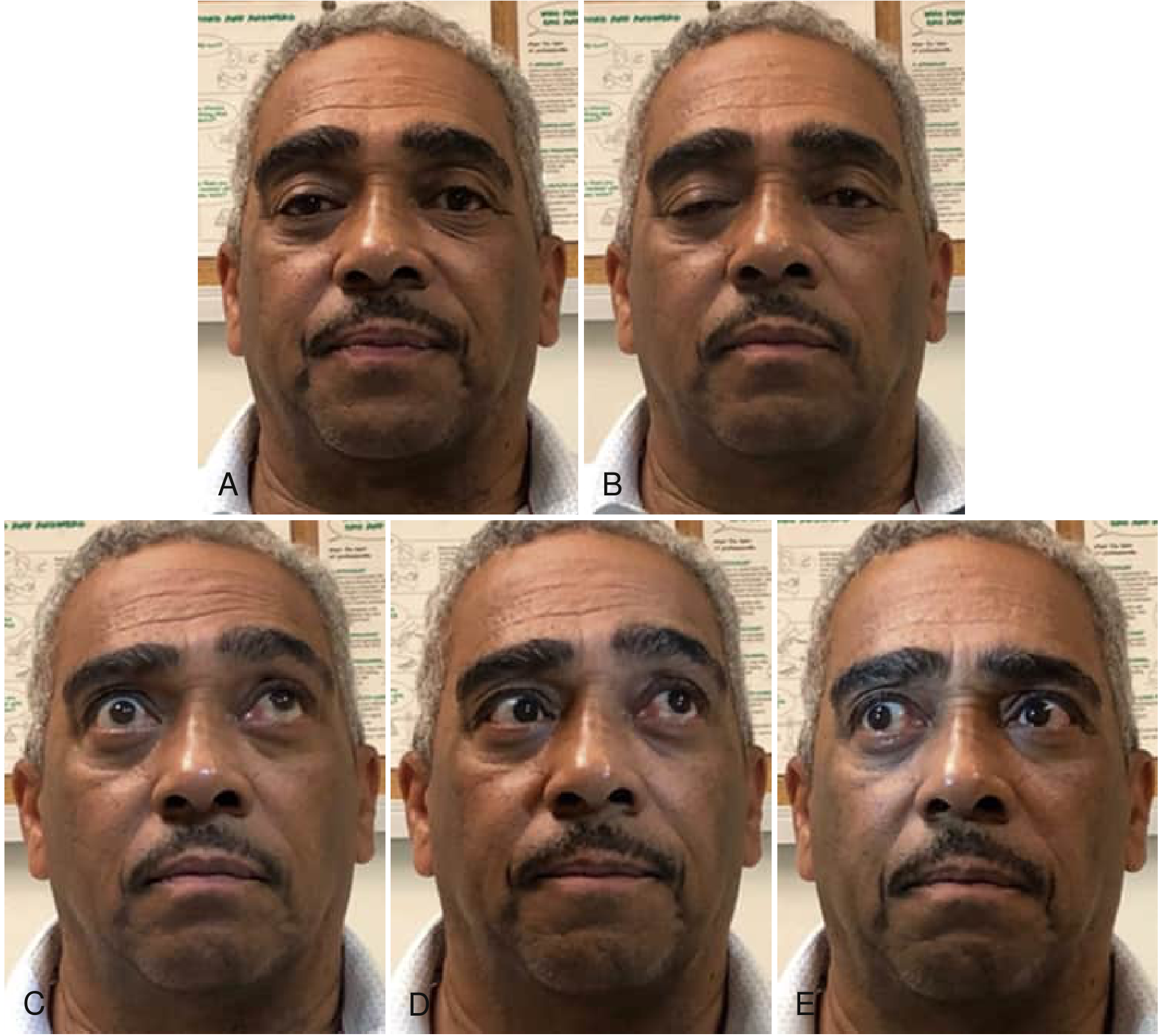
Ocular motility abnormalities in MG showing progressive right ptosis and incomplete eye movements in multiple gaze directions
Fig: Ocular motility abnormalities in MG - progressive right lid ptosis during sustained forward gaze (A→B), incomplete upward gaze (C), skew deviation on left lateral gaze (D), and incomplete movement on right lateral gaze (E). - Bradley and Daroff's Neurology

Generalized MG

  • Progresses to facial, oropharyngeal, and limb muscles in most patients
  • Maximum weakness within the first year in 2/3 of patients
  • Bulbar symptoms: dysphagia (oral, pharyngeal, esophageal), dysarthria, dysphonia
    • Aspiration (often silent) in ≥35% of MG patients with dysphagia
  • Facial weakness: expressionless, "snarling" smile
  • Neck weakness: difficulty holding head up
  • Limb weakness: proximal > distal

Factors that worsen MG

Emotional upset, systemic illness (especially viral respiratory infections), hypothyroidism or hyperthyroidism, pregnancy, menstrual cycle, surgery, fever, and certain medications (aminoglycosides, fluoroquinolones, beta-blockers, magnesium, neuromuscular blocking agents)

Myasthenic Crisis

Life-threatening respiratory failure from respiratory muscle weakness. Dysphagia is a major precipitant in ~56% of crises. Requires ICU admission, mechanical ventilation, and urgent immunotherapy (plasma exchange or IVIG).

Diagnosis

1. Clinical pattern - fatigable, fluctuating weakness in characteristic distribution
2. Serological testing
  • Anti-AChR antibodies (binding, blocking, modulating)
  • Anti-MuSK (if AChR negative)
  • Anti-LRP4 (if both negative)
3. Electrophysiology
  • Repetitive nerve stimulation (RNS): decremental response (>10% decrement at 3 Hz) - characteristic of postsynaptic NMJ disorder
  • Single-fiber EMG (SFEMG): most sensitive test - increased jitter and blocking
4. Edrophonium (Tensilon) test - short-acting AChE inhibitor; transient improvement in ptosis/diplopia confirms NMJ disorder (less used now due to side effects)
5. Imaging - CT/MRI chest for thymoma in all patients

Treatment

1. Symptomatic: Acetylcholinesterase (AChE) Inhibitors

  • Pyridostigmine (Mestinon) - first-line symptomatic treatment
  • Inhibits AChE → prolongs ACh availability at the reduced number of receptors
  • Caution: overdose causes cholinergic crisis (overstimulation → prolonged depolarization → Na+ channel inactivation → synaptic blockade)

2. Immunosuppression (long-term disease modification)

  • Corticosteroids (prednisone) - most commonly used first-line immunosuppressant
  • Azathioprine - slow onset (6-12 months), effective steroid-sparing agent
  • Mycophenolate mofetil
  • Methotrexate
  • Cyclosporine/Tacrolimus
  • Rituximab - particularly effective in anti-MuSK MG (see recent evidence below)

3. Rapid Immunomodulation (crisis/perioperative)

  • Plasma exchange (PLEX/plasmapheresis) - removes circulating antibodies; effect within days
  • IV Immunoglobulin (IVIG) - similar efficacy to PLEX; safer in patients with hemodynamic instability

4. Novel Biologics (expanding rapidly)

  • Eculizumab (anti-C5 complement inhibitor) - approved for AChR+ generalized MG
  • Efgartigimod (FcRn antagonist - reduces IgG levels including AChR antibodies)
  • Rozanolixizumab, Nipocalimab (other FcRn antagonists)
  • Zilucoplan (anti-C5)

5. Thymectomy

  • Recommended for all patients with thymoma
  • Also beneficial in non-thymomatous generalized AChR+ MG (especially age <60, within 5 years of onset) - improves long-term outcomes

MG Classification (Osserman/MGFA)

ClassDescription
IOcular only
IIMild generalized
IIIModerate generalized
IVSevere generalized
VMyasthenic crisis (intubation)

Recent Evidence (2024-2026)

A 2025 Cochrane systematic review on rituximab for MG and a 2025 network meta-analysis comparing novel biologics show expanding evidence for targeted therapies - particularly rituximab in MuSK+ MG and FcRn antagonists (efgartigimod, rozanolixizumab) showing strong efficacy for generalized MG with favorable safety profiles. These newer agents are increasingly being positioned earlier in the treatment algorithm for refractory or severe cases.

Key Differentials

FeatureMGLambert-Eaton (LEMS)Botulism
Antibody targetPostsynaptic AChRPresynaptic VGCCPresynaptic (toxin)
ReflexesNormalReduced (improve post-exercise)Reduced
Autonomic featuresNoYesYes
Associated cancerThymomaSCLCNo
Response to repetitive stimDecrementIncrementDecrement

Sources: Bradley and Daroff's Neurology in Clinical Practice; Medical Physiology (Boron & Boulpaep)
attachment
attachment

?

This is a Clinical Head & Neck - Complete high-yield summary chart by PakMedics. Here's a structured breakdown of everything it covers:

1. SCALP INJURIES

Layers (SCALP mnemonic):
  1. Skin
  2. Connective tissue
  3. Aponeurosis
  4. Loose areolar layer
  5. Pericranium
  • Danger area = Loose areolar layer - infection spreads via emissary veins to intracranial sinuses
  • Clinical: Profuse bleeding (rich vascular supply), Black eye (blood tracking)

2. ORBITAL CLINICALS

  • Blow-out fracture → Inferior wall fracture → Diplopia + enophthalmos
  • Orbital cellulitis → Spread from sinuses → Painful eye + proptosis
  • Cavernous sinus thrombosis → Infection from face ("danger triangle") → CN III, IV, V₁, V₂, VI affected

3. NOSE & SINUS

  • Little's area (Kiesselbach plexus) → Common site of epistaxis
  • Sinusitis:
    • Maxillary → tooth pain
    • Frontal → forehead pain
  • Can spread → orbit/brain

4. EAR CLINICALS

  • Otitis media → Infection via Eustachian tube → Common in children
  • Mastoiditis → Spread to mastoid air cells
  • Facial nerve risk → Runs in middle ear → paralysis possible

5. CRANIAL NERVE LESIONS

NerveLesionClinical Sign
IIIOculomotor palsyDown & out eye
VTrigeminal neuralgiaSevere facial pain
VIIBell's palsyFacial paralysis
IXGlossopharyngealLoss of gag reflex
XVagusHoarseness
XIAccessoryWeak shoulder
XIIHypoglossalTongue deviation

6. FACIAL NERVE (CN VII)

  • Bell's palsy = LMN lesion → Whole face affected (including forehead)
  • UMN lesionForehead spared (bilateral cortical representation of forehead)

7. PAROTID GLAND

  • Parotitis (Mumps)
  • Tumor → facial nerve compression
  • Surgery risk → Facial nerve damage

8. ORAL CAVITY

  • Ludwig's angina → Submandibular infection → Airway obstruction risk
  • Tonsillitis → Pain + fever
  • Can spread → deep neck spaces

9. PHARYNX & LARYNX

  • Laryngitis → Hoarseness
  • Epiglottitis → Thumb sign (X-ray) → Emergency airway
  • Recurrent laryngeal nerve injury → Hoarse voice

10. NECK TRIANGLES (Clinical)

  • Anterior triangle → Carotid surgery landmark
  • Posterior triangle → Spinal accessory nerve risk

11. THYROID GLAND

  • Goiter → swelling
  • Hyperthyroidism → weight loss
  • Hypothyroidism → weight gain
  • Surgery risks: Recurrent laryngeal nerve + External laryngeal nerve injury

12. LYMPHATIC DRAINAGE

  • Jugulodigastric node → drains tonsil
  • Virchow's node (left supraclavicular) → Abdominal malignancy

13. DANGER TRIANGLE OF FACE

  • Area: Nose to upper lip
  • Infection spreads → cavernous sinus → Life-threatening

14. MENINGEAL & VASCULAR

  • Epidural hematoma → Middle meningeal artery → Lucid interval
  • Subdural hematoma → Bridging veins

15. IMPORTANT CLINICAL SIGNS

SignIndicates
Battle signSkull base fracture
Raccoon eyesAnterior skull fracture
TrismusPterygoid spasm
DysphagiaCN IX, X lesion

HIGH YIELD QUICK REVISION

  • Face danger area → cavernous sinus
  • CN VII → facial expression
  • CN X → voice
  • Thyroid surgery → nerve injury
  • Epiglottitis → emergency

This is an excellent exam-prep chart covering the most tested clinical anatomy points for the head and neck. Would you like me to go deeper on any specific section?

Sartorius muscle

Finding Sources
Finding Sources
Reading File
Reading File
Reading File
Finding Sources
Finding Sources
Excellent - I have all I need. Here is the comprehensive answer:

Sartorius Muscle

Overview

The sartorius is the longest muscle in the human body (~51 cm). It is a long, strap-like muscle that runs obliquely across the anterior thigh, crossing two joints (hip and knee). Its name comes from the Latin sartor (tailor) - because it helps position the leg in the cross-legged "tailor's sitting" posture.
  • Gray's Anatomy for Students; Imaging Anatomy Atlas Vol. 3

Attachments

Detail
OriginAnterior Superior Iliac Spine (ASIS)
InsertionMedial surface of the proximal shaft of the tibia (medial to tibial tuberosity)
Insertion typeFlat aponeurotic insertion - part of the pes anserinus

Pes Anserinus ("Goose's Foot")

The sartorius inserts onto the tibia in a three-pronged pattern alongside two other muscles - their combined tendons form the pes anserinus:
TendonNerve Supply
Sartorius (anterior)Femoral nerve
Gracilis (middle)Obturator nerve
Semitendinosus (posterior)Tibial nerve (sciatic)
The anserine bursa lies deep to these three tendons and superficial to the medial collateral ligament - a common site of bursitis (especially in obese, osteoarthritic patients).
  • Goldman-Cecil Medicine; Rosen's Emergency Medicine

Actions

JointAction
Hip jointFlexion, Abduction, External (lateral) rotation
Knee jointFlexion, Internal (medial) rotation
Mnemonic - FABER at the hip: Flexion, ABduction, External Rotation
The classic movement it produces is placing the foot on the opposite knee while seated - the typical tailor's/cross-legged position.
Key anatomical note: At the level of the knee, the sartorius has migrated posteriorly with the pes anserinus to a point posterior to the flexion-extension axis of the knee - which is why despite being in the anterior compartment, it flexes (not extends) the knee.
  • THIEME Atlas of Anatomy

Innervation & Blood Supply

  • Nerve: Femoral nerve (L2, L3 - some sources cite L1-L4)
  • Blood supply: 6-8 vascular pedicles from:
    • Lateral circumflex femoral artery (proximal)
    • Superficial femoral artery (middle)
    • Descending genicular artery (distal)
    • Two major pedicles: proximal (18-25 cm from ASIS) and distal (35-44 cm from ASIS)
  • Imaging Anatomy Atlas Vol. 3

Key Anatomical Relations & Clinical Roles

1. Femoral Triangle (Scarpa's Triangle)

  • Mnemonic: SAIL - Sartorius, Adductor longus, Inguinal ligament
    • Sartorius = lateral boundary
    • Adductor longus = medial boundary
    • Inguinal ligament = superior boundary (base)
  • Contents (lateral to medial): Femoral Nerve, Artery, Vein, Empty space, Lymphatics (NAVEL)
  • Roberts and Hedges' Clinical Procedures in Emergency Medicine

2. Adductor Canal (Hunter's Canal / Subsartorial Canal)

  • In the middle third of the thigh, the sartorius forms the anterior (roof) wall of the adductor canal
  • The canal transmits: femoral artery, femoral vein, saphenous nerve, nerve to vastus medialis
  • The saphenous nerve courses beneath the sartorius muscle throughout the adductor canal

3. Surgical / Reconstructive Use

  • The sartorius can be transposed as a muscle flap for groin defects (proximal half) or complicated knee defects (distal half)
  • Used in sartorius muscle transposition to cover vascular grafts or groin wounds after femoral artery surgery
  • However, because of its many equal-importance vascular pedicles, only partial segments are suitable for free flap transfer

Summary Table

FeatureDetail
CompartmentAnterior thigh
Longest muscle~51 cm
OriginASIS
InsertionMedial proximal tibia (pes anserinus)
Hip actionsFlexion, Abduction, External rotation
Knee actionsFlexion, Internal rotation
NerveFemoral nerve (L2, L3)
Femoral triangleLateral boundary
Adductor canalAnterior wall (roof)
Pes anserinus partnersGracilis, Semitendinosus
Sources: Gray's Anatomy for Students; THIEME Atlas of General Anatomy and Musculoskeletal System; Imaging Anatomy Vol. 3

Create a visual anatomy poster of the femoral triangle

Generating Image

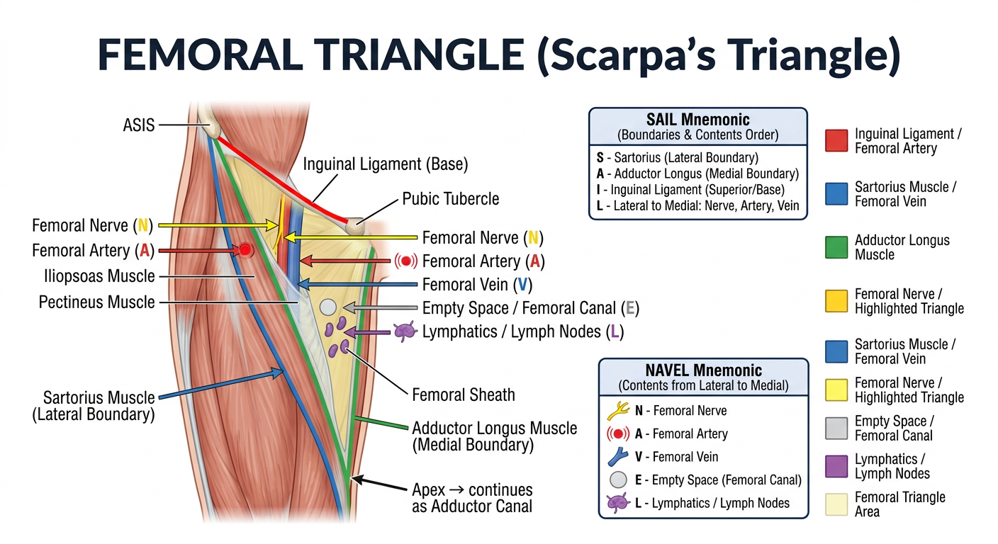
Medical anatomy education poster of the femoral triangle (Scarpa's triangle) of the anterior thigh. Clean, professional, high-detail illustration on a white background. Show: 1. A clear anterior view of the right thigh with the femoral triangle highlighted and shaded in light yellow. 2. The three boundaries clearly labeled with colored lines: - Superior boundary (base): Inguinal ligament (red line) connecting ASIS to pubic tubercle - Lateral boundary: Sartorius muscle (blue, running obliquely) - Medial boundary: Adductor longus muscle (green) 3. The floor of the triangle labeled: Iliopsoas (lateral) and Pectineus (medial) 4. Contents of the femoral triangle shown with colored arrows from lateral to medial spelling out NAVEL mnemonic: - Femoral Nerve (yellow) - Femoral Artery (red, pulsating dot) - Femoral Vein (blue) - Empty space / femoral canal (grey) - Lymphatics / Lymph nodes (purple) 5. The femoral sheath labeled (enclosing artery, vein, and canal but NOT nerve) 6. At the bottom: the apex of the triangle pointing inferiorly, labeled "Apex → continues as Adductor Canal" 7. A separate small inset box showing the SAIL mnemonic: S - Sartorius (lateral) A - Adductor longus (medial) I - Inguinal ligament (superior/base) L - Lateral to medial: Nerve, Artery, Vein 8. Another inset showing the NAVEL mnemonic with icons for each structure. Title at top: "FEMORAL TRIANGLE (Scarpa's Triangle)" in bold dark navy text. Color-coded legend on the side. Medical textbook illustration style, precise anatomical labels, clean sans-serif font.

Femoral Triangle Anatomy Poster
Here's your femoral triangle anatomy poster! It covers all the key high-yield points:
Boundaries (SAIL mnemonic):
  • S - Sartorius (lateral)
  • A - Adductor longus (medial)
  • I - Inguinal ligament (superior/base)
  • L - Lateral-to-medial contents
Contents (NAVEL mnemonic):
  • N - Femoral Nerve
  • A - Femoral Artery
  • V - Femoral Vein
  • E - Empty space (femoral canal)
  • L - Lymphatics
Floor: Iliopsoas (lateral) + Pectineus (medial)
Apex → continues inferiorly as the Adductor Canal (Hunter's Canal), with the sartorius forming its roof.
Would you like a poster of the adductor canal, or any other anatomical region?
This is a shared conversation. Sign in to Orris to start your own chat.