
What are dna oncogenic virus in pathology
Reading File
Now let me search the medical library for DNA oncogenic viruses in pathology.
Finding Sources
Finding Sources
Finding Sources
Finding Sources
Finding Sources
Finding Sources
Reading File
Finding Sources
Reading File
Finding Sources
Excellent! Found the key Robbins pathology section. Let me read that and the Harrison's section on HPV.
Reading File
Reading File
Reading File
Loading Image
I now have comprehensive information from Robbins Pathology. Let me compile the full answer.
DNA Oncogenic Viruses in Pathology
(Based primarily on Robbins, Cotran & Kumar - Pathologic Basis of Disease and Schwartz's Principles of Surgery)
Overview
DNA oncogenic viruses differ fundamentally from RNA oncogenic viruses (retroviruses). Their oncogenes are viral in origin (not derived from cellular proto-oncogenes). In permissive (productive) hosts, infection causes a lytic cycle ending in cell death and viral release. In nonpermissive cells, viral DNA integrates into the host chromosome; early viral genes are persistently expressed, transforming cells into a neoplastic state. The key shared mechanism is binding of viral oncoproteins to tumor-suppressor proteins p53 and RB.
The five major DNA oncogenic viruses implicated in human cancer are:
- Human Papillomavirus (HPV)
- Epstein-Barr Virus (EBV)
- Hepatitis B Virus (HBV)
- Merkel Cell Polyomavirus (MCPyV)
- Human Herpesvirus 8 (HHV-8 / Kaposi Sarcoma Herpesvirus)
1. Human Papillomavirus (HPV)
Type: DNA virus (double-stranded circular DNA)
High-risk types: 16, 18, 31 (carcinogenic) | Low-risk types: 6, 11 (benign warts)
Associated cancers:
- Cervical carcinoma (most important)
- Anal, vulvar, vaginal, penile carcinomas
- Oropharyngeal carcinoma (tonsil, base of tongue)
Mechanism of Transformation
In benign warts, HPV DNA is maintained as a non-integrated episome. In cancers, the HPV genome integrates into the host genome, interrupting the E1/E2 open-reading frame - this destroys the E2 repressor, leading to markedly increased expression of E6 and E7 oncoproteins.
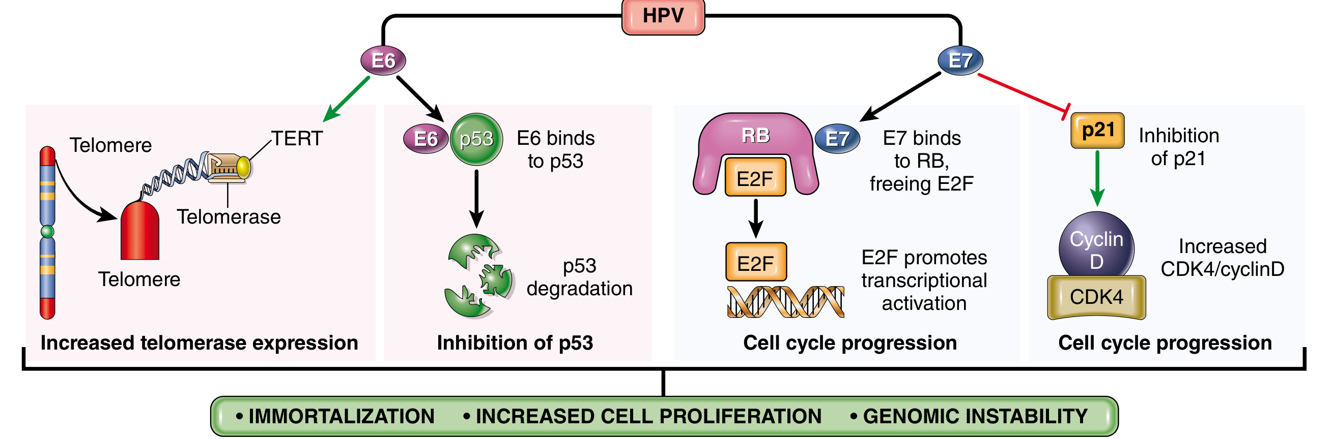
Fig. 7.44 from Robbins Pathology - Transforming effects of HPV E6 and E7 proteins
E6 oncoprotein actions:
- Binds to and mediates degradation of p53 (prevents DNA repair and apoptosis)
- Stimulates expression of TERT (telomerase reverse transcriptase) - promotes immortalization
- High-risk HPV E6 has much higher affinity for p53 than low-risk E6
E7 oncoprotein actions:
- Binds RB protein and displaces E2F transcription factors, pushing cells through the G1/S checkpoint
- Inactivates CDK inhibitors p21 and p27
- Binds and activates cyclins A and E (in high-risk types 16, 18, 31)
- High-risk E7 has higher affinity for RB than low-risk E7
Net result: Immortalization + increased cell proliferation + genomic instability
HPV infection alone is not sufficient for cancer - other genetic hits (e.g., mutant RAS), smoking, HIV co-infection, and immune failure are needed, typically over several decades.
2. Epstein-Barr Virus (EBV)
Type: Herpesvirus family (dsDNA)
Associated cancers:
- Burkitt lymphoma (first human tumor linked to EBV)
- Hodgkin lymphoma
- Nasopharyngeal carcinoma (100% EBV-associated worldwide)
- Immunosuppression-related B-cell lymphomas
- EBV-positive gastric carcinoma, thymic carcinoma
Mechanism
EBV uses its surface glycoproteins to bind CD21 (complement receptor) on B cells, establishing latent infection - no lytic replication, cells are not killed. Instead, EBV genes immortalize B cells by hijacking normal signaling pathways:
- LMP1 (Latent Membrane Protein-1): Acts as a constitutively active CD40 receptor. Activates NF-κB and JAK/STAT signaling, promotes B-cell survival and proliferation, and prevents apoptosis via BCL2 activation. In nasopharyngeal carcinoma, LMP1 also upregulates VEGF, FGF2, MMP9, and COX2.
- EBNA2: Encodes a nuclear protein that activates expression of several cellular and viral genes, including the proto-oncogene MYC.
Burkitt Lymphoma
EBV acts as a polyclonal B-cell mitogen, setting the stage for the acquisition of the t(8;14) translocation (MYC dysregulation) and other mutations. EBV is not directly oncogenic here - it expands the pool of proliferating B cells, increasing the probability of secondary mutations. Chronic malaria co-infection (in endemic areas) favors persistent EBV infection, contributing to the geographic pattern of endemic Burkitt lymphoma.
Nasopharyngeal Carcinoma
All nasopharyngeal carcinomas worldwide contain EBV (clonal viral genome, indicating infection preceded tumor). LMP1 is expressed and tumor cells often express PD-L1, enabling immune evasion.
3. Hepatitis B Virus (HBV)
Type: Partially double-stranded DNA virus (Hepadnavirus)
Associated cancer: Hepatocellular carcinoma (HCC) - HBV/HCV together account for 70-85% of HCCs worldwide; highest incidence in Far East and Africa.
Mechanism
Unlike HPV and EBV, no viral oncogene has been identified in HBV. The proposed mechanisms include:
- Chronic liver injury + regenerative hyperplasia - repeated cycles of hepatocyte death and regeneration from a persistent inflammatory response increase the probability of accumulating mutations
- HBV X protein (HBx): Transactivates various growth-regulatory genes and may impair p53 function
- Viral DNA integrates randomly into the host genome, potentially causing insertional mutagenesis (though this is random and not the primary mechanism)
- Cirrhosis acts as a permissive background for neoplastic transformation
HBV vaccination in childhood has already reduced liver cancer incidence in East Asia.
4. Merkel Cell Polyomavirus (MCPyV)
Type: Small dsDNA virus (Polyomaviridae)
Associated cancer: Merkel cell carcinoma - a rare but aggressive neuroendocrine skin cancer.
Mechanism
- Viral DNA integrates into the host genome in a clonal fashion (before tumor development)
- Expresses T antigens (large T antigen) that bind and inactivate RB and p53, similar to other polyomaviruses (e.g., SV40)
- Truncating mutations in the large T antigen prevent viral replication but preserve transforming capacity
5. Human Herpesvirus 8 (HHV-8 / KSHV)
Type: Gamma-herpesvirus (dsDNA)
Associated cancers:
- Kaposi sarcoma (classic, endemic, AIDS-related, and transplant-related forms)
- Primary effusion lymphoma
- Multicentric Castleman disease
Mechanism
- HHV-8 encodes viral homologs of cellular genes, including v-cyclin D, v-FLIP (inhibits apoptosis), and v-IL-6
- v-cyclin D binds CDK6 and drives cells through the G1/S checkpoint
- v-FLIP activates NF-κB, promoting cell survival
- Viral GPCR (vGPCR) activates VEGF and angiogenic pathways, contributing to the vascular nature of Kaposi sarcoma
Summary Table
| Virus | Type | Key Oncogenic Mechanism | Associated Cancer(s) |
|---|---|---|---|
| HPV 16/18 | dsDNA | E6 degrades p53; E7 inactivates RB; TERT activation | Cervical, oropharyngeal, anogenital carcinomas |
| EBV | dsDNA (Herpes) | LMP1 mimics CD40 (NF-κB, JAK/STAT); EBNA2 activates MYC | Burkitt lymphoma, Hodgkin lymphoma, nasopharyngeal carcinoma |
| HBV | Partial dsDNA | Chronic inflammation + regeneration; HBx protein | Hepatocellular carcinoma |
| MCPyV | dsDNA | T antigen inactivates RB and p53 | Merkel cell carcinoma |
| HHV-8 | dsDNA (Herpes) | v-cyclin D, v-FLIP (NF-κB), vGPCR/VEGF | Kaposi sarcoma, primary effusion lymphoma |
Key Shared Concept
"The binding of viral oncoproteins to cellular tumor-suppressor proteins p53 and RB is fundamental to the carcinogenesis induced by most DNA viruses."
- Robbins, Cotran & Kumar - Pathologic Basis of Disease
Viral carcinogenesis is always a multistep process - the virus creates a permissive environment and early genetic changes, but additional hits (genetic mutations, immune suppression, environmental co-factors) are required for full malignant transformation. This explains why most infected individuals never develop cancer, and why cancers typically arise decades after initial infection.
Gangrene
Finding Sources
Finding Sources
Reading File
Reading File
Reading File
Reading File
Reading File
Reading File
Reading File
Reading File
Reading File
Loading Image
I now have comprehensive information from multiple authoritative surgical textbooks. Here is the full structured answer:
Gangrene
Definition: Gangrene is the death (necrosis) of macroscopic portions of tissue, characterised by blackening of the affected part due to breakdown of haemoglobin and formation of iron sulphide. It typically affects the most distal part of a limb because of arterial obstruction.
- Bailey and Love's Short Practice of Surgery, 28th Ed.
Signs of Gangrene
- Change of colour - pale → bluish-purple → finally black
- Loss of temperature (cold to touch)
- Loss of sensation (anaesthesia)
- Loss of peripheral pulsation
- Loss of function
Clinical Types
1. Dry Gangrene
- Cause: Gradual, slow occlusion of arterial circulation - most commonly atherosclerosis (hence "senile gangrene")
- The blood supply diminishes slowly, allowing the tissue to desiccate before infection sets in
- Features: Part becomes dry, shrivelled, hard, mummified, and discoloured
- A clear line of demarcation forms between viable and dead tissue within days
- Separation is clean with minimal infection; the stump tends to be conical when bone is involved
- No offensive smell (no putrefaction)
2. Wet (Moist) Gangrene
- Cause: Sudden arterial blockage (e.g., embolus) OR gangrene superimposed on inflammation
- The tissue does not have time to desiccate before bacteria invade
- Features: Oedema, blistering (blebs), foul smell from putrefaction, spreading infection
- The line of demarcation forms more proximally than in dry gangrene due to infection extending into adjacent living tissue
- A surgical emergency - requires urgent debridement or amputation
3. Gas Gangrene
- Cause: Clostridium perfringens (Gram-positive, anaerobic, spore-bearing bacillus)
- Spores are widespread in soil and faeces; thrive in anaerobic conditions (devascularised, necrotic tissue)
- Common in: war/military wounds (high-velocity missiles cause massive tissue devitalisation), immunocompromised patients, diabetics, patients with malignancy
- Features:
- Severe local wound pain (early and disproportionate)
- Crepitus on palpation (gas in tissues - may also be visible on plain X-ray)
- Thin, brown, sweet-smelling exudate - Gram stain reveals organisms
- Oedema and spreading gangrene from collagenase, hyaluronidase, other proteases, and alpha toxin
- Rapid systemic deterioration: circulatory collapse, multi-organ failure
- Treatment: Large-dose IV penicillin + aggressive surgical debridement; early amputation may be life-saving; hyperbaric oxygen as adjunct
Specific Types by Cause
Diabetic Gangrene
A triad of contributing factors:
- Ischaemia - macrovascular atherosclerosis (typically crural vessels) + microvascular dysfunction (shunting)
- Peripheral sensorimotor neuropathy (PSN) - sensory loss (stocking distribution) leads to unnoticed injury and neglect; motor involvement causes foot deformity and abnormal pressure loading → callosities
- Immunosuppression - hyperglycaemia impairs immune function → predisposes to infection
Ischaemia and PSN act synergistically. Superadded infection spreads rapidly in subfascial planes → fulminant foot sepsis and death.
Treatment: Revascularisation (angioplasty/surgery) + drainage of pus + liberal debridement + antibiotics; primary amputation in systemic sepsis.
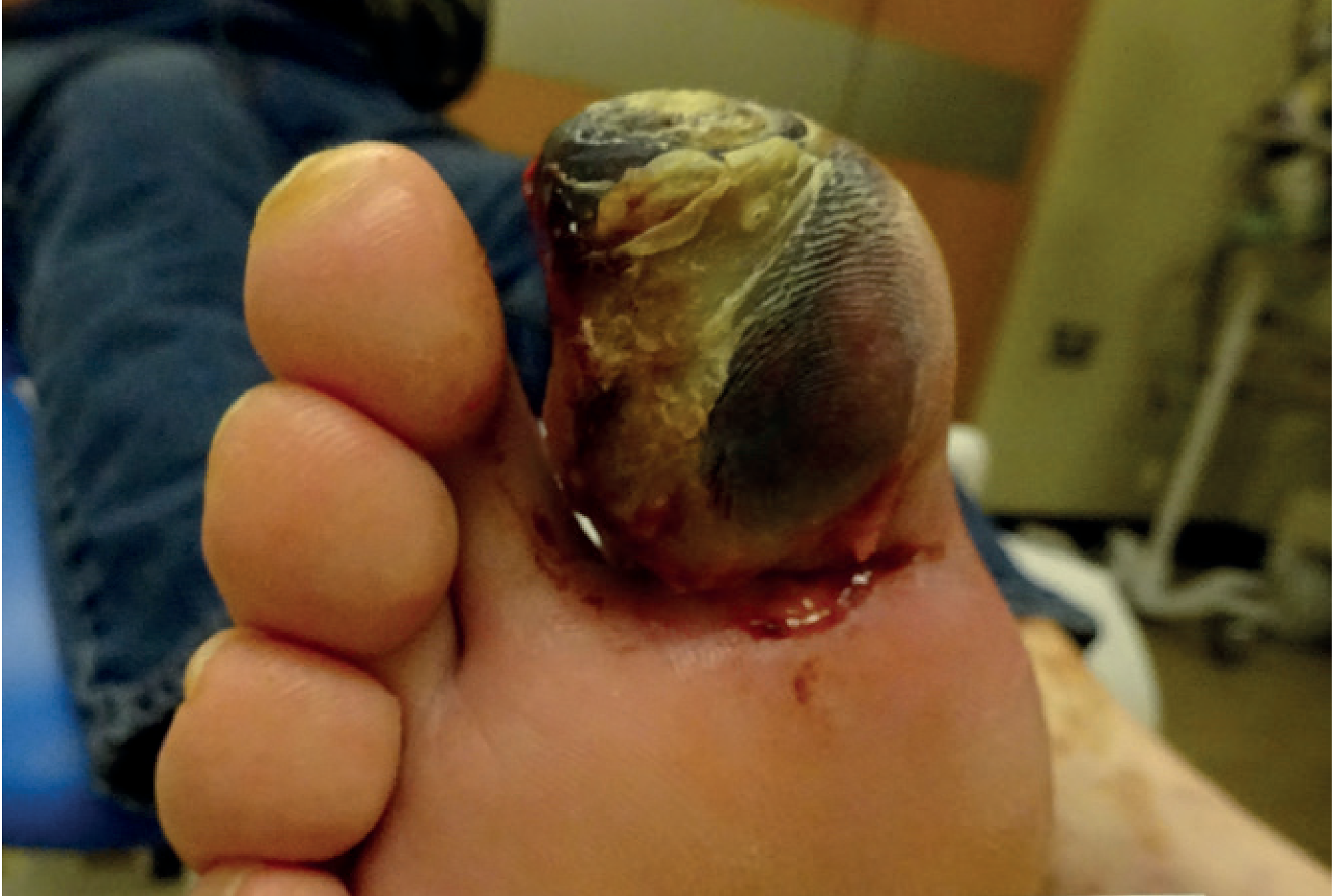
Diabetic gangrene - Bailey and Love's Short Practice of Surgery
Synergistic Gangrene (Necrotising Fasciitis)
- Cause: Synergistic action of non-haemolytic streptococci and staphylococci (classically); usually polymicrobial (mixed aerobic and anaerobic)
- Spreads rapidly through the deep fascia (hence "fasciitis")
- More common in diabetics, debilitated, or immunocompromised patients - but can occur in healthy individuals
- Features: Systemic septic shock, high fever, foul smell, crepitus (gas-producing organisms), subcutaneous necrosis spreading into chest/axilla/thigh/perineum
- Diagnosis: Gold standard is surgical exploration - easy dissection along fascial planes, grey/dusky/edematous fascia, brown stringy exudate
- Treatment: Immediate broad-spectrum IV antibiotics + early, aggressive surgical debridement (repeated until all necrotic tissue cleared) + skin grafting for survivors; hyperbaric oxygen advocated
Fournier's Gangrene
- A subtype of necrotising fasciitis localised to the genitalia, perineum, anus, and occasionally lower abdomen
- Peak age: 50-60 years; most have diabetes mellitus or immunocompromise
- Source: perirectal/perianal infection, untreated perirectal abscess, severe UTI, genital trauma
- Nearly always polymicrobial (mixed aerobic and anaerobic)
- CT scan to delineate extent; treatment is immediate surgical debridement ± diverting colostomy for rectal involvement
Causes of Gangrene - Summary
| Category | Examples |
|---|---|
| Cardiovascular | Atherosclerosis (senile gangrene), embolism, Buerger's disease, Raynaud's, cervical rib, syphilitic endarteritis |
| Traumatic | Direct arterial injury, crushing injuries |
| Infective | Gas gangrene, necrotising fasciitis, carbuncle, cancrum oris |
| Metabolic | Diabetic gangrene |
| Neurogenic | Tabes dorsalis, syringomyelia, leprosy, peripheral neuritis |
| Physical | Frostbite, burns, radiation, pressure (bedsores) |
Separation of Gangrene
A zone of demarcation - a layer of granulation tissue - develops between dead and living parts:
- In dry gangrene: line appears in days, separation is neat with minimal infection
- In moist gangrene: infection extends into adjacent living tissue, so the demarcation line is more proximal; separation is slower and messier
- If the proximal arterial supply is poor, no line forms at all - gangrene continues to spread unless revascularisation is achieved
Senile (Atherosclerotic) Gangrene - Clinical Picture
- Elderly patients (>50 years), lower limbs
- Progression: intermittent claudication → rest pain → trophic changes → dry gangrene
- Investigation: ABI (ankle-brachial index), oscillometry, Doppler, arteriography (shows site of block, calcification)
- Leriche's syndrome: bilateral internal iliac occlusion causing buttock claudication + impotence
Buerger's Disease (Thromboangiitis Obliterans)
- Young men <40 years; strong association with cigarette smoking
- Inflammatory reaction in small/medium arteries with involvement of adjacent vein and nerve → segmental thrombosis
- Starts in tibial arteries, foot vessels (foot claudication - at the arch, NOT the thigh/buttock)
- Characteristic arteriography: smooth normal larger arteries + extensive occlusion of smaller vessels with "corkscrew" collaterals
- Leads to digital ulceration and gangrene of toes/fingers
(Bailey and Love's Short Practice of Surgery, 28th Ed.; Das - Manual on Clinical Surgery, 13th Ed.; Fitzpatrick's Dermatology)
Gangrene in pathology
Finding Sources
Finding Sources
Finding Sources
Finding Sources
Finding Sources
Finding Sources
Finding Sources
Finding Sources
Found the key Robbins sections. Let me read both:
Reading File
Reading File
Reading File
Finding Sources
Now let me fetch the key pathology image from Robbins showing necrosis patterns:
Loading Image
Loading Image
I now have everything needed for a comprehensive pathology-focused answer on gangrene. Here it is:
Gangrene in Pathology
(Robbins, Cotran & Kumar - Pathologic Basis of Disease; Robbins & Kumar Basic Pathology)
What is Gangrenous Necrosis?
"Gangrenous necrosis is not a specific pattern of cell death, but the term is commonly used in clinical practice. It is usually applied to a limb, generally the lower leg, that has lost its blood supply and has undergone necrosis (typically coagulative necrosis) involving multiple tissue planes."
- Robbins, Cotran & Kumar Pathologic Basis of Disease
This is the key pathological concept: gangrene is a clinical term, not a unique histological pattern. It describes massive tissue necrosis, but the underlying cellular mechanism is one of the established patterns of necrosis.
Gangrene in the Context of Patterns of Tissue Necrosis
Robbins lists the following patterns of tissue necrosis (Key Concepts, Chapter 2):
| Pattern | Key Feature |
|---|---|
| Coagulative | Architecture preserved; eosinophilic ghost cells |
| Liquefactive | Tissue dissolved into viscous liquid; pus formation |
| Gangrenous | Coagulative ± liquefactive; multi-tissue-layer involvement in limbs |
| Caseous | Cheese-like; structureless granular debris; tuberculosis |
| Fat | Chalky white calcium soap deposits; pancreatitis |
| Fibrinoid | Bright pink amorphous deposits in vessel walls; vasculitis/immune reactions |
Pathological Basis of the Two Main Types
Dry Gangrene = Predominantly Coagulative Necrosis
- Caused by gradual, slow arterial occlusion (e.g., atherosclerosis)
- Blood supply diminishes slowly - the tissue desiccates before bacterial invasion can occur
- Coagulative necrosis: the injury denatures structural proteins AND enzymes simultaneously, blocking proteolysis of dead cells
- Result: the architecture of dead tissue is preserved for days - intensely eosinophilic ghost cells with indistinct or "reddish" nuclei persist
- Gross appearance: dry, shrivelled, mummified, hard tissue; turns black (from iron sulphide formed by breakdown of haemoglobin)
- A clear line of demarcation forms between viable and dead tissue
- No offensive smell (no significant putrefaction)
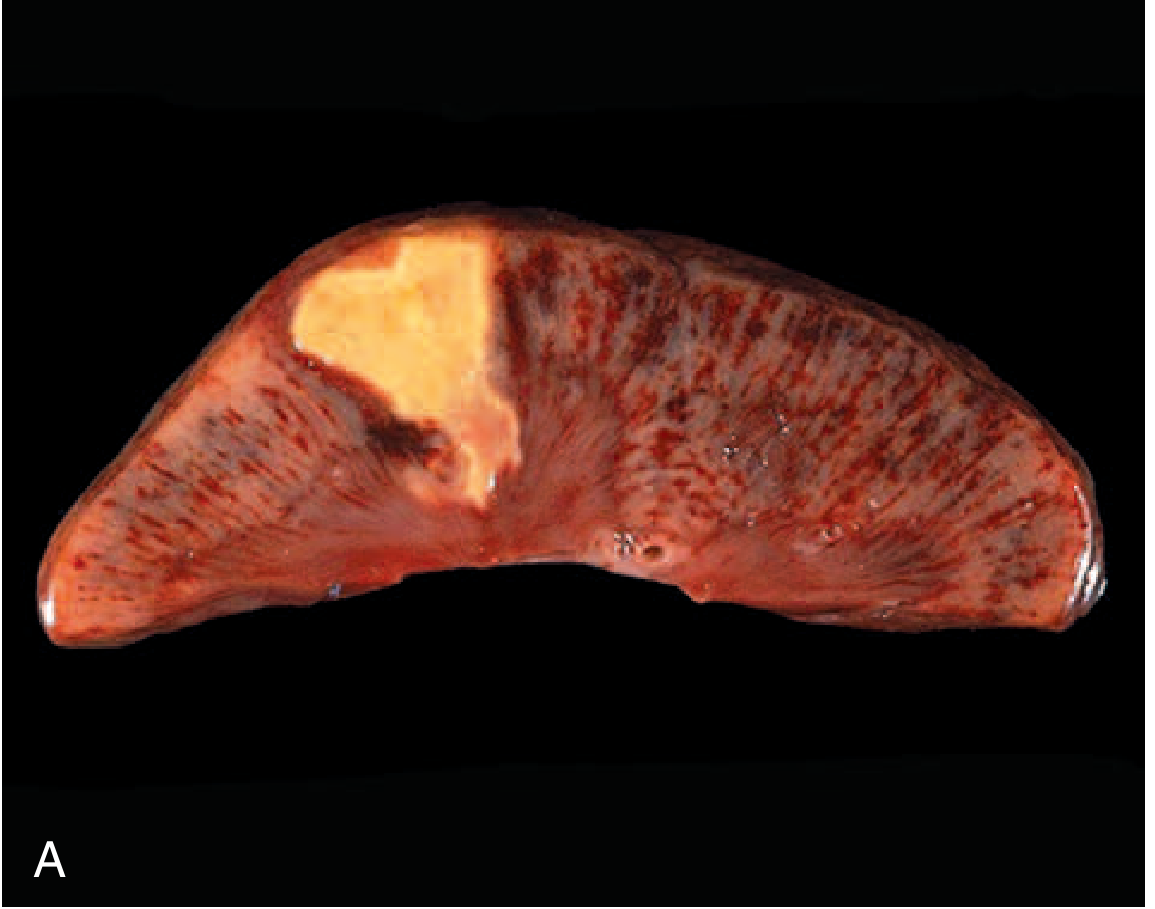
Coagulative necrosis: wedge-shaped infarct in kidney showing preserved architecture - the same process underlying dry gangrene (Robbins Basic Pathology)
Wet (Moist) Gangrene = Coagulative + Superimposed Liquefactive Necrosis
- Caused by sudden arterial occlusion (embolus) OR gangrene with bacterial superinfection
- When bacteria invade, liquefactive necrosis is superimposed:
- Leukocytes are recruited → release lysosomal enzymes
- Bacterial degradative enzymes (proteases, collagenase, hyaluronidase) destroy tissue
- Dead cells are completely digested → tissue transforms into a viscous liquid
- Gross appearance: oedematous, blebs/bullae, foul smell (putrefaction), pus
- The line of demarcation is indistinct and forms more proximally because infection spreads into adjacent living tissue
- The necrotic material is creamy yellow from leukocytes (pus)
- No preserved tissue architecture remains - cellular outlines are lost
Gas Gangrene = Liquefactive Necrosis + Gas Production
- Clostridium perfringens (anaerobic organism) in devascularised tissue
- Produces alpha toxin (phospholipase C), collagenase, hyaluronidase → extensive tissue destruction
- Gas (CO₂, H₂, H₂S) produced by bacterial metabolism accumulates in tissues → crepitus
- Histology: widespread myonecrosis with relatively sparse inflammatory response (toxins kill leukocytes)
- Spreads rapidly through tissue planes
Why Does Gangrene Turn Black?
The black discolouration is a specific pathological change:
- Loss of blood supply → haemoglobin leaks from lysed red cells
- Haemoglobin is broken down by bacteria and tissue enzymes
- Haemoglobin → iron sulphide (FeS) - a black pigment
- This is not melanin and not carbon
Cellular Mechanism: Why Ischaemia Causes Necrosis
The fundamental basis (from Robbins cell injury chapter):
- Ischaemia → ATP depletion → failure of Na⁺/K⁺-ATPase → cellular swelling
- Mitochondrial dysfunction (irreversible) → failure of oxidative phosphorylation
- Cytosolic Ca²⁺ rises → activates phospholipases, proteases, endonucleases → membrane damage
- Lysosomal membrane rupture → enzymatic dissolution of the cell (necrosis)
- Cellular contents leak out → local inflammatory response (this is why necrosis, unlike apoptosis, induces inflammation)
Two criteria define the point of no return (irreversible injury):
- Inability to reverse mitochondrial dysfunction even after restoring oxygen
- Profound disturbances in membrane function
Fate of Necrotic Tissue in Gangrene
After tissue death:
- Leukocyte infiltration → enzymatic digestion of necrotic cells → phagocytosis of debris
- Zone of demarcation forms: granulation tissue develops between dead and living parts
- If necrotic cells are not cleared promptly → calcium salts deposit → dystrophic calcification
- In dry gangrene: autoamputation can occur as the demarcation line matures
- In wet gangrene: infection spreads → the demarcation forms more proximally → requires surgical intervention
Summary Table: Pathological Comparison
| Feature | Dry Gangrene | Wet Gangrene | Gas Gangrene |
|---|---|---|---|
| Necrosis type | Coagulative | Coagulative + Liquefactive | Liquefactive + myonecrosis |
| Cause | Gradual ischaemia (atherosclerosis) | Sudden occlusion + bacterial infection | C. perfringens toxins |
| Architecture | Preserved (ghost cells) | Destroyed | Destroyed |
| Inflammatory infiltrate | Minimal (late) | Prominent (pus) | Sparse (toxins kill leukocytes) |
| Gas/Crepitus | Absent | Absent | Present (CO₂, H₂, H₂S) |
| Demarcation line | Sharp and early | Proximal and late | Absent / rapidly advancing |
| Colour | Black (iron sulphide) | Black + greenish (putrefaction) | Bronze-brown, bubbly |
| Smell | None | Foul (putrefaction) | Sweet-smelling (characteristic) |
(Robbins, Cotran & Kumar Pathologic Basis of Disease, Ch. 2; Robbins & Kumar Basic Pathology, Ch. 1)
This is a shared conversation. Sign in to Orris to start your own chat.