
Pathophysiology of enteric fever in details
"enteric fever" AND pathophysiology
typhoid fever enteric fever pathophysiology Peyer patches ileum ulcer
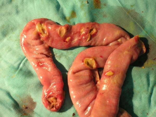
This clinical photograph shows a surgical specimen of the distal ileum placed on a green surgical drape. The anatomical structure exhibits multiple typhoid-related intestinal perforations. The ileal wall is predominantly pinkish-red, appearing moist and edematous. Approximately six to seven distinct perforations are visible, ranging in morphology from small pinholes to larger, irregular ovoid defects. The edges of these perforations are ragged and hyperemic, suggesting significant transmural inflammation. Yellowish slough or intestinal contents are visible within and surrounding several of the holes, indicative of leakage and associated peritonitis. This image illustrates a severe complication of enteric fever (Typhoid fever), specifically Peyer's patch necrosis leading to multiple intestinal perforations, which typically occur within 30 cm of the ileocecal valve. It serves as an educational reference for surgical pathology and complications of infectious gastrointestinal diseases.
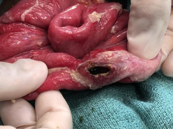
This clinical photograph is an intra-operative view depicting a typhoid intestinal perforation in the terminal ileum. The image shows a segment of small bowel held by gloved hands against a sterile surgical drape. A single, well-defined oval-shaped perforation is visible on the antimesenteric border, oriented along the longitudinal axis of the bowel. The perforation site displays dark, necrotic edges and is surrounded by an area of blanched, pale, and thickened tissue, indicating localized ischemia and inflammatory exudate. The adjacent intestinal wall appears erythematous and edematous, consistent with generalized peritonitis or severe enteritis. Small yellowish inflammatory plaques are visible on the serosal surface. This lesion is a pathognomonic finding of Typhoid Intestinal Perforation (TIP), resulting from the necrosis of Peyer's patches. It is a critical educational resource for understanding the surgical complications of Salmonella typhi infection in general surgery and infectious disease specialties.
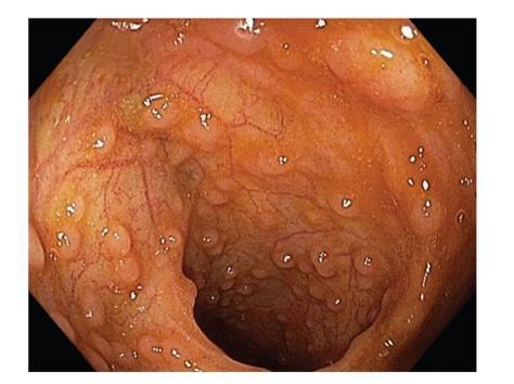
This endoscopic image shows the distal ileum of a 20-year-old male, captured during an ileocolonoscopy. The visual focuses on Peyer's patches (PPs), which are organized lymphoid follicles characteristic of the gut-associated lymphoid tissue (GALT). The Peyer's patches are visible as multiple raised, discrete, oval-shaped nodular structures with a smooth surface texture. In this distal region of the small intestine, they exhibit a high density and are arranged in a distinctive circumferential or lymphoid ring pattern. The surrounding ileal mucosa is reddish-orange in color with visible fine, branching subepithelial blood vessels. The image illustrates the normal physiological peak of lymphoid development in young adulthood, demonstrating the characteristic anatomical distribution and morphology of follicular aggregates in the terminal ileum. Key educational features include the contrast between the raised lymphoid follicles and the flatter surrounding mucosa, and the identification of the lymphoid ring as a normal anatomical variant in the distal ileum.
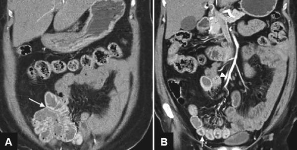
Diagnostic imaging consisting of two coronal sections from a Computed Tomography Angiography (CTA) of the abdomen and pelvis. Image A highlights the right lower quadrant, where a white arrow points to the terminal ileum showing significant bowel wall thickening and hyperenhancement, consistent with inflammatory changes. Image B demonstrates the vascular anatomy, where a white arrowhead indicates the ileocolic artery, a branch of the superior mesenteric artery, exhibiting bright contrast opacification. Multiple loops of the small intestine and colon are visible, with some segments showing mild dilation and mural thickening. The liver, gallbladder, and stomach are partially visualized in the upper abdomen. These findings illustrate the diagnostic utility of CTA in identifying the source of lower gastrointestinal bleeding and mesenteric vascular abnormalities, specifically in the context of enteric fever (typhoid fever) complications such as ileal ulceration and hemorrhage.
Pathophysiology of Enteric Fever - Detailed Overview
1. Inoculum and Gastric Passage
- S. Typhi is resistant to gastric acid, unlike most gram-negative enteric bacteria, allowing it to survive the stomach environment and reach the small intestine.
- Reduced gastric acidity (from antacid use, H. pylori-induced atrophic gastritis, or malnutrition) significantly lowers the infectious threshold and facilitates infection.
2. Intestinal Entry via M Cells (Week 1 - Incubation Phase)
- The bacteria preferentially invade M cells (microfold cells) - the specialized antigen-sampling epithelial cells that overlie Peyer's patches in the terminal ileum.
- Unlike S. Enteritidis (which triggers frank enterocolitis), S. Typhi passes through M cells largely without causing immediate inflammatory diarrhea, enabling silent transepithelial migration.
- After crossing the epithelium, bacteria are engulfed by subepithelial macrophages within the lamina propria of Peyer's patches.
3. Intracellular Survival and the Salmonella-Containing Vacuole
- Vi (virulence) antigen: A polysaccharide capsule unique to S. Typhi that inhibits complement activation and opsonization, and dampens the oxidative burst of phagocytes.
- Salmonella Pathogenicity Islands (SPI-1 and SPI-2): SPI-2 encodes a Type III Secretion System (T3SS) that injects bacterial effector proteins into the macrophage, preventing phagosome-lysosome fusion and creating a protected intracellular replication niche - the Salmonella-containing vacuole (SCV).
- Inside the SCV, bacteria actively replicate and suppress host innate immune signaling.
4. Primary Bacteremia and Seeding of the Reticuloendothelial System (End of Week 1)
- This produces a primary, low-grade bacteremia - often subclinical, corresponding to the incubation period (mean 10-14 days; range 5-21 days depending on inoculum size and host immunity).
- Bacteria-laden macrophages disseminate haematogenously to the liver, spleen, and bone marrow - the organs of the reticuloendothelial system (RES).
- In the spleen: the red pulp expands due to prominent phagocyte hyperplasia, with obliteration of follicular markings.
- In the liver: small foci of parenchymal necrosis appear in which hepatocytes are replaced by macrophage aggregates - the so-called "typhoid nodules" (typhoid hepatitis). These nodules are also found in the bone marrow and lymph nodes.
- Bacteria continue to replicate intracellularly throughout the RES during this phase.
5. Secondary Bacteremia and Sustained Fever (Week 1-2)
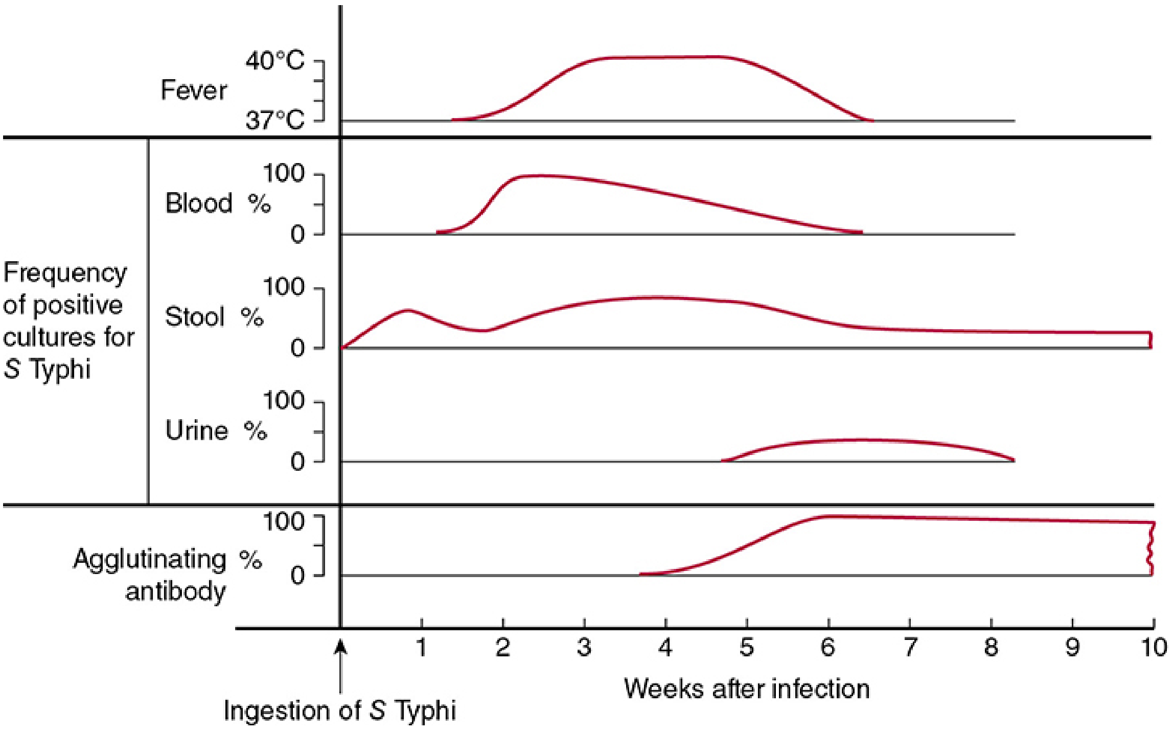
- Fever is typically 39-40.5°C (103-105°F), rising in a characteristic stepwise fashion over the first 3-5 days before becoming sustained.
- The fever is driven by lipopolysaccharide (LPS/endotoxin) of S. Typhi, which stimulates macrophages to release pyrogenic cytokines: IL-1β, TNF-α, and IL-6 - these act on the hypothalamus to raise the thermoregulatory set-point.
- Relative bradycardia (Faget's sign): A slow pulse despite high fever is a pathognomonic feature. This paradoxical response is thought to be related to endotoxin-mediated cardiac effects or direct myocardial involvement.
6. Intestinal Re-Seeding and Peyer's Patch Necrosis (Week 2-3)
- Peyer's patches, already primed from initial bacterial contact, mount a massive immune response - hyperplasia and infiltration by macrophages, lymphocytes, neutrophils, and plasma cells.
- Peyer's patches in the terminal ileum enlarge into sharply demarcated, plateau-like elevations up to 8 cm in diameter.
- Progressive ischemia and inflammation lead to mucosal shedding, creating oval ulcers oriented longitudinally along the long axis of the ileum (parallel to the bowel's axis - distinguishing typhoid ulcers from transverse ulcers of TB).
- The mesenteric lymph nodes also enlarge substantially due to reactive hyperplasia and accumulation of bacteria-laden phagocytes.
7. Systemic Inflammatory Response and Organ Involvement
Liver
- Hepatomegaly in ~50% of patients; elevated liver enzymes
- Typhoid nodules (focal macrophage aggregates replacing hepatocytes)
- Occasionally frank hepatitis or hepatic abscess
Spleen
- Splenomegaly in ~50%; spleen is soft and congested due to red pulp expansion
- Risk of spontaneous splenic rupture (rare but catastrophic)
Skin - "Rose Spots"
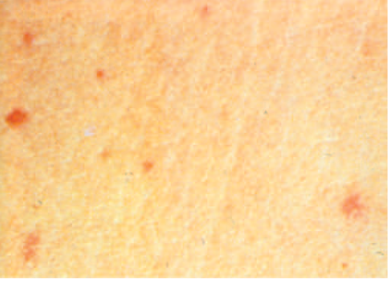
- Appear in ~30% of patients at the end of week 1 to week 2, primarily on the trunk/chest
- Salmon-colored, blanching, maculopapular lesions - 2-4 mm
- Represent foci of bacterial emboli within skin capillaries, with surrounding macrophage infiltration
- S. Typhi can be cultured from punch biopsies of these lesions
- More difficult to visualize in patients with darker skin
Central Nervous System
- Neurologic manifestations in 2-40% of patients
- "Muttering delirium" or "coma vigil" (typhoid encephalopathy) - picking at bedclothes or imaginary objects
- Meningitis, Guillain-Barré syndrome, neuritis, seizures
- Mechanism: endotoxemia, direct bacterial invasion, and cytokine-mediated neuroinflammation
Bone Marrow
- Hyperplasia with macrophage infiltration; typhoid nodules
- Leukopenia and neutropenia (in ~20% of patients) - likely due to bone marrow suppression and splenic sequestration
- Thrombocytopenia may occur, contributing to hemorrhagic complications
8. Complications (Week 3-4 and Beyond)
Intestinal Perforation (1-5%)
- Results from full-thickness necrosis of Peyer's patches in the terminal ileum/proximal colon
- Typically occurs in the 3rd to 4th week of untreated illness
- Perforation is typically on the antimesenteric border within 30 cm of the ileocecal valve
- Leads to peritonitis - a life-threatening surgical emergency
Gastrointestinal Hemorrhage (6%)
- Ulcer erosion into mesenteric vessels
- More common than perforation; presents with melena or hematochezia
Disseminated Intravascular Coagulation (DIC)
- Endotoxin activates the coagulation cascade
- Presents with bleeding tendency, petechiae, and organ ischemia
Other Metastatic Infections
- Biliary tree: gallbladder infection → chronic carrier state (2-5% of patients; reservoir in gallbladder biofilms on stones)
- Bone and joints: osteomyelitis (especially in sickle cell disease patients)
- Meninges: bacterial meningitis
- Heart: endocarditis, myocarditis, pericarditis
- Lungs: pneumonia
- Urinary tract: pyelonephritis, glomerulonephritis
9. Chronic Carrier State
- 2-5% of patients develop chronic asymptomatic carriage (shedding S. Typhi in stool or urine for >1 year)
- The gallbladder is the primary reservoir - bacteria persist by:
- Forming biofilms on gallstones
- Invading gallbladder epithelial cells
- Risk factors for chronic carriage: female sex, infants, biliary abnormalities, concurrent Schistosoma haematobium bladder infection
- Chronic carriers have an increased risk of gallbladder cancer
10. Immune Response and Resolution
- Cell-mediated immunity (macrophage activation by IFN-γ from T cells) is the primary defense
- Specific IgM antibodies against O and H antigens rise by week 2-3 (basis of the Widal test)
- In uncomplicated untreated cases, illness gradually resolves by the end of week 4
- Relapse occurs in up to 10% of patients, typically 2-3 weeks after defervescence, with the same strain - caused by residual intracellular bacteria that were not completely eradicated
Summary Flowchart of Pathogenesis
Ingestion of S. Typhi
        ↓
Acid resistance → Survival in stomach
        ↓
Invasion via M cells in terminal ileum
        ↓
Engulfed by macrophages in Peyer's patches
        ↓  [Vi antigen + SPI-2 T3SS prevent killing]
Intracellular replication in SCV
        ↓
Lymphatic spread → Mesenteric LN → Thoracic duct
        ↓
PRIMARY BACTEREMIA (silent, incubation period ~10-14 days)
        ↓
Seeding of liver, spleen, bone marrow (RES)
        ↓
Replication → SECONDARY BACTEREMIA
        ↓
Sustained fever (stepwise rise, then plateau), rose spots,
hepatosplenomegaly, relative bradycardia
        ↓
Gallbladder colonization → biliary re-excretion into gut
        ↓
Peyer's patch hyperplasia → ulceration → necrosis
        ↓
Complications: perforation, hemorrhage, DIC, metastatic foci
        ↓
Resolution (week 4) or Chronic carrier state
- Harrison's Principles of Internal Medicine, 22nd ed. (2025) - Chapter 171, pp. 1359-1361
- Goldman-Cecil Medicine, International Ed. - Salmonella Infections (Enteric Fever)
- Robbins & Kumar Basic Pathology and Robbins, Cotran & Kumar Pathologic Basis of Disease - GI Infections chapters
- Sherris & Ryan's Medical Microbiology, 8th ed. - Chapter 33, p. 1325
- Medical Microbiology, 9th ed. - Chapter 25
Diagnosis of enteric fever according to lange microbiology and sherrys