
Clavicle for MBBS 1 st prof
- S-shaped bone connecting sternum to acromion
- Only long bone that lies horizontally
- Medial 2/3 convex forward, lateral 1/3 concave forward
- Important for transmitting forces from upper limb to axial skeleton
clavicle anatomy bone diagram superior inferior surface
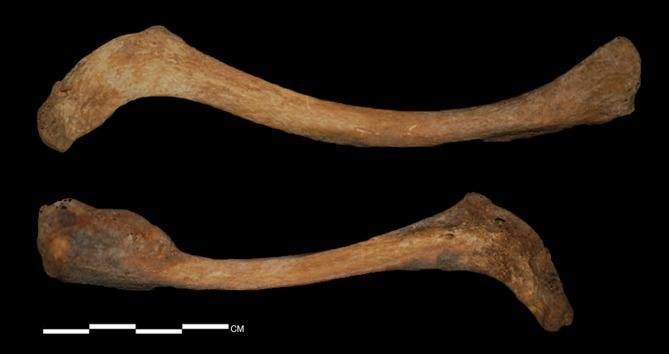
A clinical photograph of human skeletal remains showing the superior surface of two clavicles (upper bone: left side; lower bone: right side) against a black background. Both specimens display the characteristic S-shaped curvature. Significant bilateral asymmetry is visible between the two bones. The left clavicle (upper) is relatively slender with a smoother surface texture and expanded articular ends. The right clavicle (lower) is notably thicker and wider at the medial portion, appearing more flattened and angulated along the postero-inferior midshaft. The cortical surface of the right clavicle shows increased porosity, rougher texture, and focal areas of discoloration compared to the left. On the lateral end of the left clavicle, a distinct foramen is visible with evidence of localized periosteal reaction. These morphological findings are consistent with pathological asymmetry and possible systemic or localized skeletal disorders in a bio-archaeological context.
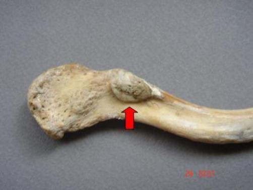
Clinical photograph of a dry bone specimen featuring the inferior surface of the acromial end of a left human clavicle. The visual focus is an anatomical variation: an anomalous clavicular facet for the coracoclavicular joint, indicated by a red arrow. This facet presents as a distinct, triangular bony outgrowth located approximately 5 cm medial to the acromial edge, near the conoid tubercle. The outgrowth's base is attached to the clavicle, with its apex directed inferiorly. The articular surface of the facet appears relatively smooth compared to the surrounding cortical bone, which exhibits a characteristic porous, beige, and slightly rough texture typical of an osteological specimen. This image demonstrates a diarthrotic coracoclavicular joint, a rare anatomical variant that can be clinically relevant in cases of shoulder pain or restricted range of motion. The photograph is intended for educational use in gross anatomy, orthopedics, and osteology to illustrate variations in the coracoclavicular syndesmosis.
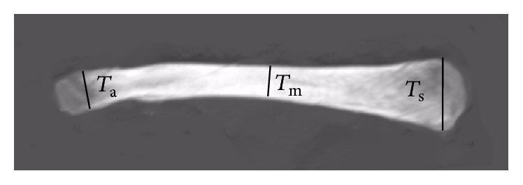
A clinical image of a human clavicle in the coronal plane, used to demonstrate quantitative morphometric analysis. The diagram illustrates the varying thickness of the bone across its longitudinal axis. Three specific measurement sites are indicated by vertical black lines perpendicular to the superior surface: 'Ts' represents the maximum thickness at the medial (sternal) end, which appears as the thickest portion; 'Ta' indicates the maximum thickness at the lateral (acromial) end; and 'Tm' identifies the minimum thickness located within the middle segment of the shaft. The image highlights the characteristic S-shaped curvature and the relative thinning of the clavicular midshaft compared to the expanded articular ends, a key anatomical feature relevant to orthopedic surgery, fracture management, and biomechanical modeling of the shoulder girdle.
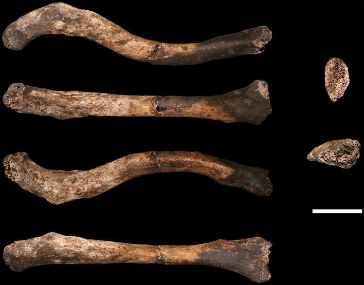
This clinical specimen image displays six views of a right human clavicle (U.W. 102a-021). The left column provides superior, anterior, inferior, and posterior longitudinal views, while the right column shows the medial and lateral articular surfaces. The specimen demonstrates a typical S-shaped curvature and measures approximately 10-12 cm in length, indicated by a 2 cm scale bar. Visible pathology and taphonomic features include a midshaft fracture where two pieces have been conjoined, and missing articular surfaces at the sternal (medial) end, which exposes the underlying trabecular bone structure. The acromial (lateral) end shows minor damage. The bone surface exhibits significant mineral staining, with dark coatings on the anterior-sternal half and patchy black staining on the acromial half. Fine, hairline longitudinal cracks are present across the acromial portion, consistent with post-mortem sediment loading and moisture fluctuations. This anatomical specimen is used in medical education to illustrate hominin skeletal morphology and taphonomic assessment of fossilized human remains.
clavicle fracture clinical anatomy MBBS
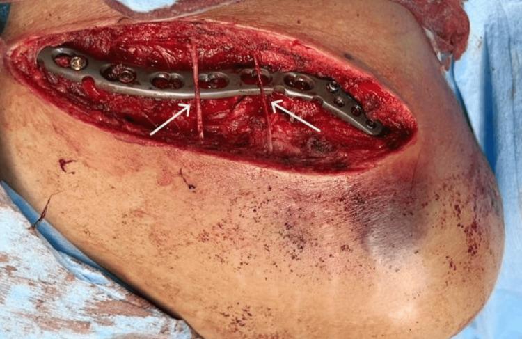
This clinical photograph is an intraoperative view of a clavicle fracture fixation. The image depicts a superior surgical approach with a sabre-shaped incision, exposing the underlying musculoskeletal anatomy of the shoulder region. A 3.5-mm precontoured metallic locking compression plate (LCP) is visible, securely fixed to the bone with multiple cortical and locking screws. A key educational feature of this image is the demonstration of fine surgical dissection to preserve neurovascular structures; specifically, two branches of the supraclavicular nerves are identified by white arrows as they cross superficially over the hardware and surgical site. The surrounding field shows mobilized skin and subcutaneous tissue with areas of ecchymosis, consistent with acute trauma. This visual serves as a primary example of orthopedic internal fixation techniques and the importance of peripheral nerve preservation during upper extremity surgery. It is intended for medical students and orthopedic residents to understand surgical anatomy, hardware placement, and nerve-sparing protocols in trauma surgery.
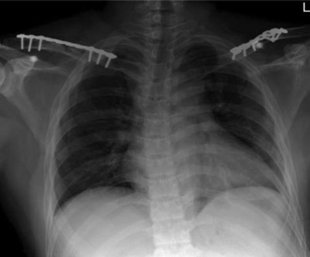
This diagnostic image is an anteroposterior (AP) chest and shoulder radiograph showing postoperative results of bilateral clavicle fracture fixation. The primary visible anatomy includes the thorax, ribs, clavicles, and proximal humeri. The central focus is the presence of surgical hardware: bilateral 3.5mm metallic reconstruction plates secured along the superior aspect of both the left and right clavicles. These radiopaque plates are fixed to the bone using multiple bicortical screws to bridge the fracture sites. The skeletal alignment of the shoulder girdle appears restored. The underlying lung fields are clear, the cardiomediastinal silhouette is within normal limits, and the trachea is midline. This image serves as a clinical example of orthopedic internal fixation for trauma management of bilateral upper extremity injuries.
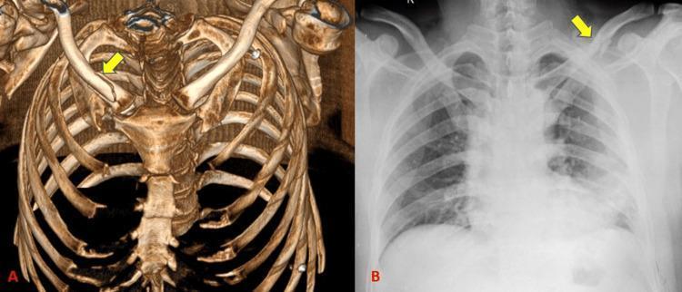
This comparison image showcases a left clavicle fracture using two different imaging modalities: (A) a 3D volume-rendered computed tomography (CT) reconstruction and (B) a standard posterior-anterior (PA) chest radiograph. Both images highlight the fracture site with yellow arrows. In the 3D CT reconstruction (A), the skeletal anatomy of the thorax, including the sternum, ribs, and clavicles, is rendered with high spatial resolution and depth. The mid-shaft clavicular fracture is clearly visualized as a complete discontinuity with inferior displacement of the distal segment. In contrast, the chest radiograph (B) displays a 2D projection where the fracture is visible but partially obscured by the superimposition of the underlying ribs and lung parenchyma. The comparison emphasizes the clinical utility of 3D CT for precisely evaluating fracture morphology, degree of misalignment, and complex spatial relationships in thoracic trauma, whereas the radiograph provides a standard screening view with lower sensitivity for subtle osseous detail.
Clavicle - Complete MBBS 1st Prof Anatomy Notes
1. Introduction
2. Type of Bone
- Long bone (despite lying horizontally)
- Ossifies in membrane (intramembranous ossification) - an important distinction
- First bone to begin ossification in fetal life (5th-6th week of intrauterine life)
- Last bone to complete ossification - the medial epiphysis fuses around 25 years of age
Exam point: The clavicle is the only long bone to ossify in membrane (not cartilage). It has two primary ossification centers, one for each half, which fuse early. The medial epiphysis is the last epiphysis in the body to appear and fuse.
3. Shape and Orientation
| Part | Curvature | Cross-section |
|---|---|---|
| Medial 2/3 | Convex forward | Cylindrical/rounded |
| Lateral 1/3 | Concave forward | Flat/flattened |
4. Surfaces and Markings
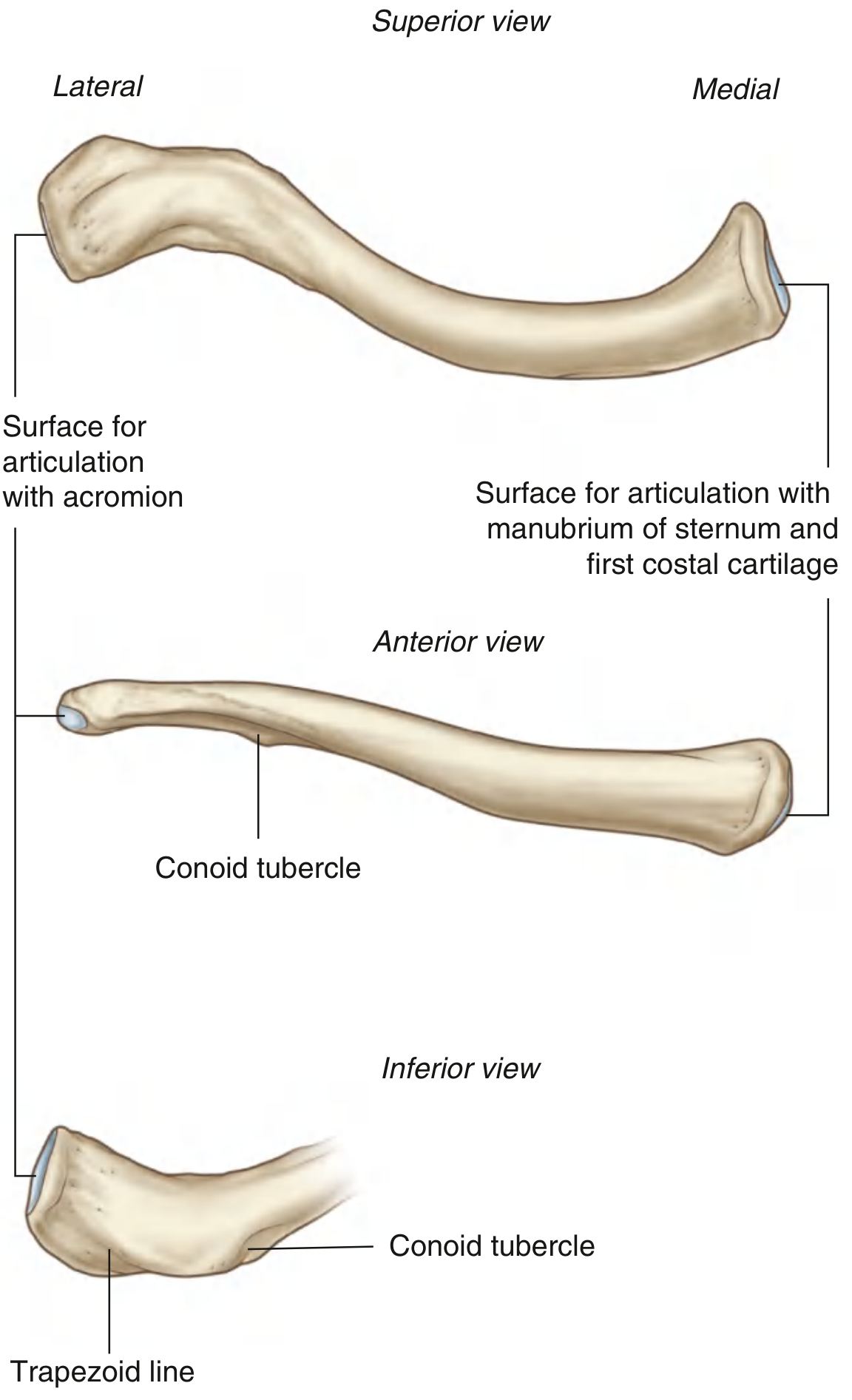
Superior Surface
- Smooth (almost bare of muscular attachment)
- Subcutaneous throughout
Inferior Surface (most important for attachments)
- Subclavian groove - for subclavius muscle (medial 2/3)
- Conoid tubercle - for conoid ligament (part of coracoclavicular ligament)
- Trapezoid line/ridge - for trapezoid ligament (part of coracoclavicular ligament)
- Impression for costoclavicular ligament - at medial end, for the rhomboid (costoclavicular) ligament
The inferior surface is roughened and more complex than the superior surface.
5. Ends
Sternal (Medial) End
- Larger, more robust, quadrangular in shape
- Has a large facet for articulation with the manubrium sterni and partly with the 1st costal cartilage
- Forms the sternoclavicular (SC) joint
Acromial (Lateral) End
- Flat and smaller
- Small oval facet for articulation with the acromion of scapula
- Forms the acromioclavicular (AC) joint
6. Muscle Attachments
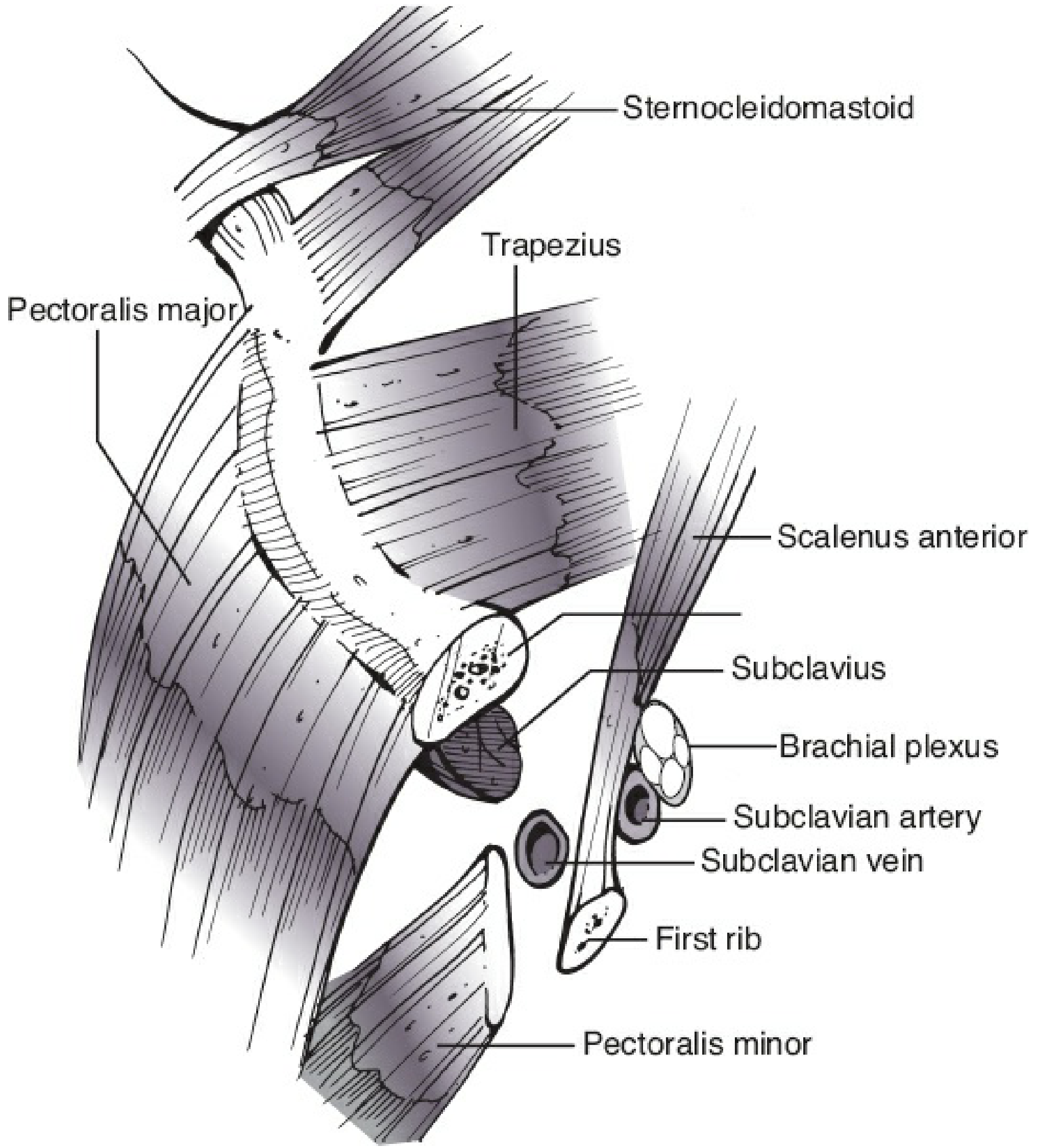
| Muscle | Region of Attachment | Surface |
|---|---|---|
| Sternocleidomastoid (SCM) | Medial 1/3 | Superior surface (insertion) |
| Pectoralis major (clavicular head) | Medial 2/3 | Anterior surface (origin) |
| Deltoid | Lateral 1/3 | Anterior/inferior surface (origin) |
| Trapezius | Lateral 1/3 | Posterior/superior surface (insertion) |
| Subclavius | Subclavian groove (medial 2/3) | Inferior surface (insertion) |
Memory trick for superior surface: "Some Damn Traitors Place Medals" → SCM (medial), Deltoid (lateral), Trapezius (lateral), Pectoralis major (medial).
7. Ligament Attachments
| Ligament | Location |
|---|---|
| Costoclavicular (rhomboid) ligament | Inferior surface, medial end - connects to 1st rib cartilage |
| Conoid ligament | Conoid tubercle, inferior surface - connects to coracoid process |
| Trapezoid ligament | Trapezoid line, inferior surface - connects to coracoid process |
8. Articulations
- Sternoclavicular joint - with manubrium sterni (+ 1st costal cartilage)
- Only bony joint between the upper limb and axial skeleton
- Synovial joint with an articular disc
- Acromioclavicular joint - with acromion of scapula
- Synovial plane joint
9. Structures Behind / Relations
- Subclavian vein (most anterior, directly under subclavius)
- Subclavian artery (posterior to vein)
- Brachial plexus (most posterior, close to clavicle midportion)
- Scalenus anterior separates the vein from the artery medially
Clinical point: Clavicle fractures can injure the subclavian vessels and brachial plexus.
10. Ossification (High-Yield for MBBS Exams)
| Feature | Details |
|---|---|
| Type | Intramembranous (endochondral at medial end only) |
| Primary centers | 2 centers (medial & lateral), appear at 5th-6th week IUL |
| Secondary center | 1 center at the sternal (medial) end |
| Appears | 15-17 years |
| Fuses | 25 years (last epiphysis to fuse in the body) |
11. Fracture of Clavicle (Clinical Anatomy)
- Medial fragment - pulled upward by sternocleidomastoid
- Lateral fragment - pulled downward and medially by weight of arm + deltoid + pectoralis major
- Birth trauma - fracture during difficult delivery; heals well
- Greenstick fracture - in children (clavicle is more flexible)
12. Side Determination (How to Tell Left from Right)
| Feature | Implication |
|---|---|
| Convex curvature | Faces anteriorly/medially |
| Flat end (acromial) | Faces laterally |
| Conoid tubercle + trapezoid ridge | On inferior surface of lateral third |
| Subclavian groove | On inferior surface of medial 2/3 |
13. Functions of the Clavicle
- Acts as a strut - holds the upper limb away from the trunk, allowing free movement
- Transmits forces from the upper limb to the axial skeleton
- Protects the neurovascular bundle (subclavian vessels + brachial plexus) below it
- Provides attachment for muscles and ligaments
- Transmits weight of upper limb to the sternum via sternoclavicular joint
14. Exam High-Yield Points Summary
| Topic | Key Fact |
|---|---|
| Only long bone to lie horizontally | Clavicle |
| Only long bone to ossify in membrane | Clavicle |
| First bone to begin ossification | Clavicle (5th-6th week IUL) |
| Last epiphysis to fuse | Medial end of clavicle (25 yrs) |
| Only bony link, limb to axial skeleton | Sternoclavicular joint |
| Most common fracture site | Middle 1/3 (junction of curves) |
| Muscle elevating medial fragment | Sternocleidomastoid |
| Ligament at conoid tubercle | Conoid ligament (coracoclavicular) |
| Structure directly below clavicle | Subclavian vein (then artery, then brachial plexus) |