
Explain me 10 common heart diseases with ECG changes and ECG images of those diseases
Reading File
Now let me search the library in parallel for ECG-related content across multiple heart diseases:
Finding Sources
Finding Sources
Good - found several relevant sources. Let me now read the key ECG sections and search for more disease-specific ECG content in parallel:
Reading File
Finding Sources
Finding Sources
Excellent content found. Now let me read the key ECG sections and grab images in parallel:
Reading File
Reading File
Finding Sources
Finding Sources
Now let me read more key content and search for more ECG images from the textbooks:
Reading File
Reading File
Finding Sources
Finding Sources
Now let me fetch the key ECG images from the textbooks and read a few more sections in parallel:
Loading Image
Loading Image
Loading Image
Reading File
Excellent! Now let me fetch a few more key ECG images - MI, WPW, heart block, and search for more content:
Loading Image
Reading File
Finding Sources
Let me now fetch the heart block ECG images and the MI ECG image, and read the STEMI ECG content:
Loading Image
Loading Image
Loading Image
Reading File
I now have extensive content. Let me fetch 2 more critical images (MI and Wellens/anterior ischemia) and get the atrial flutter image:
Loading Image
Reading File
I now have comprehensive content and ECG images for a thorough answer. Let me also get one more MI ECG image and a Wellens sign image:
Loading Image
Here is a comprehensive overview of 10 common heart diseases with their ECG changes and images, drawn from Harrison's Principles of Internal Medicine 22E, Ganong's Review of Medical Physiology, Tintinalli's Emergency Medicine, and Guyton and Hall Textbook of Medical Physiology.
10 Common Heart Diseases: ECG Changes and Images
1. ST-Elevation Myocardial Infarction (STEMI)
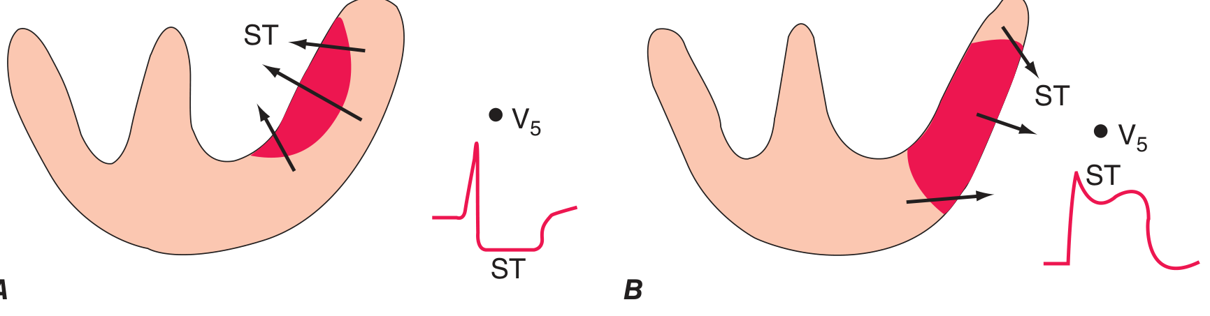
Mechanism: Acute transmural ischemia due to complete coronary artery occlusion. Ischemia lowers resting membrane potential and shortens action potential duration, generating a "current of injury."
ECG Changes (sequential evolution):
- Hyperacute phase (minutes): Tall, broad, positive "hyperacute" T waves over the ischemic zone
- Acute phase (hours): ST-segment elevation in leads overlying the infarct zone; reciprocal ST depression in opposite leads
- Evolving phase (hours-days): T-wave inversions develop; pathological Q waves appear (>40 ms wide, >25% R-wave depth)
- Chronic phase (weeks-months): Persistent Q waves; ST may normalize
Localization by leads:
| Territory | Culprit artery | Leads with ST elevation |
|---|---|---|
| Anterior | LAD | V1-V4, I, aVL |
| Inferior | RCA | II, III, aVF |
| Lateral | LCx | I, aVL, V5-V6 |
| Posterior | RCA/LCx | Reciprocal ST depression V1-V3 |
| Right ventricle | RCA | V3R, V4R |
Current of injury diagram - subendocardial vs transmural ischemia:
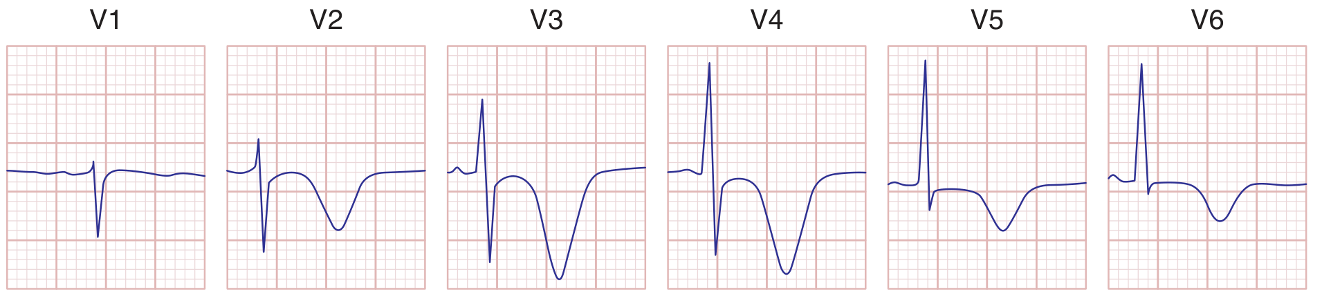
Anterior STEMI - Wellens pattern (deep T inversions V1-V4):
2. Non-ST Elevation Myocardial Infarction (NSTEMI) / Unstable Angina
Mechanism: Subtotal occlusion or subendocardial ischemia. The ST injury vector shifts toward the subendocardium and ventricular cavity.
ECG Changes:
- ST-segment depression (horizontal or downsloping) in anterior/lateral leads
- T-wave inversions - may be deep and symmetric
- No pathological Q waves (by definition, no transmural necrosis)
- ST elevation in aVR with widespread ST depression suggests left main or proximal LAD disease
- The ECG may be normal in up to 6% of confirmed NSTEMIs
3. Atrial Fibrillation (AF)
Mechanism: Multiple re-entrant wavelets circulating chaotically through atrial myocardium. Atrial rate >600 bpm; AV node filters to produce an irregular ventricular response.
ECG Changes (classic hallmarks):
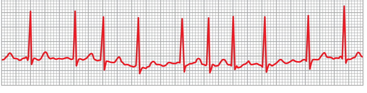
- Absence of distinct P waves - replaced by irregular fibrillatory baseline (most visible in V1)
- Irregularly irregular RR intervals (no two RR intervals are the same)
- QRS complexes are usually narrow (unless aberrant conduction or bundle branch block is present)
- Ventricular rate typically 120-170 bpm if AV node is unaffected
- Very rapid rate (>200 bpm) suggests an accessory pathway (WPW + AF)
ECG - Atrial Fibrillation (Guyton and Hall):
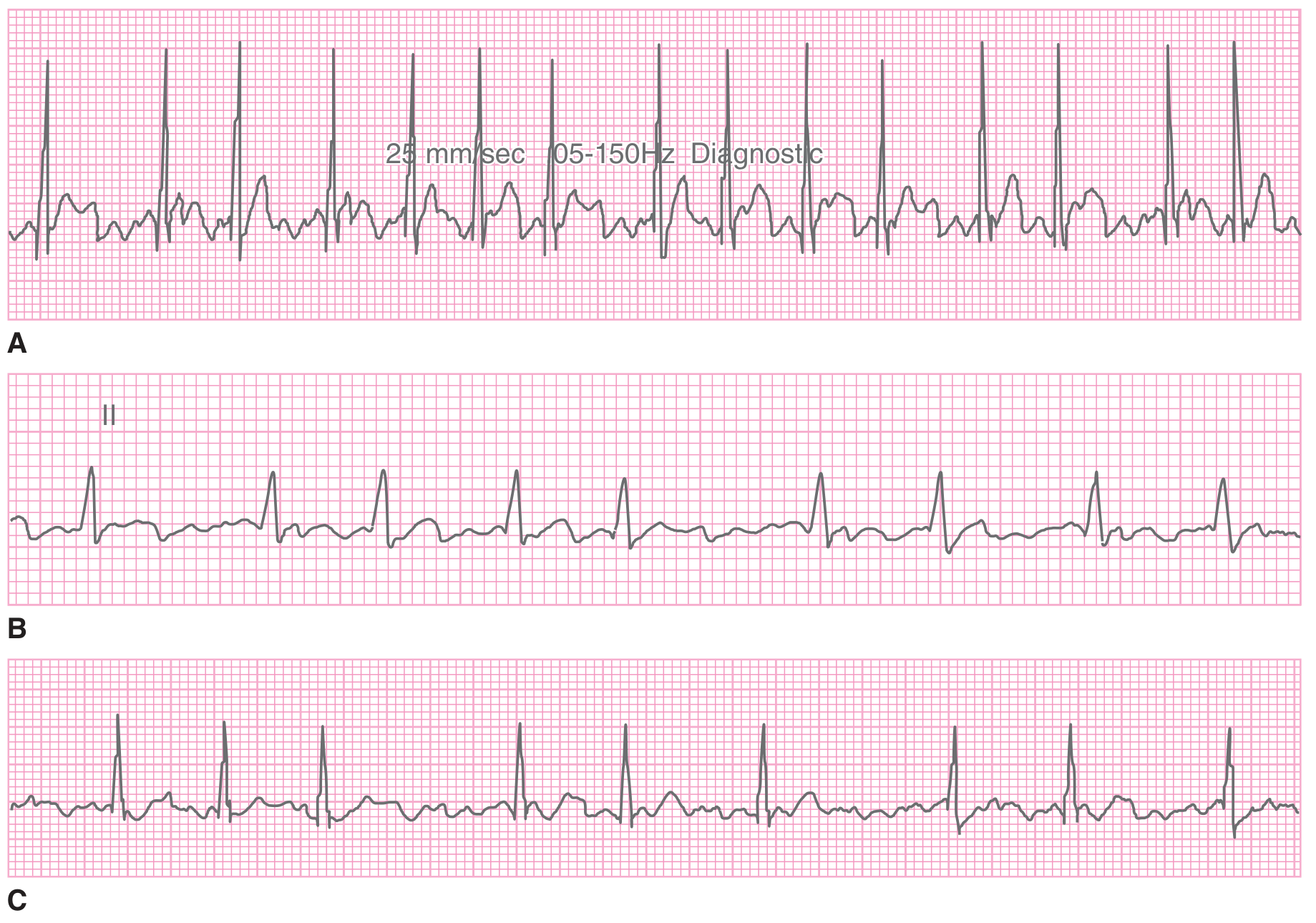
Three real-patient AF tracings:
4. Heart Block (AV Conduction Disease)
Mechanism: Impaired conduction through the AV node or His-Purkinje system, due to ischemia, fibrosis, infiltration, or drugs.
ECG Changes by degree:
First-degree AV Block:
- PR interval prolonged > 200 ms (0.20 s), but every P wave conducts
- QRS is normal; rhythm is regular
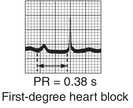
Second-degree AV Block - Mobitz I (Wenckebach):
- Progressive PR lengthening with each beat until one P wave is not conducted (dropped QRS)
- The cycle then resets
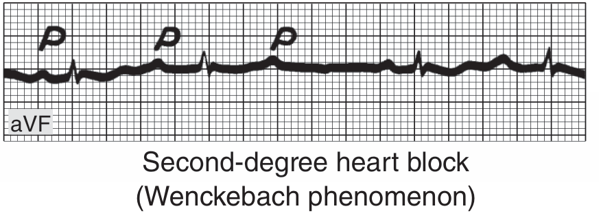
Third-degree (Complete) AV Block:
- P waves and QRS complexes beat independently - AV dissociation
- P-P intervals regular; RR intervals regular; but PR has no fixed relationship
- Escape QRS rate: 40-60 bpm (junctional escape) or 20-40 bpm (ventricular escape, wide QRS)
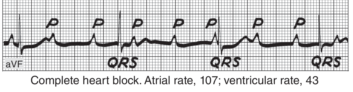
5. Wolff-Parkinson-White (WPW) Syndrome
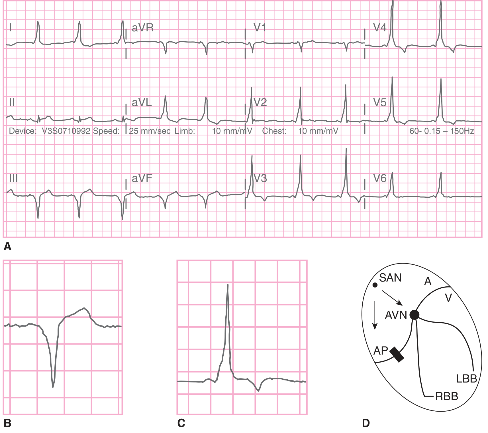
Mechanism: Accessory pathway (Bundle of Kent) bypasses the AV node, causing ventricular preexcitation. Dual conduction via the accessory pathway and normal AV node sets up re-entrant arrhythmias.
ECG Changes (in sinus rhythm):
- Short PR interval (< 120 ms) - bypass of AV node delay
- Delta wave - slurred upstroke at the beginning of QRS (cell-to-cell conduction via accessory pathway)
- Widened QRS complex (>120 ms) due to fusion of normal and accessory pathway depolarization
- Pseudo-infarct pattern - delta waves can cause negative deflections mimicking Q waves
- ST-T changes secondary to abnormal depolarization sequence
- During AF with WPW: wide, bizarre QRS at >200 bpm (life-threatening)
Full 12-lead WPW with diagram:
6. Bundle Branch Block
Mechanism: Interruption of right (RBBB) or left (LBBB) bundle branch forces conduction to spread slowly through ventricular myocardium (cell-to-cell), widening the QRS.
ECG Changes - RBBB:
- QRS duration ≥ 120 ms
- rSR' pattern ("rabbit ears") in V1 - terminal broad R'
- Broad, slurred S wave in V5-V6 and lead I
- Secondary T-wave inversions in V1-V2 (discordant from the last QRS deflection)
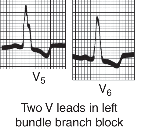
ECG Changes - LBBB:
- QRS duration ≥ 120 ms
- Broad, notched R wave in V5-V6, I, aVL (no septal q waves)
- Deep QS or rS in V1
- Secondary T inversions in V5-V6 (discordant from last QRS deflection)
- Important: LBBB masks STEMI on ECG - use Sgarbossa criteria
RBBB vs LBBB comparison (V1 and V5):
7. Hypertrophic Cardiomyopathy (HCM)
Mechanism: Genetic sarcomere mutations cause massive septal hypertrophy and disorganized myocyte architecture, leading to diastolic dysfunction, LVOTO, and risk of sudden death.
ECG Changes (present in >90% of patients):
- Left ventricular hypertrophy (LVH) pattern: Increased voltage - Sokolow-Lyon (SV1 + RV5/6 ≥ 35 mm)
- Left axis deviation
- Deep, narrow "dagger-like" Q waves in inferior (II, III, aVF) and lateral leads (I, aVL, V4-V6) - caused by septal hypertrophy, not infarction
- ST depression and T-wave inversions particularly in lateral leads
- WPW-like pseudo-preexcitation pattern in some patients
- Giant negative T waves in apical HCM (Yamaguchi syndrome) - deep T inversions in V3-V6
8. Dilated Cardiomyopathy (DCM)
Mechanism: Ventricular dilation with systolic dysfunction (reduced EF), from ischemic, viral, toxic, or idiopathic causes.
ECG Changes:
- LBBB is very common (present in up to 25-30% of patients with DCM) and worsens dyssynchrony
- Sinus tachycardia at rest due to compensatory sympathetic activation
- Ventricular ectopy / ventricular tachycardia - risk of sudden death
- First-degree AV block or non-specific intraventricular conduction delay
- Low voltage if associated with pericardial effusion
- Atrial fibrillation (very common due to atrial dilation)
- Poor R-wave progression in precordial leads
- Non-specific ST-T changes
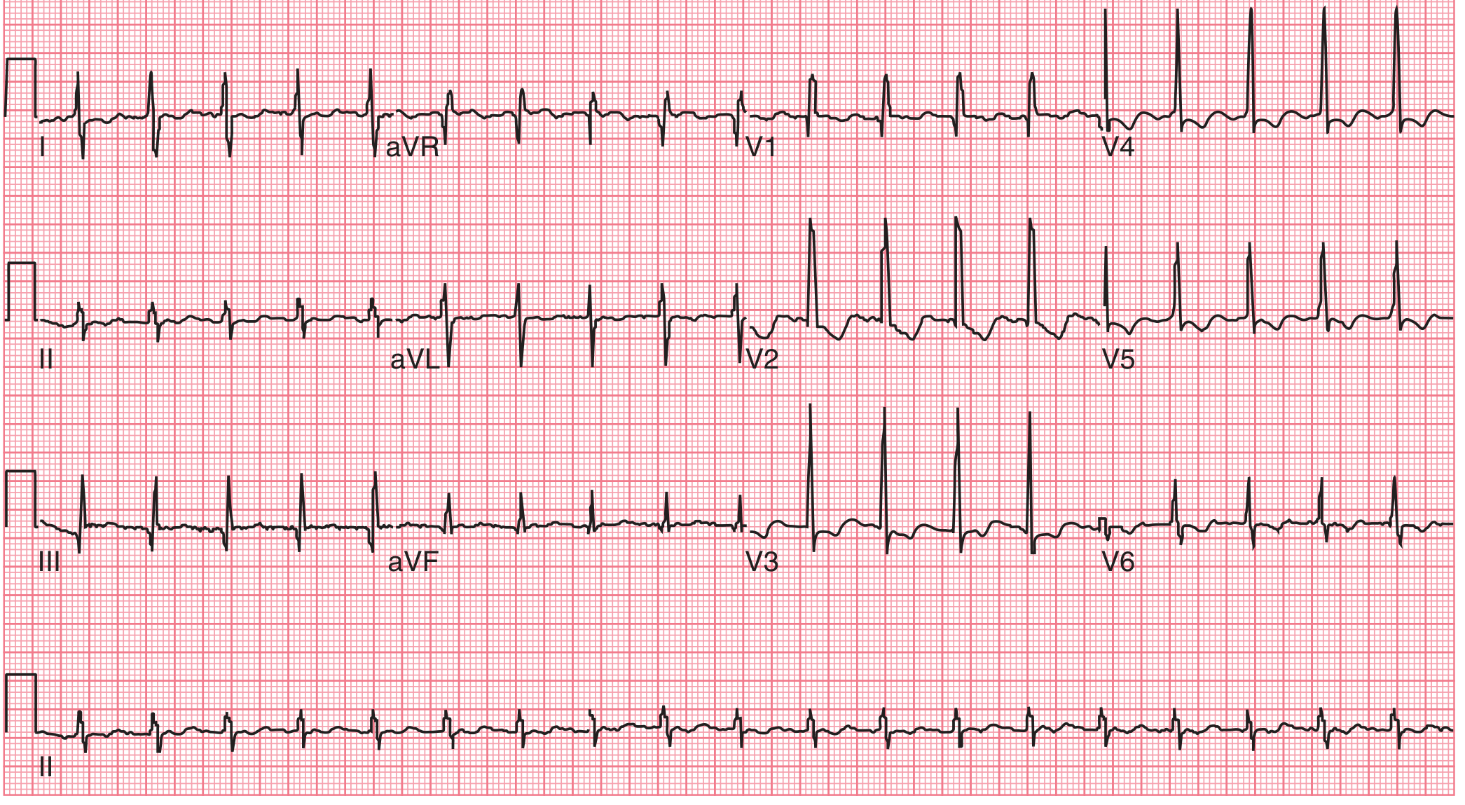
9. Atrial Flutter
Mechanism: A single re-entrant circuit rotates around the tricuspid annulus (typical flutter), producing regular atrial depolarization at ~300 bpm. The AV node blocks most impulses, producing a 2:1, 3:1, or 4:1 ventricular rate.
ECG Changes:
- Sawtooth flutter waves at ~300 bpm - best seen in inferior leads (II, III, aVF) and V1
- Flutter waves are negatively deflected (inverted) in inferior leads in typical flutter
- Regular ventricular rhythm at a fraction of 300 bpm: 2:1 block → ~150 bpm; 4:1 block → ~75 bpm
- No isoelectric baseline between flutter waves
- Regular narrow QRS unless aberrant conduction or BBB is present
- A regular narrow-complex tachycardia at exactly ~150 bpm should always raise suspicion for atrial flutter with 2:1 block
AF with atrial flutter context - 12-lead ECG:
10. Long QT Syndrome (LQTS)
Mechanism: Congenital (ion channel mutations in SCN5A, KCNQ1, KCNH2, etc.) or acquired (drugs, hypokalemia, hypomagnesemia) prolongation of ventricular repolarization. Risk of triggered ventricular arrhythmia - torsades de pointes (TdP).
ECG Changes:
- Prolonged QTc interval - corrected QT (Bazett formula: QT / √RR)
- Normal QTc: < 440 ms (men), < 460 ms (women)
- Borderline: 440-480 ms
- Abnormal/high risk: ≥ 500 ms
- T-wave morphology abnormalities: broad/notched T waves; bifid T waves; T-U wave fusion
- Torsades de pointes on ECG: rapid (~200-250 bpm) polymorphic ventricular tachycardia where the QRS axis rotates around the isoelectric line - appears as a "twisting of the points"
- Each subtype has a characteristic T-wave morphology:
- LQT1: broad-based T waves
- LQT2: notched/bifid T waves
- LQT3: long, flat ST segment with late-peaking T wave
Summary Table
| Disease | Key ECG Finding | Leads Affected |
|---|---|---|
| STEMI | ST elevation + Q waves → T inversion | Territory-specific (II/III/aVF, V1-V4, etc.) |
| NSTEMI/UA | ST depression, T inversion, no Q waves | Anterior/lateral |
| Atrial Fibrillation | No P waves, irregularly irregular RR | All leads |
| Heart Block (3°) | P-QRS dissociation, slow escape rhythm | All leads |
| WPW | Short PR, delta wave, wide QRS | All leads |
| Bundle Branch Block | QRS ≥ 120 ms, rSR' (RBBB) or broad R (LBBB) | V1 vs V5-V6 |
| HCM | LVH, deep septal Q waves, ST-T changes | Lateral, inferior |
| Dilated CM | LBBB, AF, low voltage, poor R progression | All leads |
| Atrial Flutter | Sawtooth waves ~300 bpm, regular ventricular rate | II, III, aVF, V1 |
| Long QT Syndrome | Prolonged QTc ≥ 500 ms, torsades de pointes | All leads |
Sources: Harrison's Principles of Internal Medicine 22E (2025), Ganong's Review of Medical Physiology 26E, Guyton and Hall Textbook of Medical Physiology, Tintinalli's Emergency Medicine - A Comprehensive Study Guide.Here is a complete overview of all 10 conditions. Key points to remember:
Images included directly from textbooks:
- Current of injury diagram (Harrison's) - shows mechanistically why STEMI causes ST elevation vs depression
- Wellens T-wave sign (Harrison's) - deep anterior T inversions indicating LAD disease
- Atrial fibrillation (Guyton & Hall + Tintinalli's) - absent P waves with irregular RR intervals
- First-degree heart block (Ganong's) - prolonged PR = 0.38 s
- Second-degree Wenckebach block (Ganong's) - progressive PR lengthening then dropped beat
- Complete heart block (Ganong's) - fully independent P and QRS rhythms
- WPW syndrome (Tintinalli's) - full 12-lead with delta wave and pathway diagram
- 12-lead AF ECG (Tintinalli's)
Clinical pearls:
- LBBB can completely mask a STEMI - always apply Sgarbossa criteria
- A regular narrow tachycardia at exactly 150 bpm = atrial flutter with 2:1 block until proven otherwise
- WPW + AF is life-threatening - never give AV nodal blockers (adenosine, verapamil, diltiazem)
- QTc ≥ 500 ms = high risk for torsades de pointes - check and correct all offending drugs and electrolytes
Explain me sandhi vata with specific reference and management with treatment
Reading File
Searching the Web
Sandhi Vata Ayurveda disease definition symptoms management treatment classical references
Good. Now let me search the medical library for Ayurveda/Sandhi Vata content and also search for clinical evidence in parallel:
Finding Sources
Finding Sources
Reading Web Page
https://pdfs.semanticscholar.org/4aff/97362585d63444416abc10…
Searching PubMed
Sandhigata Vata osteoarthritis Ayurvedic treatment
The medical library doesn't have Ayurveda textbooks in its collection. Let me get more detailed classical references and treatment details through targeted web extraction and a broader PubMed search:
Searching the Web
Sandhigata Vata Charaka Samhita Sushruta classical reference nidana samprapti lakshana chikitsa shloka
Searching the Web
Sandhi Vata Panchakarma treatment Basti Janubasti Abhyanga Swedana formulations Guggulu Rasayana
Searching PubMed
Ayurveda osteoarthritis knee treatment Panchakarma randomized
Excellent - rich content gathered. Let me now extract detailed classical reference content and get the key RCT abstract:
Reading Web Page
https://wjpr.s3.ap-south-1.amazonaws.com/article_issue/01b1d…
Reading Article
PMID: 29426006
You've run out of free credits. Sign up for a free account to continue using Orris.
This is a shared conversation. Sign in to Orris to start your own chat.