
Desribe the compartments of mediastinum, their normal contents and boundaries with appropriate diagrams Write the imaging differential diagnosis of an anterior mediastinal mass lesion with findings -50 Marks
mediastinum compartments diagram boundaries anatomy
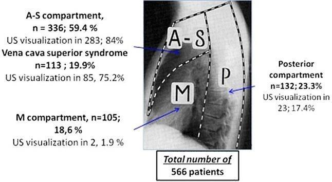
An educational anatomical diagram overlaid on a lateral chest radiograph, illustrating the surgical division of the mediastinum into three primary compartments: Anterior-Superior (A-S), Middle (M), and Posterior (P). The boundaries of these compartments are demarcated by white dashed lines. Annotations provide statistical data from a study of 566 patients regarding the distribution of mediastinal lesions and the success rate of ultrasound (US) visualization within each zone. The A-S compartment contains 59.4% of lesions (n=336) with an 84% US visualization rate. The M compartment contains 18.6% (n=105) with the lowest US visualization at 1.9%. The P compartment contains 23.3% (n=132) with a 17.4% visualization rate. Specifically noted is 'Vena cava superior syndrome' occurring in 19.9% of cases, with 75.2% US visualization. This infographic serves as a clinical guide for thoracic imaging, highlighting the feasibility of US-guided core needle biopsy based on anatomical localization.
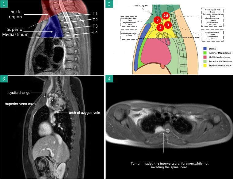
A four-panel medical figure illustrating the surgical anatomy and diagnostic imaging of superior mediastinal masses. Panel 1: A sagittal T2-weighted magnetic resonance imaging (MRI) view highlighting the superior mediastinum (shaded blue) and its anatomical boundaries, specifically the region between the thoracic inlet and the T4-T5 intervertebral disc level. Panel 2: A schematic diagram color-coded by mediastinal compartments (anterior, middle, posterior, and superior) showing the distribution and pathological types of identified lesions, including schwannoma, ganglioneuroma, bronchogenic cyst, and cavernous hemangioma. Panel 3: A sagittal contrast-enhanced MRI demonstrating a large heterogeneous mass in the superior mediastinum with cystic changes. The image labels its close proximity to the superior vena cava and the arch of the azygos vein. Panel 4: An axial MRI slice showing a tumor invading the intervertebral foramen. The annotation notes that while the tumor occupies the neural foramen, it does not invade the spinal cord. This collection is used to teach preoperative assessment of neurovascular involvement and resectability of mediastinal tumors using the da Vinci robotic surgical system.
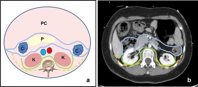
This dual-panel educational material illustrates the anatomy of the retroperitoneal space using a comparative approach. Panel (a) is a schematic anatomical diagram in axial cross-section. It delineates the three main retroperitoneal compartments: the anterior pararenal space (outlined in blue), containing the pancreas (P) and ascending/descending colon (C); the perirenal space (outlined in green), housing the kidneys (K), adrenal glands, and perirenal fat; and the posterior pararenal space (outlined in yellow dashed lines), located between the posterior renal fascia and the transversalis fascia. The peritoneal cavity (PC) is shown anteriorly. Panel (b) presents a corresponding axial CT scan with intravenous contrast, applying the same color-coded boundary lines to clinical imaging. The CT demonstrates the contrast-enhanced kidneys within the perirenal space and the relative positions of the pancreas and bowel. This comparison aids in understanding fascial planes and the compartmentalization of potential retroperitoneal pathology, such as hematomas or abscesses, which are often confined by these anatomical boundaries.
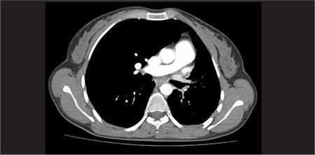
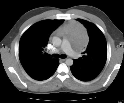
This diagnostic image is an axial cross-section from a Computed Tomography (CT) scan of the human chest, at the level of the mid-thorax. The image demonstrates a transverse view of the mediastinum and pulmonary parenchyma. Key anatomical structures visible include the thoracic vertebral body located posteriorly, the sternum anteriorly, and the rib cage forming the lateral boundaries. Centrally, the mediastinum contains high-density (bright) vascular structures consistent with the heart and great vessels, including the ascending and descending aorta. The bilateral lungs appear as low-density (dark), air-filled regions flanking the central mediastinal structures. No gross masses, significant pleural effusions, or lobar consolidations are immediately apparent in this single slice. This imaging modality is used in clinical medicine to evaluate thoracic anatomy, assess pulmonary disease, and, when combined with functional imaging like SPECT, localize radiopharmaceutical tracer activity within specific anatomical compartments such as the ileal lumen or renal systems.
This composite medical illustration depicts the surgical and anatomical aspects of compartment syndrome in the gluteal region. The image is divided into two primary sections: a surgical clinical illustration and an anatomical diagram. On the left, a surgical illustration shows a decompressive fasciotomy of the right buttock with the patient in a lateral decubitus position. A large, linear incision through the skin and fascia exposes bulging muscle tissue, which appears edematous and reddish with focal pale areas, suggesting pressure-induced ischemia or muscle fiber disruption. On the right, a color-coded anatomical diagram identifies the three non-distensible fascial compartments of the gluteal region: the tensor fascia lata compartment (positioned anteriorly in red), the gluteus medius and minimus compartment (located centrally with a layered appearance), and the gluteus maximus compartment (positioned posteriorly in green). White arrows indicate the specific spatial boundaries and contents of each compartment. This visual serves as an educational tool for orthopedic surgery and emergency medicine, demonstrating the surgical management (fasciotomy) and the underlying regional anatomy relevant to diagnosing and treating acute gluteal compartment syndrome.
This medical anatomical diagram is a posterior-lateral sketch of the human torso, illustrating the musculature and boundaries of the lumbar triangle, also known as the Triangle of Petit. The diagram highlights the clinical significance of this region for procedures such as the transversus abdominis plane (TAP) block. The anatomical boundaries are clearly labeled: the posterior border is formed by the latissimus dorsi muscle, the anterior border by the external oblique muscle, and the base (inferior border) by the iliac crest. Other visible anatomical landmarks include the rectus abdominis muscle, located medially and anteriorly, and the trapezius muscle superiorly. The illustration demonstrates the convergence of these muscular structures to form a small, triangular area of relative weakness in the abdominal wall, which is a potential site for lumbar hernias. This diagram is designed for educational use in anatomy, regional anesthesia, and general surgery to help students and clinicians identify surface landmarks and deep structural relationships in the lumbar region.
anterior mediastinal mass thymoma lymphoma teratoma CT imaging differential diagnosis
**Imaging Modality:** Non-contrast axial Computed Tomography (CT) of the chest. **Anatomical Region:** Anterior mediastinum at the level of the aortic arch and pulmonary trunk. **Observed Pathology:** A large, well-circumscribed, soft-tissue density mass is present in the anterior mediastinum. The mass is predominantly left-sided but crosses the midline, exerting a mass effect on adjacent vascular structures. It appears relatively homogeneous in attenuation. **Characteristic Visual Features:** * **Location:** Anterior to the ascending aorta and pulmonary artery, posterior to the sternum. * **Morphology:** Lobulated borders with a solid internal composition. * **Surrounding Structures:** The mass is in close proximity to the great vessels; however, no gross invasion of the lung parenchyma or pleural effusion is visualized in this single slice. * **Vascular findings:** A hyperdense (calcified or metallic) focus is noted in the right hilar/paratracheal region, likely representing a calcified lymph node or surgical clip. **Key Diagnostic Features:** The presence of a large anterior mediastinal mass in an adult necessitates the differential diagnosis of the "4 Ts": Thymoma, Teratoma (germ cell tumors), Thyroid (substernal goiter), and "Terrible" Lymphoma. The homogeneity and location are highly suggestive of a thymic epithelial tumor or lymphoma.
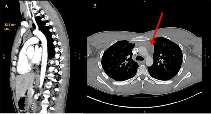
This figure presents contrast-enhanced computed tomography (CT) scans of the chest in sagittal (A) and axial (B) planes, focused on the mediastinum. Panel A shows a sagittal reconstruction illustrating a soft tissue mass within the anterior mediastinal space, specifically located in the left lobe of the thymus. A digital measurement indicates the lesion's long axis is approximately 32.6 mm. Panel B provides an axial view at the level of the great vessels, where a red arrow highlights a well-circumscribed, heterogeneous nodular mass. The lesion demonstrates areas of central low density, suggestive of cystic changes or necrosis within the thymic tissue. Key anatomical landmarks visible include the sternum, ascending aorta, and vertebral bodies. This diagnostic imaging is characteristic of an anterior mediastinal tumor, clinically diagnosed in this case as a primary mediastinal seminoma. The images are essential for teaching the differential diagnosis of anterior mediastinal masses (the 'four Ts': thymoma, teratoma, 'terrible' lymphoma, and thyroid) and demonstrating the use of multi-planar CT imaging for tumor localization and characterization.
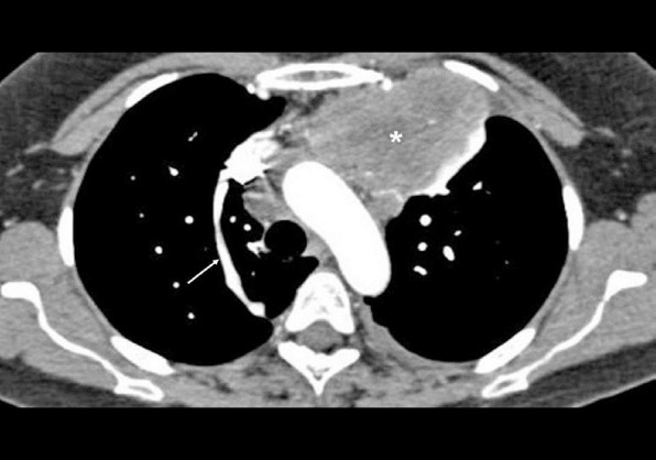
**Imaging Modality:** Contrast-enhanced computed tomography (CT) of the chest, axial section. **Anatomical Region:** Mediastinum at the level of the aortic arch and superior vena cava (SVC). **Observed Pathology:** A large, heterogeneous, mildly enhancing soft-tissue mass is centered in the anterior mediastinum. The mass demonstrates irregular borders and exerts a mass effect on adjacent vascular structures. Based on clinical context, this is consistent with a mediastinal germ cell tumor. **Characteristic Visual Features:** * **Mass:** The lesion (marked with an asterisk) occupies the prevascular space, displacing the mediastinal pleura laterally. * **Vascular Anatomy:** The superior vena cava and aortic arch are visible. A prominent, linear vascular structure (marked with an arrow) is identified as the azygos vein, seen here in a longitudinal plane as it courses anteriorly to drain into the posterior aspect of the SVC. * **Attenuation:** The mass shows internal areas of varying attenuation, suggesting a complex solid component. **Key Diagnostic Features:** The location in the anterior mediastinum (one of the "4Ts": Thymoma, Teratoma/Germ cell tumor, Thyroid, Terrible Lymphoma) and the patient's age are primary indicators for the differential diagnosis. The image highlights the relationship between the anterior mass and the normal posterior vascular anatomy, specifically the azygos vein arch.
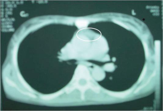
This diagnostic axial CT scan of the chest reveals a soft tissue mass located in the anterior mediastinum, indicated by a white elliptical annotation. The mass exhibits a lobulated morphology and is situated immediately posterior to the sternum and anterior to the great vessels and heart base. In this contrast-enhanced image, the mass shows minimal enhancement relative to the surrounding cardiovascular structures, suggesting low vascularity. Other visible anatomical landmarks include the thoracic vertebrae, bilateral lung fields, and the chest wall. Clinically, this imaging finding is representative of a differential diagnosis for anterior mediastinal tumors, such as thymoma, lymphoma, or a teratoma. The image is a primary resource for students learning to identify mediastinal compartment pathologies and radiological presentations of chest masses.
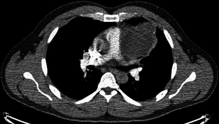
This diagnostic image is an axial contrast-enhanced CT scan of the chest at the level of the pulmonary artery bifurcation. It demonstrates a large, well-defined mass located in the anterior mediastinum, consistent with a mature teratoma. The mass exhibits heterogeneous density with both cystic (hypodense) and solid (isodense to soft tissue) components, a hallmark of germ cell tumors. Spatially, the lesion is situated posterior to the sternum and anterior to the heart and great vessels, causing notable mass effect and lateral displacement of the adjacent mediastinal structures and the left lung. The presence of varying internal attenuation suggests different tissue types, such as fat, fluid, or soft tissue. This clinical finding is significant for differential diagnosis of anterior mediastinal masses, which includes the '4 Ts': Thymoma, Teratoma, Thyroid (retrosternal goiter), and 'Terrible' lymphoma.
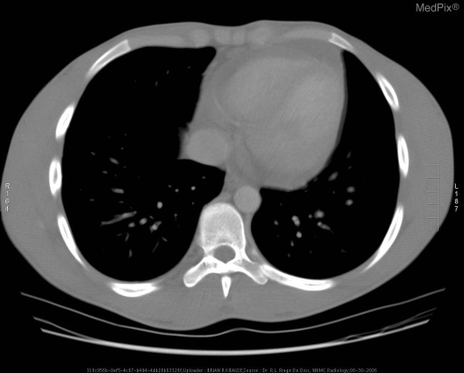
**Imaging Modality:** Computed Tomography (CT), axial section. **Anatomical Region:** Thorax at the level of the heart and lower mediastinum. **Observed Pathology:** The image demonstrates a significant soft-tissue attenuation mass located in the anterior mediastinum. The lesion is characterized by a homogeneous appearance and demonstrates a mass effect, subtly displacing adjacent cardiovascular structures. **Characteristic Visual Features:** * **Mediastinal Widening:** There is an expansive, well-circumscribed soft-tissue density occupying the anterior mediastinal space, consistent with lymphadenopathy or a primary mediastinal tumor. * **Cardiovascular Landmarks:** The mass is situated anterior to the heart and great vessels. The descending aorta and vertebral body appear unremarkable. * **Pulmonary Parenchyma:** The lung fields are largely clear without overt evidence of large pulmonary nodules or consolidations at this level. * **Pleural Space:** No significant pleural effusions are visualized in this section. **Clinical Context/Differentiating Features:** The location and homogeneous morphology in the anterior mediastinum are highly suggestive of lymphoma (e.g., Hodgkin’s or Diffuse Large B-cell Lymphoma), though the differential diagnosis for an anterior mediastinal mass includes thymoma, teratoma, and thyroid lesions.
MEDIASTINUM: COMPARTMENTS, CONTENTS, BOUNDARIES & ANTERIOR MEDIASTINAL MASS IMAGING
PART 1: COMPARTMENTS OF THE MEDIASTINUM
Definition
Classification
- Superior mediastinum - above the sternal angle/TIV-TV plane
- Inferior mediastinum - below the sternal angle/TIV-TV plane, further subdivided into three parts by the pericardium
- Anterior mediastinum (prevascular space)
- Middle mediastinum (visceral pericardial space)
- Posterior mediastinum (paravertebral space)
KEY DIAGRAMS
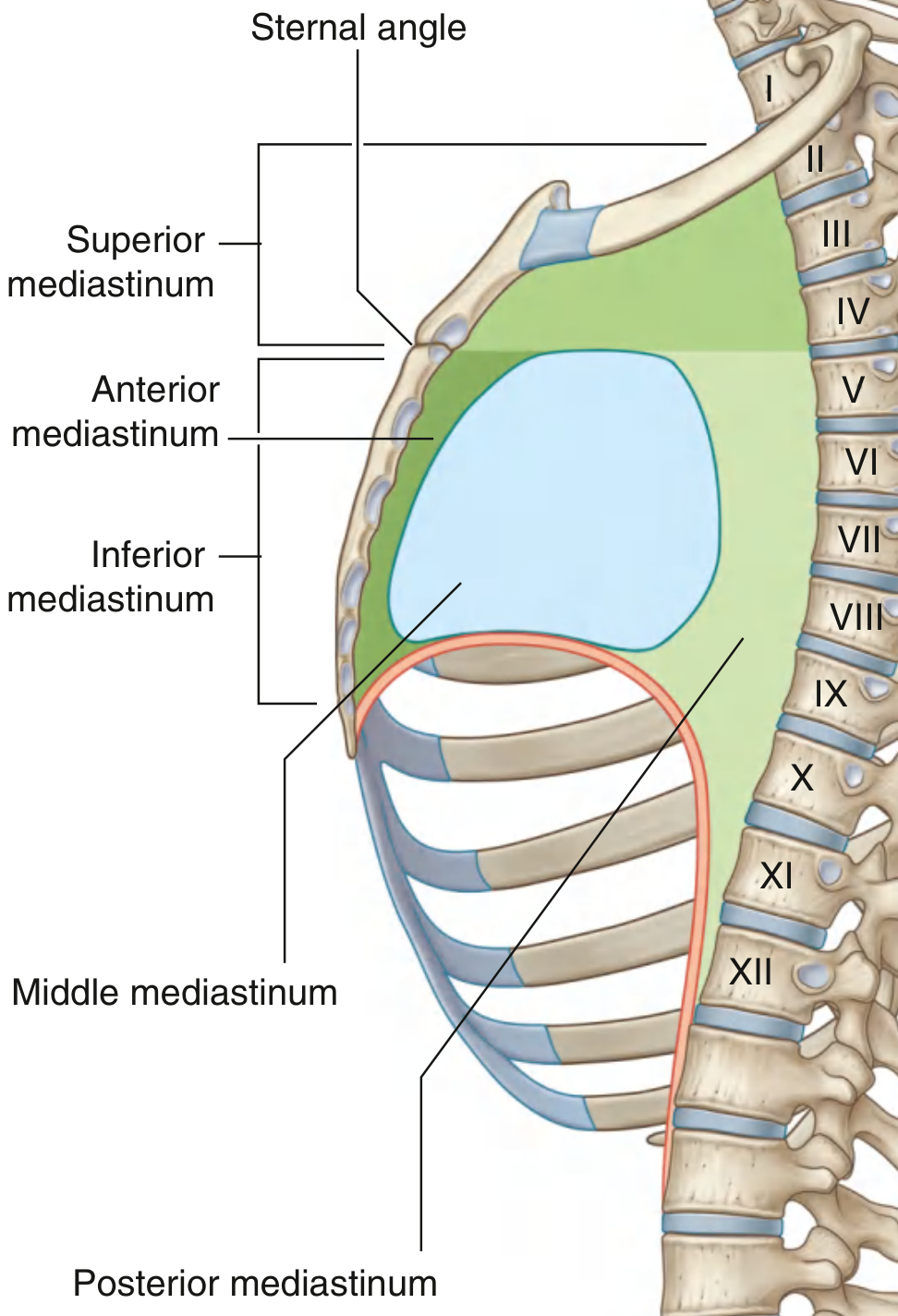
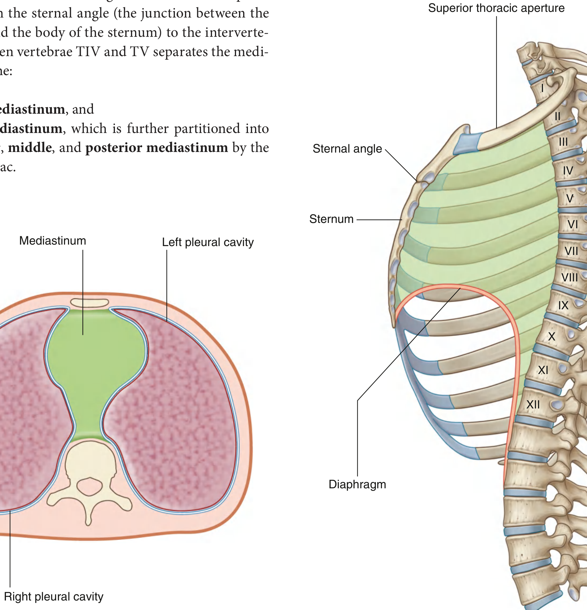
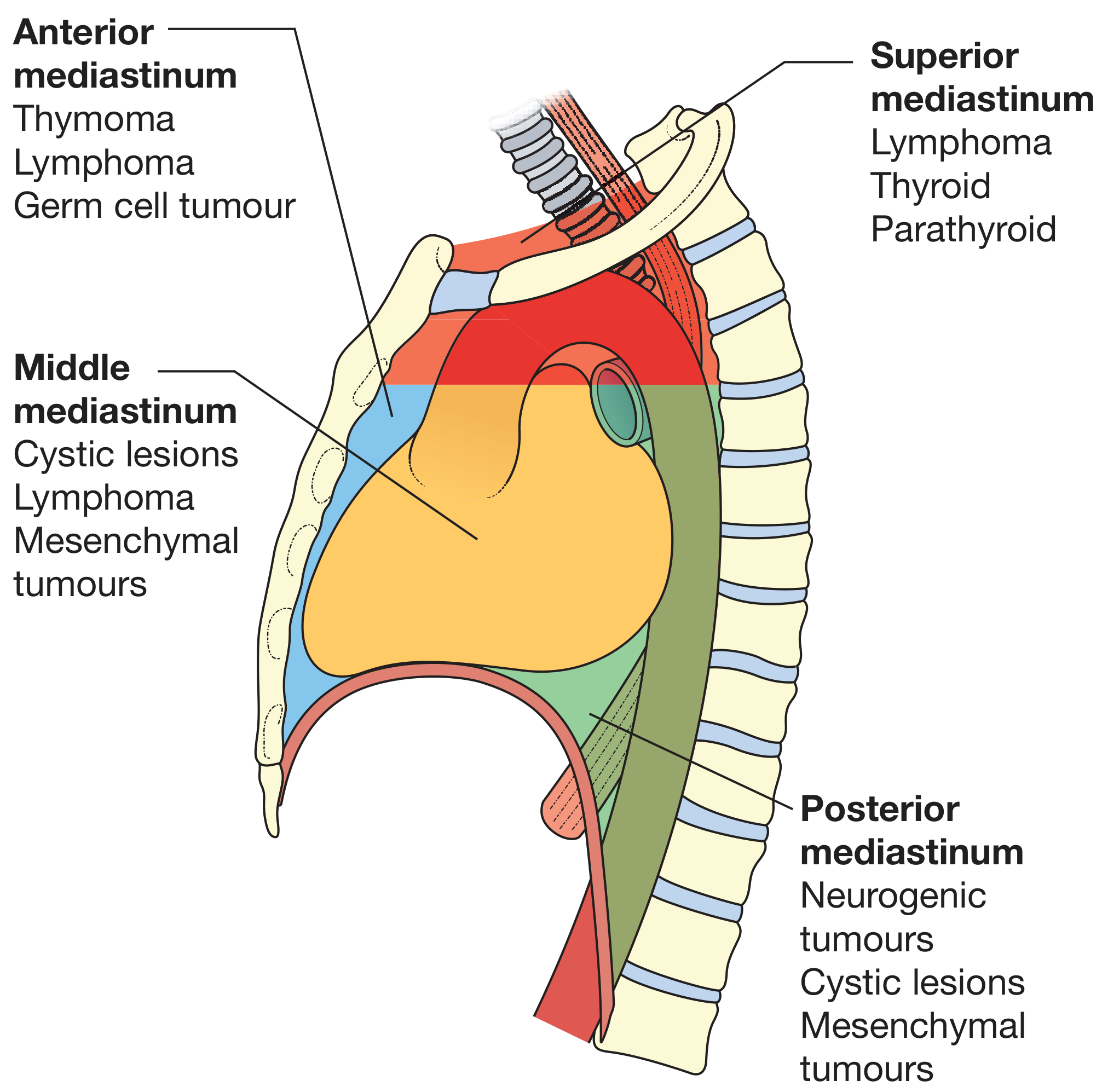
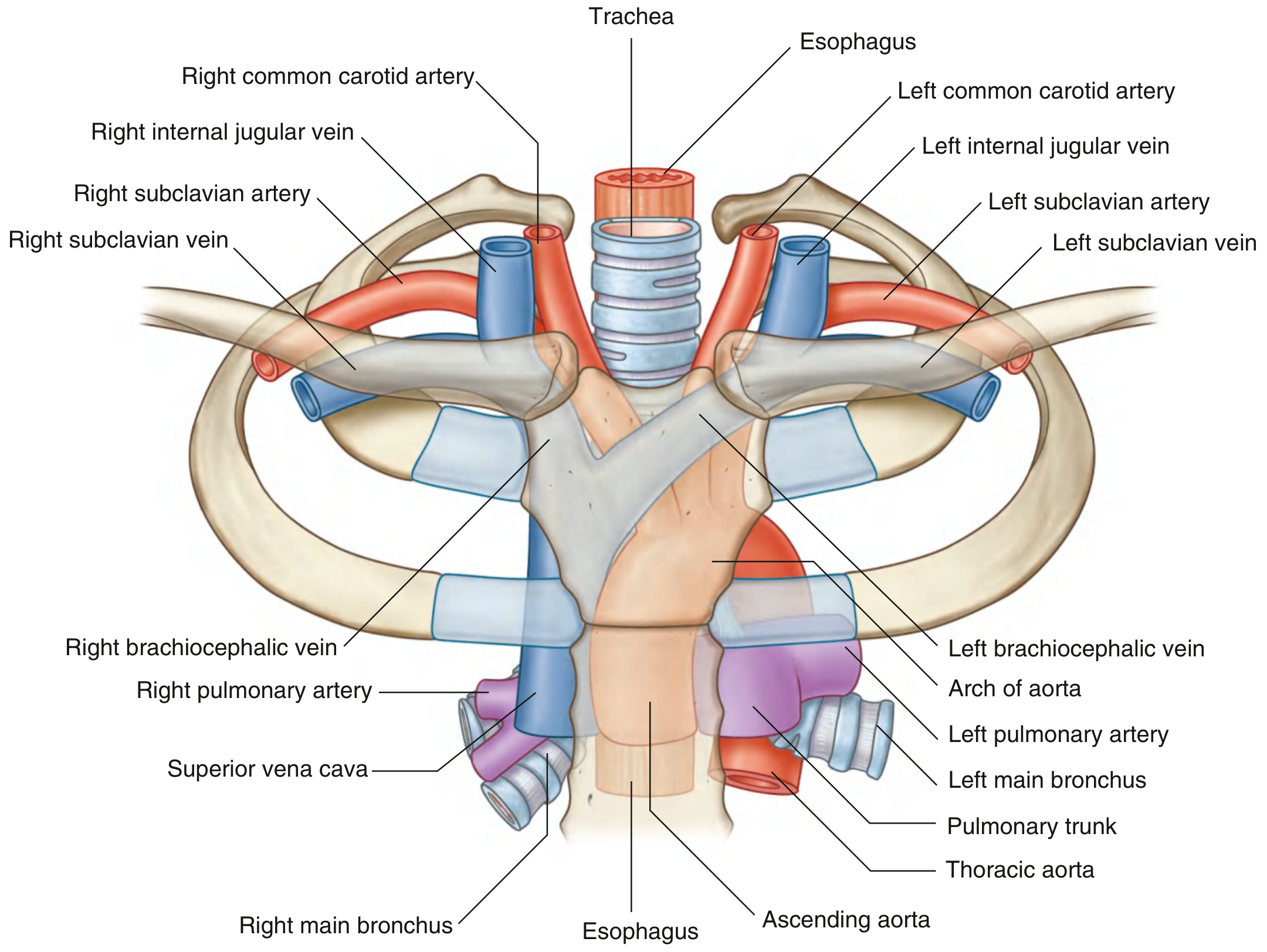
1. SUPERIOR MEDIASTINUM
| Feature | Detail |
|---|---|
| Superior boundary | Oblique plane from jugular notch upward/posteriorly to superior border of T1 (thoracic inlet) |
| Inferior boundary | Transverse plane through the sternal angle to TIV/TV intervertebral disc |
| Anterior boundary | Posterior surface of manubrium sterni |
| Posterior boundary | Bodies of T1-T4 vertebrae |
| Lateral boundaries | Mediastinal parietal pleura bilaterally |
| Structure | Details |
|---|---|
| Thymus | Most anterior component, involutes with age |
| Brachiocephalic veins | Right and left, converge to form SVC |
| Superior vena cava (SVC) | Formed by union of brachiocephalic veins |
| Left superior intercostal vein | Drains into left brachiocephalic vein |
| Arch of the aorta | With three branches: brachiocephalic trunk, left common carotid, left subclavian artery |
| Trachea | Bifurcates at carina at level of TIV/TV (sternal angle) |
| Esophagus | Posterior and slightly to the right of trachea |
| Phrenic nerves | Run on either side |
| Vagus nerves | Left recurrent laryngeal branch loops under aortic arch |
| Thoracic duct | Ascends on the left side |
| Small nerves, blood vessels, lymphatics | Deep cardiac plexus, lymph nodes |
2. ANTERIOR MEDIASTINUM (Prevascular Space)
| Feature | Detail |
|---|---|
| Superior boundary | Transverse plane through sternal angle to TIV-TV disc (separates from superior mediastinum) |
| Inferior boundary | Diaphragm |
| Anterior boundary | Posterior surface of body of sternum |
| Posterior boundary | Anterior surface of the pericardial sac |
| Lateral boundaries | Mediastinal parietal pleura bilaterally |
| Structure | Details |
|---|---|
| Inferior extension of thymus | Major structure; in adults largely replaced by fat |
| Fat and connective tissue | Prevascular fat pad |
| Lymph nodes | Internal mammary chain |
| Internal thoracic (mammary) vessels | Mediastinal branches |
| Sternopericardial ligaments | Connect sternum to fibrous pericardium |
Note: In adults the thymus involutes and its space is filled by fat; in children it is a prominent soft tissue structure.
3. MIDDLE MEDIASTINUM (Visceral/Pericardial Space)
| Feature | Detail |
|---|---|
| Boundaries | Defined by the fibrous pericardium itself |
| Position | Centrally located in the thoracic cavity |
| Structure | Details |
|---|---|
| Pericardium (fibrous + serous) | Encloses the heart and roots of great vessels |
| Heart | All four chambers |
| Ascending aorta | Root up to arch |
| Pulmonary trunk and main pulmonary arteries | Bifurcates into right and left |
| Superior and inferior vena cava | Drain into right atrium |
| Pulmonary veins | 4 veins drain into left atrium |
| Tracheal bifurcation and main bronchi | At carina |
| Phrenic nerves | Pass through to diaphragm |
| Lymph nodes | Subcarinal, paratracheal, hilar groups |
| Various nerves and smaller vessels |
4. POSTERIOR MEDIASTINUM (Paravertebral Space)
| Feature | Detail |
|---|---|
| Superior boundary | Transverse plane through sternal angle to TIV-TV disc |
| Inferior boundary | Diaphragm |
| Anterior boundary | Posterior surface of pericardial sac and posterior surface of diaphragm |
| Posterior boundary | Bodies of mid and lower thoracic vertebrae (TV-TXII) |
| Lateral boundaries | Mediastinal parietal pleura bilaterally |
| Structure | Details |
|---|---|
| Esophagus and esophageal nerve plexus | Passes through esophageal hiatus at TX |
| Thoracic aorta and its branches | Posterior intercostal arteries, subcostal artery |
| Azygos system of veins | Azygos, hemiazygos, accessory hemiazygos |
| Thoracic duct | Right of midline inferiorly, crosses left superiorly |
| Sympathetic trunks | Run lateral to vertebral bodies |
| Thoracic splanchnic nerves | Greater, lesser, least (T5-T12) |
| Lymph nodes | Posterior mediastinal group |
SUMMARY TABLE: Mediastinal Compartments at a Glance
| Compartment | Ant. Boundary | Post. Boundary | Key Contents |
|---|---|---|---|
| Superior | Manubrium | T1-T4 bodies | Thymus, SVC, aortic arch + 3 branches, trachea, esophagus, both phrenic, both vagus, thoracic duct |
| Anterior | Body of sternum | Pericardium | Thymic remnant, fat, lymph nodes, internal thoracic vessels |
| Middle | Pericardium | Pericardium | Heart, pericardium, ascending aorta, pulmonary trunk, SVC/IVC, main bronchi, phrenic nerves |
| Posterior | Pericardium | T5-T12 bodies | Thoracic aorta, esophagus, azygos system, thoracic duct, sympathetic trunks, splanchnic nerves |
PART 2: IMAGING DIFFERENTIAL DIAGNOSIS OF AN ANTERIOR MEDIASTINAL MASS
The "4 Ts" Mnemonic (Classic Teaching)
- Thymoma (most common primary mediastinal tumor - 25%)
- Teratoma / Germ Cell Tumor (GCT)
- Thyroid (retrosternal goiter / ectopic thyroid)
- Terrible Lymphoma (Hodgkin's / DLBCL)
Imaging Approach
1. THYMOMA
- 40-45% have myasthenia gravis (virtually diagnostic combination)
- May also associate with pure red cell aplasia, agammaglobulinemia
- One-third are asymptomatic (incidental finding)
- Presence of AChR antibodies clinches the diagnosis
| Modality | Findings |
|---|---|
| CXR | Well-defined, lobulated anterior mediastinal mass, often asymmetric; may calcify at the rim |
| CT (key modality) | Well-circumscribed, solid, lobulated mass in prevascular space; homogeneous soft-tissue attenuation; no fat or fluid components (distinguishes from teratoma); may show rim calcification; can invade adjacent pericardium, vessels (Masaoka staging) |
| MRI | T1: isointense to muscle; T2: intermediate to high signal; can assess vascular invasion and pericardial involvement better than CT |
- Solid, well-encapsulated mass, anterior to the great vessels
- Homogeneous enhancement
- No fat, calcium deposits, or cystic areas (if present, think teratoma)
- Obliteration of fat planes with pericardium = invasive thymoma (Masaoka stage III)
- Pleural nodules ("drop metastases") = stage IVa
2. LYMPHOMA (Hodgkin's Disease / Primary Mediastinal B-Cell Lymphoma)
- B symptoms: fever, night sweats, weight loss
- Peripheral lymphadenopathy elsewhere (neck, axilla)
- SVC syndrome (facial plethora, arm edema)
- Rapid onset of symptoms
- Elevated LDH
| Modality | Findings |
|---|---|
| CXR | Lobulated widening of superior mediastinum; "lobulated" or "polycyclic" anterior mediastinal contour; bilateral hilar enlargement (especially in Hodgkin's) |
| CT | Multiple enlarged confluent lymph nodes forming a bulky anterior/superior mediastinal mass; homogeneous on CT unless large (then low-density necrotic areas); may encase great vessels (but rarely invade); bilateral and symmetric; often extends to involve other nodal groups (paratracheal, subcarinal, hilar) |
| MRI | T1: intermediate signal; T2: high signal; post-treatment assessment with PET-MRI |
| FDG-PET/CT | Highly avid; essential for staging and post-treatment response |
3. GERM CELL TUMORS (Teratoma / Seminoma / Non-seminomatous GCT)
- Elevated AFP (non-seminomatous GCT), beta-hCG (seminoma, choriocarcinoma)
- Testes examination mandatory to exclude primary gonadal GCT
- Can rupture into lung, pericardium (hair in sputum = pathognomonic of teratoma)
| Modality | Findings |
|---|---|
| CXR | Well-defined, often large anterior mass; may show calcification (teeth, bone - virtually diagnostic) |
| CT (pathognomonic) | Heterogeneous mass containing multiple tissue densities in the SAME lesion: fat (-100 to -50 HU), soft tissue, fluid (cystic areas), calcium or calcified teeth/bone. The combination of fat + calcium + soft tissue in one anterior mediastinal mass = virtually diagnostic of mature teratoma |
| MRI | Fat signal on T1 (high signal, saturates on fat suppression); cystic areas T2 bright; fibrous components low T1/T2 |
- Large, lobulated, heterogeneous mass
- Areas of necrosis and hemorrhage
- Less well-defined borders than mature teratoma
- No fat component
4. RETROSTERNAL GOITER (Intrathoracic Thyroid)
- Dysphagia, stridor, orthopnea (respiratory symptoms on lying flat)
- Tracheal deviation/compression
- Neck swelling palpable in most cases
- Thyroid function may be normal, hypo-, or hyperthyroid
- Elevated TSH or T4
| Modality | Findings |
|---|---|
| CXR | Superior mediastinal widening; tracheal deviation (usually to the right); extends from the neck into the thorax |
| CT | Continuity with the cervical thyroid gland (pathognomonic); high attenuation on non-contrast CT (due to iodine content of thyroid - 100-150 HU, vs soft tissue 30-50 HU); heterogeneous with cystic areas and calcifications (in multinodular goiter); avid enhancement; tracheal displacement/compression |
| MRI | T1: heterogeneous; T2: high signal; characterization of cystic vs solid components |
| Tc-99m Pertechnetate or I-123 scan | Uptake in mediastinal mass = diagnostic |
5. THYMIC CYST
| Modality | Findings |
|---|---|
| CT | Thin-walled, unilocular or multilocular cystic mass in typical thymic location; homogeneous fluid attenuation (0-20 HU); no enhancement of internal contents (wall may enhance); no solid nodules (if present = thymoma with cystic change) |
| MRI | T1: low signal (unless hemorrhagic); T2: uniformly high signal; no restricted diffusion |
6. THYMIC CARCINOMA
- Large, irregular, poorly defined anterior mediastinal mass
- Heterogeneous with necrosis
- Invasion of mediastinal fat, vessels, chest wall
- Mediastinal lymphadenopathy and pleural/pericardial effusions common
- PET-CT: highly FDG avid (unlike thymoma which is mildly avid)
- Differentiating from invasive thymoma requires histopathology
7. CASTLEMAN DISEASE (Giant Lymph Node Hyperplasia)
- Well-defined, markedly enhancing mass (hyaline vascular type: intense homogeneous enhancement due to marked vascularity - "lightning bolt" enhancement)
- Can mimic carcinoid or vascular tumor
- CT: intense, homogeneous enhancement post-contrast (arterial phase)
- Central "spoke-wheel" calcification pattern on CT in some cases
8. LIPOMA / LIPOMATOSIS
- Homogeneous fat attenuation (-100 to -150 HU) on CT
- No enhancement
- Well-circumscribed (lipoma) vs diffuse fat deposition (lipomatosis - steroid-induced, Cushing's)
SUMMARY TABLE: Imaging Features of Anterior Mediastinal Masses
| Diagnosis | Age/Sex | Key CT Features | Differentiating Clue |
|---|---|---|---|
| Thymoma | 35-70 yr, M=F | Solid, homogeneous, well-circumscribed; no fat/calcium/cyst | Myasthenia gravis; AChR-Ab+ |
| Lymphoma | <40 yr (Hodgkin's) | Confluent nodal mass, homogeneous (unless necrotic), bilateral | B symptoms; bilateral hilar nodes; PET avid |
| Mature Teratoma | 2nd-3rd decade | MIXED attenuation: fat + calcium + fluid + soft tissue in one mass | Pathognomonic: tooth or bone on CT |
| Malignant GCT | Young males | Heterogeneous, large, poorly defined; necrosis/hemorrhage | AFP + / beta-hCG + |
| Retrosternal Goiter | Elderly females | High attenuation non-contrast CT; continuity with cervical thyroid | Tc-99m scan; tracheal deviation |
| Thymic Cyst | Any age | Thin-walled, water-attenuation (0-20 HU), no solid component | Incidental; no wall enhancement |
| Thymic Carcinoma | Middle age | Irregular, invasive, necrotic; mediastinal LN | PET highly avid; histology needed |
| Castleman Disease | Any | Marked homogeneous enhancement; spoke-wheel calcification | "Lightning-bolt" enhancement pattern |
| Lipoma | Adults | Pure fat density (-100 HU), no enhancement | Homogeneous fat = diagnostic |
Radiological Localization: 3-Compartment vs 4-Compartment Models
- Anterior (prevascular) - anterior to the SVC and pericardium
- Middle (visceral) - vessels, trachea, bronchi, pericardium
- Posterior (paravertebral) - posterior to the trachea and pericardium, paravertebral region
Key Points for the Examination
- Most common anterior mediastinal mass overall in adults = Thymoma (25% of all mediastinal masses)
- Most common in <40 years = Lymphoma
- Pathognomonic CT finding = Fat + calcium (teeth/bone) + cyst in one mass = Mature teratoma
- High CT attenuation on non-contrast + cervical thyroid continuity = Retrosternal goiter
- Marked homogeneous enhancement = Castleman disease
- Myasthenia gravis + anterior mediastinal mass = Thymoma until proven otherwise
- Elevated AFP/beta-hCG in a young male = Malignant GCT
- B symptoms + bilateral nodal enlargement = Lymphoma
- PET-CT is used for staging lymphoma and response assessment; FDG uptake is high in lymphoma and thymic carcinoma, lower in thymoma, absent in teratoma and thymic cysts
- Biopsy is generally avoided for thymoma (risk of spread) if diagnosis is clinically confident; mandatory for lymphoma and malignant GCT as treatment is non-surgical
Discuss the coronary arterial tree anatomy relevant to cardiac CT. Classify congenital coronary artery variants and anomalies - describe their imaging appearances on CT coronary angiography, emphasizing high-risk anomalies differential diagnosis and clinical implications - 50 marks
coronary artery anatomy diagram CT coronary angiography left right coronary
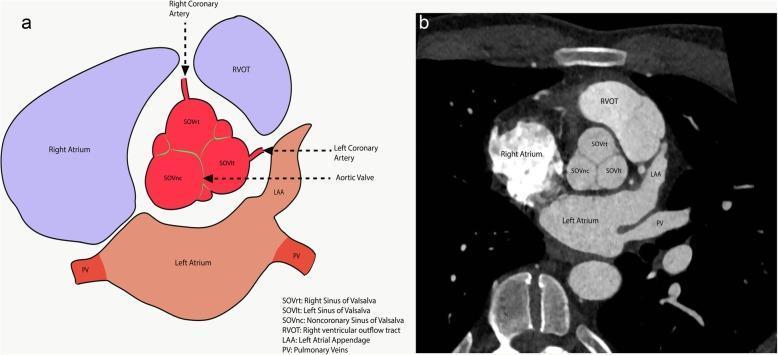
This educational image pairs an anatomical diagram (a) with a short-axis CT angiography (CTA) image (b) to illustrate the cross-sectional anatomy of the aortic root and surrounding cardiac structures. The focus is on the Sinuses of Valsalva (SOV), showing their spatial orientation and coronary artery origins. The central aortic root is divided into three distinct sinuses: the Right Sinus (SOVrt), from which the Right Coronary Artery originates; the Left Sinus (SOVlt), which gives rise to the Left Coronary Artery; and the Non-coronary Sinus (SOVnc), which lacks a coronary origin and lies adjacent to the interatrial septum. Surrounding structures are clearly labeled: the Right Ventricular Outflow Tract (RVOT) is located anteriorly and to the left; the Right Atrium is lateral to the SOVrt; the Left Atrium lies posteriorly; and the Left Atrial Appendage (LAA) and Pulmonary Veins (PV) are positioned laterally and posteriorly to the left. This material is designed for cardiovascular imaging training to aid in the precise identification of aortic root anatomy during diagnostic evaluation.
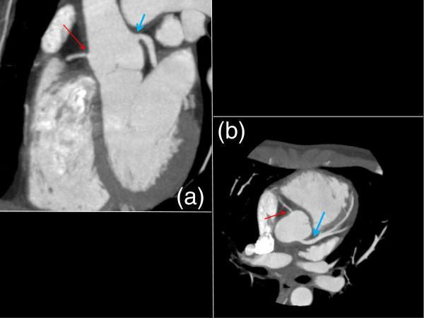
Two images from a cardiac gated CT angiography (CTA) of the chest in coronal (a) and axial (b) planes, demonstrating the coronary artery anatomy following systemic therapy for Takayasu's arteritis. In the coronal view (a), a thin red arrow identifies the right coronary artery (RCA) originating from the aortic root. Although better visualized than on previous imaging, it remains significantly stenotic with a narrowed lumen at the ostium. A blue arrow highlights the left coronary artery (LCA), which shows substantial interval improvement in patency. The axial view (b) provides a cross-sectional perspective of the same vessels, with the red arrow pointing to the persistent narrowing of the RCA ostium and the blue arrow indicating the significantly opened lumen of the LCA. These diagnostic images are used to monitor vascular wall thickening and luminal stenosis in large-vessel vasculitis, illustrating the differential response of the coronary ostia to pharmacological treatment.
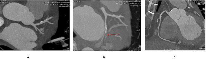
This diagnostic image set consists of three panels (A, B, C) showing 3D cardiac CT angiography (CTA) reconstructions of the coronary arterial system. Panel A illustrates the left coronary anatomy, highlighting an intact left main coronary artery bifurcating into the left anterior descending (LAD) and circumflex (LCx) arteries. Panel B displays a more detailed view of the circumflex territory where a red arrow identifies a focal area of hypocontrast, suggesting a suspected occlusion of the first marginal branch. There is a visible contrast filling defect in this vessel compared to the surrounding patent coronary branches. Panel C provides a reconstruction of the right coronary artery (RCA), demonstrating a dominant, intact vessel without significant stenosis or calcification. These reconstructions are used in preoperative screening to evaluate coronary anatomy and identify potential ischemic causes of left ventricular free wall rupture (LVFWR) or pseudoaneurysm.
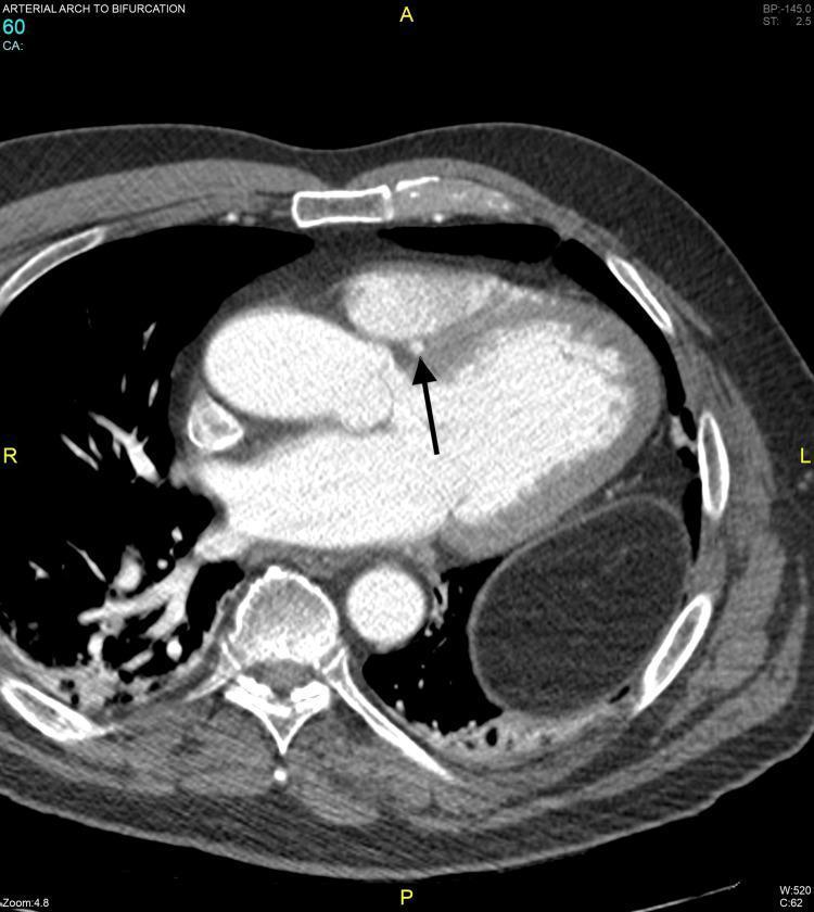
This diagnostic axial CT angiography (CTA) image provides a cross-sectional view of the mid-thoracic cavity, specifically focused on cardiac and mediastinal anatomy. The scan demonstrates the four heart chambers and the descending aorta. A primary clinical finding is an anomalous Left Main Coronary Artery (LMCA) highlighted by a black arrow. The vessel is shown following a septal course, traversing the interventricular septum between the right and left ventricular outflow regions. The image allows for assessment of the arterial caliber and relationship to surrounding myocardial structures, specifically looking for evidence of intra-arterial or intramyocardial compression. Other visible landmarks include the sternum anteriorly, vertebral bodies posteriorly, and bilateral lung parenchyma. This image is a critical educational resource for understanding coronary artery anomalies and the use of gated CTA in evaluating vascular pathways relative to cardiac anatomy.
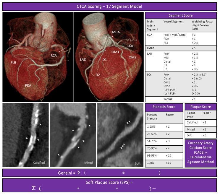
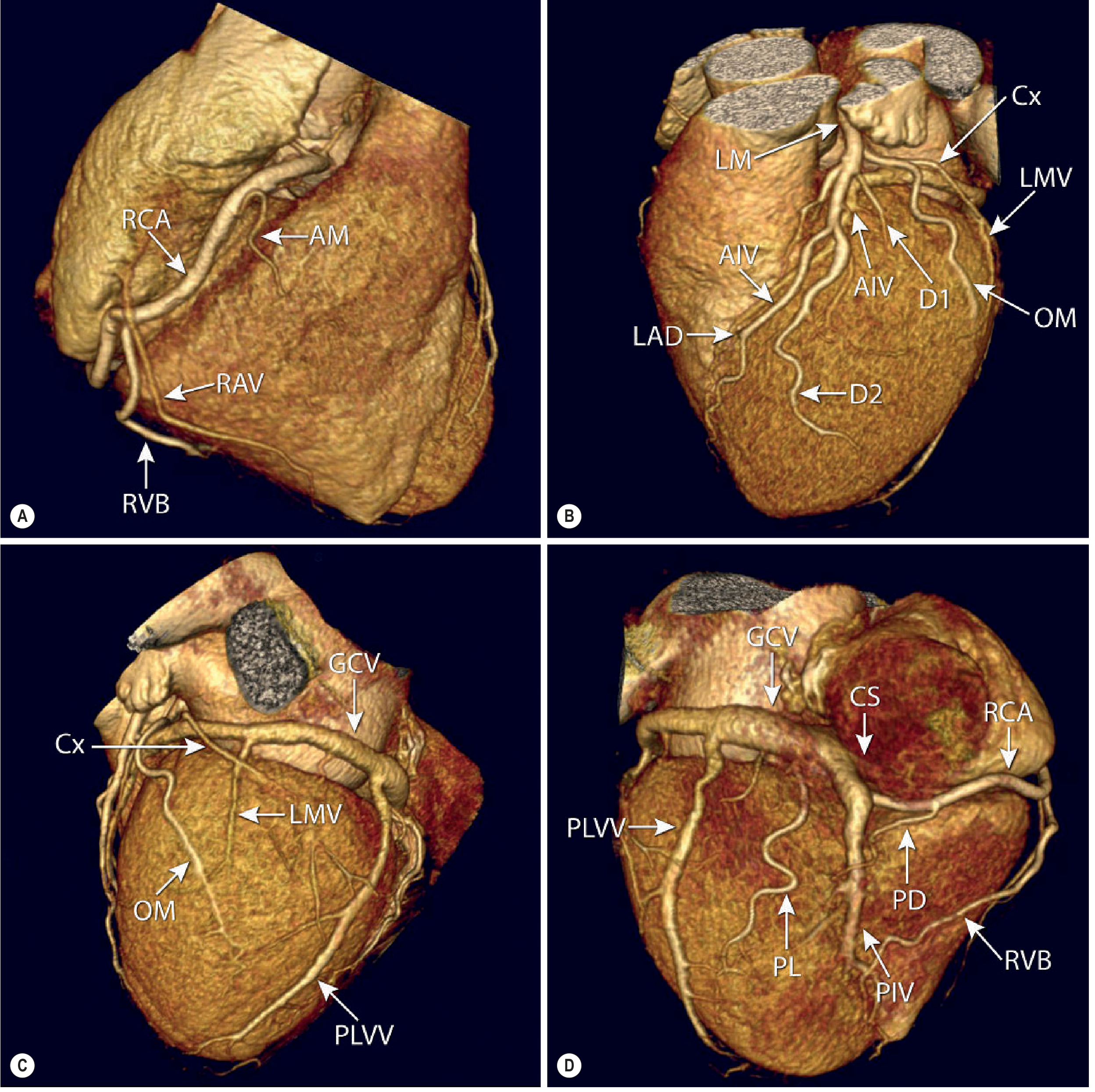
Educational infographic illustrating the CT Coronary Angiography (CTCA) semi-quantitative plaque scoring system, based on the Society of Cardiovascular Computed Tomography 17-segment model. The top section features two 3D volume-rendered heart reconstructions labeling key coronary anatomy: Right Coronary Artery (RCA), PDA, PLB, Left Main (LMCA), LAD, LCx, and branches D1/D2 and OM1/OM2. Tables define the Segment Score weighting factors for right and left dominance, and the Stenosis Score (1% to 100%). The bottom-left panels provide curved multiplanar reconstructions (cMPR) of coronary vessels showing three distinct plaque morphologies: 'Calcified' (high-attenuation/bright), 'Mixed' (heterogeneous density), and 'Soft' (low-attenuation/grey). A 'Plaque Score' table assigns multipliers (x1 for calcified, x2 for mixed, x3 for soft) to calculate the Soft Plaque Score (SPS) alongside the standard Gensini score and Agatston calcium score. This resource is designed for cardiology and radiology training on atherosclerotic burden assessment.
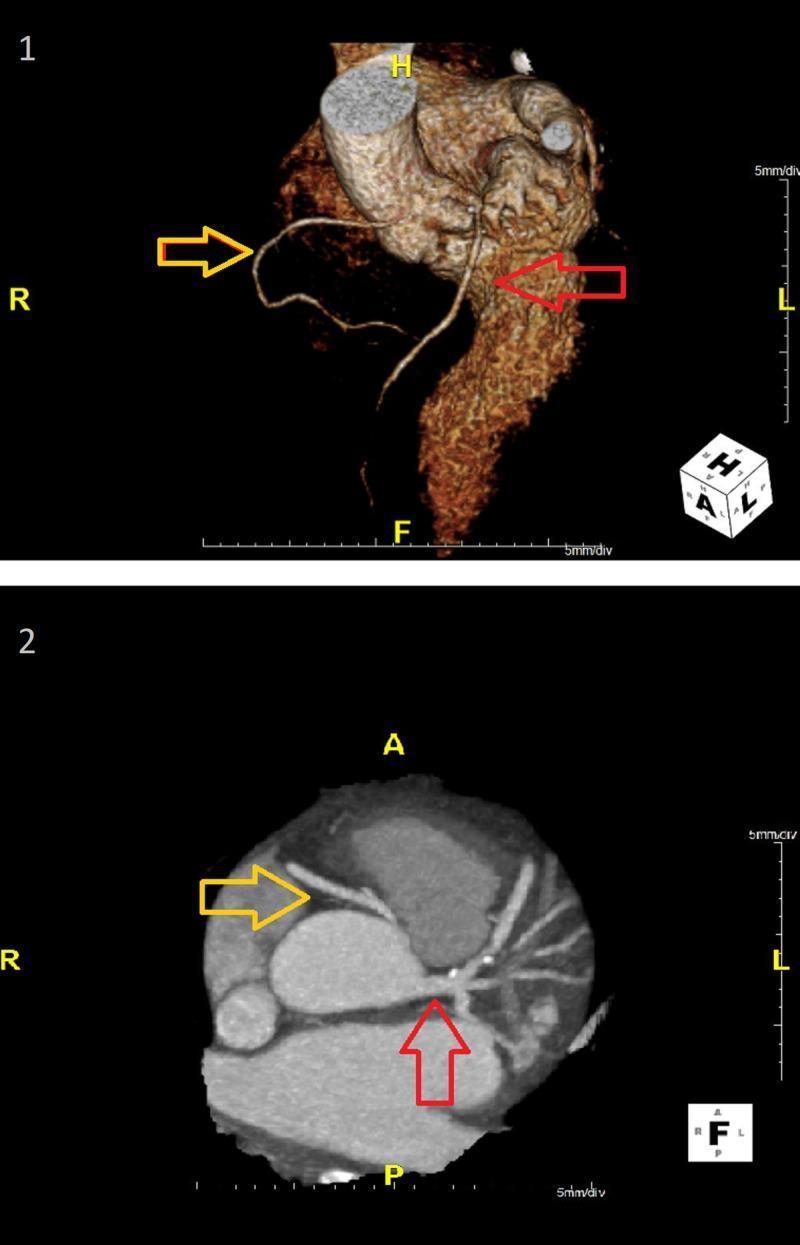
Two-panel diagnostic imaging showcasing cardiac anatomy via CT coronary angiography. Panel 1 features a 3D volume-rendered reconstruction of the heart and aorta, highlighting the spatial relationship of the coronary vasculature. Panel 2 presents an oblique maximum intensity projection (MIP) providing a cross-sectional view of the aortic root and arterial origins. The images demonstrate a congenital anomaly: an anomalous origin of the right coronary artery (RCA) from the left coronary sinus (ARCA-LCS). The yellow arrows indicate the RCA, which originates from the left sinus and follows an interarterial course between the aorta and pulmonary artery. The red arrows indicate the left coronary artery (LCA), also originating from the left sinus. This visual illustrates a high-risk coronary anomaly where the interarterial course can lead to mechanical compression of the vessel during physical exertion, which is a critical finding for cardiovascular risk assessment in clinical practice.
anomalous coronary artery origin interarterial course CT angiography high risk
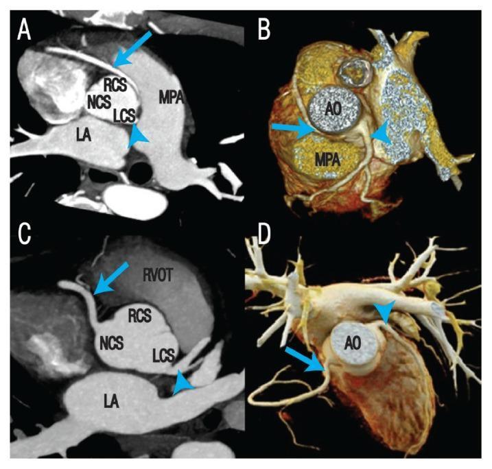
This diagnostic image composite features axial maximal intensity projection (MIP) coronary CT angiography (A, C) and 3D volume-rendered reconstructions (B, D) illustrating Anomalous Origin of the Coronary Artery from the Opposite Sinus (ACAOS). Panels A and B demonstrate an anomalous Right Coronary Artery (RCA, arrow) arising from the Left Coronary Sinus (LCS). The RCA follows a high-risk interarterial course, passing between the Aorta (AO) and the Main Pulmonary Artery (MPA). Panels C and D show a variant where the RCA (arrow) originates from the Non-Coronary Sinus (NCS), passing near the Right Ventricular Outflow Tract (RVOT). Key anatomical landmarks labeled include the Left Atrium (LA), Right Coronary Sinus (RCS), and the aortic root. This visual material is used in cardiovascular radiology to teach the identification of coronary anomalies and to assess the spatial relationship between anomalous vessels and great arteries, which is critical for determining the risk of sudden cardiac death and planning surgical interventions like coronary deroofing.
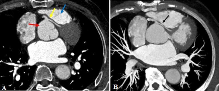
This diagnostic image consists of two Computed Tomography (CT) coronary angiogram reconstructions (A and B) demonstrating an anomalous origin and course of the left main coronary artery (LMCA). Image A is an axial reconstruction showing the LMCA (yellow arrow) taking a high-risk interarterial course between the ascending aorta (red arrow) and the pulmonary outflow tract (blue arrow). Image B utilizes Maximum Intensity Projection (MIP) reconstruction to highlight a clinically significant narrowing of the anomalous LMCA (black arrow) as it traverses between the great vessels, exhibiting an intramural or 'slit-like' appearance characteristic of malignant coronary artery anomalies. This case illustrates the diagnostic role of CT angiography in identifying congenital coronary variants and assessing for potential vascular compression. The findings are relevant for cardiology and radiology specialties, focusing on the pathophysiology of sudden cardiac death or myocardial ischemia related to anomalous coronary anatomy.
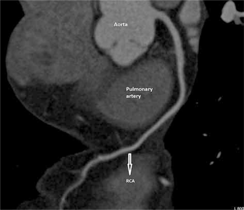
**Imaging Modality:** Computed Tomography Angiography (CTA), presented as a Maximum Intensity Projection (MIP) reconstruction of the heart and great vessels. **Anatomical Region:** Thorax, focusing on the aortic root and coronary arteries. **Observed Pathology:** Anomalous origin of the right coronary artery (RCA). **Characteristic Visual Features:** The image demonstrates the RCA arising anomalously from the left coronary sinus of the aorta, rather than the right sinus. The vessel follows an "interarterial course," passing acutely between the ascending aorta and the main pulmonary artery. Annotated labels identify the aorta and pulmonary artery, with an arrow highlighting the RCA as it traverses the space between these two major vessels before descending into the right atrioventricular groove. **Key Diagnostic Features:** - **Origin:** Left coronary sinus. - **Course:** Malignant/interarterial trajectory, which carries a risk of compression during exercise or increased cardiac output. - **Morphology:** Visible narrowing or "slit-like" appearance at the ostium and proximal segment as it passes between the great vessels. **Clinical Context:** This is a classic representation of a high-risk coronary anomaly (Anomalous Aortic Origin of a Coronary Artery - AAOCA) visualized via cardiac CT for surgical planning or risk stratification.
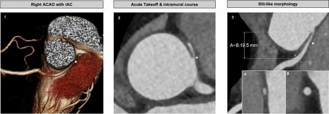
This composite diagnostic image consists of three panels demonstrating high-risk anatomical features of an anomalous origin of a coronary artery arising from the opposite sinus (ACAOS) with an interarterial course (IAC), visualized via Coronary Computed Tomography Angiography (CCTA). Panel 1 provides a 3D reconstruction showing the right coronary artery originating from the left sinus of Valsalva and traversing between the aorta and pulmonary artery. Panel 2 utilizes multiplanar axial reconstruction to highlight an acute takeoff angle and an intramural course, where the vessel is embedded within the aortic wall (indicated by an asterisk). Panel 3 demonstrates 'slit-like morphology' in a double oblique view. It quantifies the proximal luminal narrowing (Point A) compared to a distal reference point (Point B), with an interarterial course length measurement of 19.5 mm. These features are clinically significant as markers for potential myocardial ischemia or sudden cardiac death, facilitating surgical planning and risk stratification in cardiology and radiology.
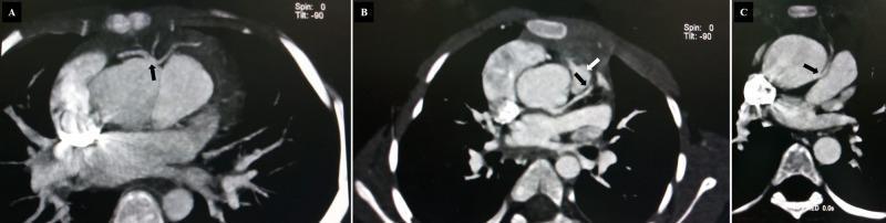
Diagnostic Image: Cardiac CT Angiography (CCTA) axial slices demonstrating various coronary artery anomalies. Panel A shows an anomalous origin of the left anterior descending (LAD) coronary artery from the right coronary artery (RCA), indicated by a black arrow. Panel B illustrates the LAD passing in close proximity to the pulmonic valve, with arrows highlighting the vessel's course. Panel C depicts the RCA originating from the left coronary cusp (LCC) with a high-risk interarterial course between the aorta and the pulmonary artery, marked by a black arrow. These images showcase congenital coronary anomalies often associated with structural heart diseases like Tetralogy of Fallot. The visual focus is on the anomalous takeoff points (ectopic origins) and the subsequent anatomical pathways (prepulmonary vs. interarterial) relative to the great vessels and heart valves. This content serves as an educational resource for cardiology and radiology specialties to identify potentially malignant coronary vessel trajectories using non-invasive imaging.
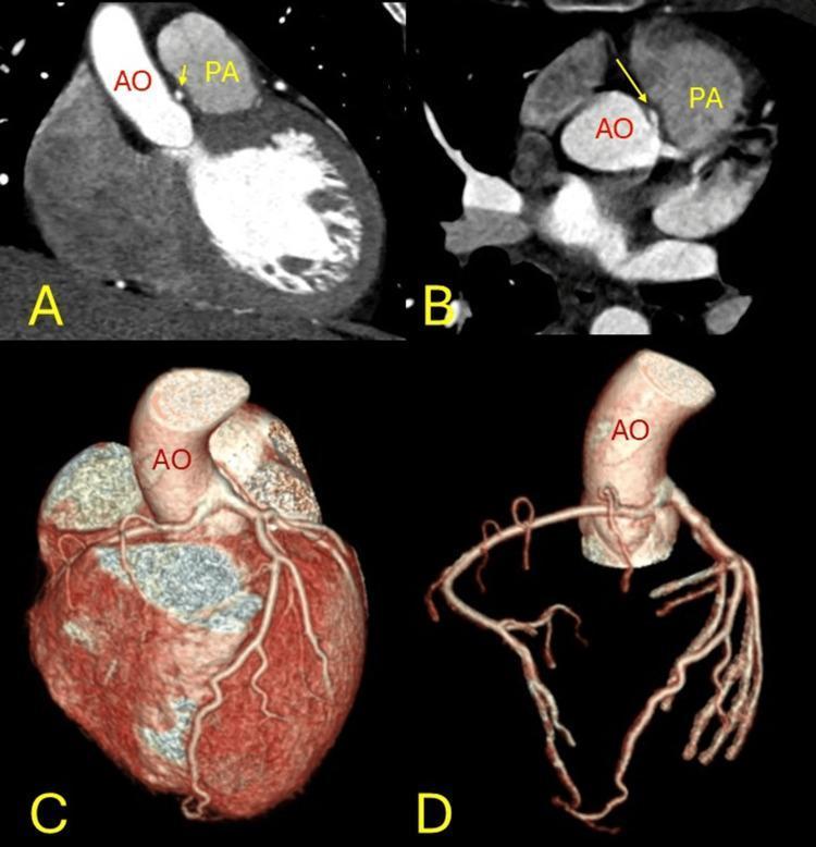
A multi-panel clinical diagnostic image set demonstrating an anomalous origin of the right coronary artery (RCA) from the left coronary sinus. Panels A and B are CT angiography (CTA) images in coronal and axial planes, respectively, showing the interarterial course of the RCA. Yellow arrows highlight the vessel passing between the aorta (AO) and the pulmonary artery (PA). Panels C and D provide three-dimensional colored volume renderings of the cardiac anatomy and coronary arterial tree. These renderings illustrate the spatial relationship of the AO and the origin of the coronary arteries, highlighting the 'malignant' course of the anomalous vessel as it exits the aorta and traverses the space between the major outflow tracts. The imagery is intended for advanced medical education regarding congenital coronary anomalies, specifically the risk of compression associated with interarterial pathways. Labels 'AO' and 'PA' identify the primary vascular landmarks, while panels C and D emphasize the superficial and deep branching patterns of the coronary circulation.
CORONARY ARTERIAL TREE: CARDIAC CT ANATOMY, VARIANTS & ANOMALIES
PART 1: CORONARY ARTERY ANATOMY RELEVANT TO CARDIAC CT
Technical Background: CT Coronary Angiography (CCTA)
- Acquisition timing: Images reconstructed at 75% of the R-R cycle (mid-diastole = cardiac rest phase) for optimal motion-free coronary imaging. High heart rates require beta-blockade (target HR <65 bpm) before scanning
- Nitrate premedication: Sublingual nitroglycerine given before scanning to dilate coronaries and improve visualisation of small branches
- Reconstruction techniques:
- Axial (transverse) multiplanar reconstructions (MPR) - primary reading tool; contain all information without reconstruction artefacts
- Curved MPR (cMPR) - allows visualisation of entire coronary artery in one image
- Maximum intensity projection (MIP) - overview of coronary tree
- 3D volume rendering (VR) - excellent for anomaly course assessment and surgical planning
- Radiation dose: Prospective ECG-triggered acquisition: ~1-5 mSv. Retrospective gating: 12-21 mSv (used when functional data needed or irregular rhythm)
- Calcium scoring: Unenhanced CT. Agatston score >400 significantly impairs lumen assessment due to "blooming artefact"
Origins: The Aortic Root and Sinuses of Valsalva
| Sinus | Also called | Coronary Origin |
|---|---|---|
| Right coronary sinus | Anterior sinus | Right coronary artery (RCA) |
| Left coronary sinus | Left posterior sinus | Left main coronary artery (LMCA) |
| Non-coronary sinus | Right posterior sinus | None (adjacent to interatrial septum) |
Left Coronary System
- Arises from the superior aspect of the left sinus of Valsalva
- Short trunk (5-20 mm long) coursing leftward posterior to the pulmonary trunk
- Absent left main (1% of population): LAD and Cx arise from a common ostium or separately from the left sinus - important CCTA finding (Fig. 12.14 in Grainger & Allison)
- Bifurcates into LAD and circumflex; trifurcation occurs in ~30% when a ramus intermedius (intermediate artery) arises between them
- Runs in the anterior interventricular groove (between LV and RV)
- Extends to the LV apex and sometimes wraps around it
- Key branches:
- Septal perforators (multiple, run straight down into the interventricular septum at 90°) - supply the anterior 2/3 of interventricular septum
- Diagonal branches (D1, D2, D3) - supply the anterolateral LV free wall; usually 1-3 in number
- On CCTA: LAD runs in the anterior interventricular groove visible on axial slices; septal branches run perpendicular to the LAD; diagonals course laterally along the LV
- Courses in the left atrioventricular (AV) groove
- Obtuse marginal branches (OM1, OM2, OM3) - supply the lateral and posterolateral LV free wall; usually 1-3 in number
- Sinus node artery: arises from the circumflex in ~40% of patients (from RCA in ~60%)
- In left dominant circulation: Cx continues to give rise to the posterior descending artery (PDA) and posterolateral branches
- On CCTA: Cx runs in the left AV groove; OM branches visible coursing laterally and posteriorly
Right Coronary System
- Arises from the superior aspect of the right (anterior) sinus of Valsalva
- Courses anteriorly between the right atrial appendage and RVOT, then inferiorly in the right AV groove
- Key branches:
- Conus branch (first branch) - supplies RVOT; in 50% arises separately from the aorta ("third coronary artery")
- Sinus node artery - arises from RCA in ~60% (proximal RCA)
- Acute marginal branches (AM) - supply the right ventricular free wall
- AV nodal artery - from the crux, supplies AV node; arises from whichever vessel crosses the crux
- Posterior descending artery (PDA) - runs in the posterior interventricular groove; defines dominance
- Posterolateral branches (PLB) - continue in the left AV groove after the crux
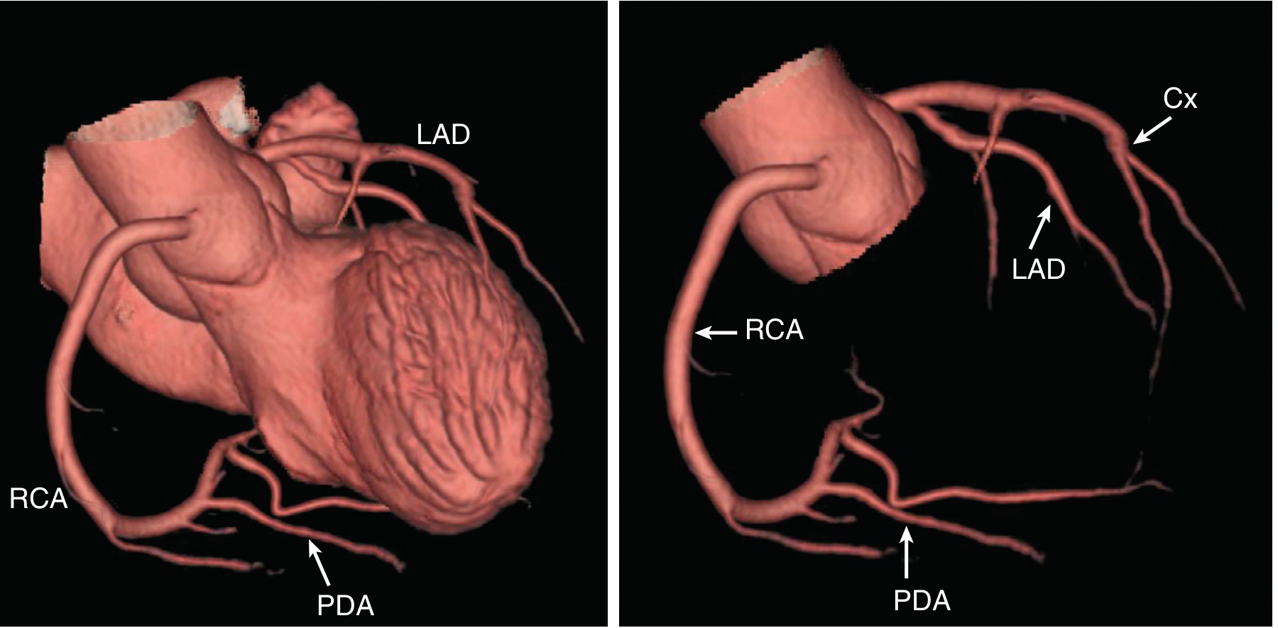
Coronary Dominance
| Dominance Pattern | Definition | Frequency |
|---|---|---|
| Right dominant | PDA arises from RCA | ~80% |
| Left dominant | PDA arises from Cx | ~10% |
| Co-dominant (balanced) | Two PDAs: one from each | ~10% |
17-Segment Model and Coronary Territories
| Territory | Segments Supplied |
|---|---|
| LAD | Anterior wall, anterior septum, apex (segments 1, 2, 7, 8, 13, 14, 17) |
| RCA | Inferior wall, inferior septum (segments 3, 4, 9, 10, 15) |
| Cx | Lateral and posterolateral wall (segments 5, 6, 11, 12, 16) |
PART 2: CLASSIFICATION OF CONGENITAL CORONARY ARTERY VARIANTS AND ANOMALIES
Classification System
- Absent left main (separate ostia for LAD and Cx)
- High/low ostial origin
- Ramus intermedius / trifurcation
- Myocardial bridging (mild)
- Anomalous LAD from RCA crossing the RVOT (in Tetralogy of Fallot - prevents safe transannular incision)
- Coronary anomalies in congenital heart disease
- Coronary arteriovenous fistula (CAVF)
- Anomalous coronary origin from the pulmonary artery (ALCAPA/ARCAPA)
- Anomalous aortic origin of a coronary artery (AAOCA) - the interarterial variant
- Myocardial bridging (symptomatic)
- Coronary artery aneurysm
PART 3: DETAILED DESCRIPTION WITH CCTA IMAGING APPEARANCES
1. ANOMALOUS AORTIC ORIGIN OF A CORONARY ARTERY (AAOCA)
Classification by Course (Mechanism of Ischemia Depends on Course):
| Course | Description | Risk |
|---|---|---|
| Interarterial | Between aorta and pulmonary artery | HIGH - malignant |
| Prepulmonary (anterior) | Anterior to pulmonary trunk | LOW |
| Retroaortic (posterior) | Posterior to aorta | LOW |
| Subpulmonic (septal/intraseptal) | Through interventricular septum | Intermediate |
Specific Variants:
- RCA arises from the left sinus of Valsalva or left main
- Passes between the aorta and pulmonary trunk = interarterial course
- Prevalence: ~0.1-0.3% of population
- Risk: Associated with sudden cardiac death, especially during exertion
- On axial views: both coronary ostia visible arising from the left sinus
- RCA courses rightward between the aorta (anterior) and the pulmonary artery (posterior)
- High-risk features on CCTA:
- Acute takeoff angle (<45°) creating slit-like ostium
- Intramural (intramural) course: vessel embedded within the aortic wall (identified as vessel traversing aortic wall without surrounding fat)
- Slit-like or elliptical lumen on cross-section (vs round in normal)
- Length of intramural/interarterial segment (>10 mm = higher risk)
- Compression of vessel between great arteries visible on axial slices
- LMCA or LAD arises from the right sinus of Valsalva
- When it follows an interarterial course = highest risk anomaly (associated with SCD in young athletes)
- CCTA Findings:
- Axial slice: LMCA seen crossing from the right sinus leftward between the aorta and pulmonary artery
- Slit-like lumen at the interarterial segment
- Intramural course (vessel within aortic wall) may be identified
Pathophysiology of Ischemia in Interarterial AAOCA:
- Acute takeoff angle creates slit-like ostium with functional stenosis, especially during exercise (aortic dilation further compresses the ostium)
- Intramural course: Vessel traverses the aortic wall - during exercise, aortic distension compresses the intramural segment
- Extrinsic compression between the aorta and pulmonary artery during exercise (both dilate under high-output states)
- These mechanisms combine to cause exercise-induced ischemia and ventricular fibrillation
CCTA High-Risk Features (surgical decision-making):
- Interarterial course confirmed
- Slit-like ostial morphology
- Acute takeoff angle
- Intramural segment length >10 mm
- Compression visible between aorta and PA on functional CT
2. ANOMALOUS CORONARY ORIGIN FROM THE PULMONARY ARTERY
A. ALCAPA (Anomalous Left Coronary Artery from Pulmonary Artery)
- Most common anomalous pulmonary origin (~1 in 300,000 live births)
- LMCA arises from the pulmonary artery instead of the aorta
- After birth and fall in pulmonary pressure: LCA perfused with deoxygenated blood at low pressure
- Coronary steal develops: collaterals from RCA shunt blood retrograde into the low-pressure PA
- Infants (6 weeks - 3 months): irritability, feeding difficulty, failure to thrive, CHF
- Adults (rare survivors): angina, mitral regurgitation (ischemic), SCD
- CXR: cardiomegaly, pulmonary edema
- ECG: lateral ischemia / infarction pattern
- No left coronary ostium in the left sinus of Valsalva
- LCA origin visible from the pulmonary artery (usually left posterior PA wall)
- Dilated, tortuous RCA (compensatory collaterals)
- Retrograde filling of LCA from collaterals (may be seen on phase-specific reconstructions)
- LV wall motion abnormality and myocardial hypoenhancement (infarction/ischemia)
- Mitral valve apparatus assessment: papillary muscle ischemia → mitral regurgitation
B. ARCAPA (Anomalous Right Coronary Artery from Pulmonary Artery)
- Very rare; RCA arises from the PA
- Usually better tolerated than ALCAPA (smaller territory)
- CCTA: RCA origin from the PA, enlarged LCA collaterals
3. CORONARY ARTERIOVENOUS FISTULA (CAVF)
| Feature | Detail |
|---|---|
| Most common origin | RCA (~55%) |
| Most common drainage | Right heart chambers (RV, RA, PA, coronary sinus) |
| Direction of shunt | Left-to-right |
| Hemodynamic effect | Volume overload of drainage chamber; coronary steal |
- Continuous machinery murmur (resembles PDA)
- CHF, angina (coronary steal), arrhythmia, endocarditis, rare SCD
- Small fistulae: often asymptomatic
- Markedly dilated, tortuous feeding coronary artery
- Fistulous connection identifiable as an abnormal vessel coursing to the drainage structure
- Drainage: opacification of the recipient structure early in arterial phase (RV, RA, coronary sinus, or PA)
- 3D VR: excellent for mapping the entire course of the fistula before intervention
- Myocardial perfusion defects distal to fistula (steal phenomenon)
4. MYOCARDIAL BRIDGING (MB)
- Anatomical (autopsy): ~25-85%
- CCTA: ~20-30% (visible as systolic compression of the tunneled segment)
- Symptomatic: much less common
- Tunneled segment: Mid-LAD dips beneath the myocardium; on axial CT the vessel is surrounded by myocardium rather than epicardial fat
- Depth measurement: Distance from epicardial surface to the tunneled vessel (deeper = more severe)
- Length measurement: Length of tunneled segment in mm
- Systolic compression: Best shown on retrospective gating with systolic phase reconstruction - "milking effect" (systolic narrowing, diastolic normalization)
- Soft plaque: Atherosclerosis typically absent in the tunneled segment but accelerated proximally to the bridge
- Stress CT myocardial perfusion: may show reversible perfusion defect in LAD territory
- Functional CT-FFR: virtual FFR calculation can assess hemodynamic significance
- Beta-blockade (first line - prolongs diastole)
- Surgery: unroofing (dividing the overlying muscle)
- PCI: stenting (higher complication rate - used selectively)
5. CORONARY ARTERY ANEURYSMS
| Cause | Features |
|---|---|
| Kawasaki disease | Most common cause in children; fusiform or saccular aneurysms, especially RCA and LMCA |
| Atherosclerosis | Most common cause in adults |
| Connective tissue disorders | Marfan, Ehlers-Danlos |
| Post-intervention | Post-PCI/stenting |
| Congenital | Rare |
- Focal or diffuse dilatation of coronary artery
- Kawasaki: multiple, bilateral, fusiform or saccular; may show calcification in chronic disease; may contain thrombus (low attenuation filling defect)
- Giant aneurysms (>8 mm): risk of thrombosis, distal embolization, rupture
- 3D VR: maps the extent and number of aneurysms for surgical planning
PART 4: HIGH-RISK ANOMALIES - DIFFERENTIAL DIAGNOSIS AND CLINICAL IMPLICATIONS
Key Differential Diagnosis on CCTA: Identifying the "Malignant" from "Benign" Coronary Anomalies
| Feature | Malignant (High-Risk) | Benign (Low-Risk) |
|---|---|---|
| Course between aorta + PA | YES - interarterial | No |
| Ostial morphology | Slit-like, acute angle | Round, wide angle |
| Intramural segment | Present | Absent |
| Associated SCD risk | HIGH | LOW |
| Examples | LMCA from RCS with interarterial course, RCA from LCS with interarterial course | Cx from right sinus with retroaortic course, RCA from non-coronary sinus with anterior course |
Practical CCTA Reporting Checklist for Coronary Anomalies
- Origin: Which sinus? Exact ostial location?
- Ostial morphology: Round vs slit-like? Takeoff angle?
- Is there an intramural segment? (vessel within aortic wall)
- Course: Interarterial / retroaortic / prepulmonary / intraseptal?
- Length of interarterial or intramural segment (in mm)
- Distal distribution: Is the anomalous vessel dominant? What territory does it supply?
- Any evidence of ischemia? Myocardial hypoenhancement, wall motion abnormality
- Any associated anomalies? Congenital heart disease?
Summary Table: Coronary Anomalies on CCTA
| Anomaly | Key CCTA Finding | Risk | Clinical Implication |
|---|---|---|---|
| ARCA-LCS (interarterial) | RCA from left sinus, courses between Ao and MPA; slit-like ostium | HIGH | SCD risk in athletes; surgical unroofing |
| ALCA-RCS (interarterial) | LMCA from right sinus between Ao and MPA | VERY HIGH | Highest SCD risk; urgent surgical correction |
| Cx from right sinus (retroaortic) | Cx arises from right sinus, courses posterior to Ao | LOW | No surgical intervention needed |
| RCA from non-coronary sinus | RCA anterior course anterior to PA | LOW | Generally benign |
| ALCAPA | No LCA ostium in left sinus; LCA from PA; dilated RCA | VERY HIGH | Fatal in infancy if untreated; surgical reimplantation |
| ARCAPA | RCA from PA; enlarged LCA collaterals | HIGH | Requires surgical repair |
| CAVF | Dilated tortuous coronary; early drainage into RV/RA/CS | MODERATE | Transcatheter/surgical closure if symptomatic |
| Myocardial bridging | LAD intramyocardial (no surrounding fat); systolic compression | LOW-MODERATE | Beta-blockade; unroofing if refractory |
| Coronary aneurysm | Focal/diffuse coronary dilation; +/- thrombus | VARIABLE | Anticoagulation; surgery for giant aneurysms |
| Absent left main | Separate LAD + Cx ostia from left sinus | LOW | No intervention; CABG planning implication |
| Ramus intermedius | Trifurcation of left main | NONE | Incidental; CABG planning consideration |
Clinical Implications Summary
- AAOCA (particularly ALCA-RCS with interarterial course) is the 2nd most common cause of SCD in young athletes (after hypertrophic cardiomyopathy)
- CCTA is the investigation of choice for definitive delineation of course and high-risk features
- Invasive coronary angiography cannot reliably demonstrate the course of an anomalous vessel - CCTA is superior
- Expert consensus guideline (AHA/ACC): symptomatic patients and those with high-risk CCTA features undergo surgical unroofing; asymptomatic patients with low-risk features may be managed conservatively with activity restriction and close follow-up
- Young patient with exertional syncope, chest pain, or palpitations
- Pre-operative assessment before congenital heart disease surgery
- Athlete evaluation after cardiac arrest or ventricular arrhythmia
- Incidental coronary anomaly detected on echocardiography or other imaging
Discuss the principles, image interpretation, and clinical applications af Diffusion-Weighted Imaging (DWI) in MRI, with emphasis on ADC mapping and its diagnostic significance