
Anatomy of head and neck
Reading File
The query is broad - "anatomy of head and neck" covers many sub-topics. Let me clarify what the user needs before diving in.
Asking for Preferences
Which aspect of head and neck anatomy are you most interested in? · What is the context?
The user skipped - I'll provide a comprehensive overview of head and neck anatomy covering all major systems. Let me search the medical library in parallel for the key sub-topics.
Finding Sources
Finding Sources
Finding Sources
Finding Sources
Finding Sources
Good - Gray's Anatomy for Students has extensive head & neck content. Let me read key sections in parallel.
Reading File
Reading File
Finding Sources
Reading File
Reading File
Reading File
Reading File
I now have excellent content. Let me fetch a few key diagrams to include in the response.
Loading Image
Loading Image
Loading Image
Excellent! I now have excellent textbook content and diagrams. Let me also fetch the dural venous sinuses image and cervical nerves diagram.
Loading Image
Loading Image
I now have comprehensive content and high-quality diagrams from Gray's Anatomy for Students. Let me compile the full response.
Anatomy of the Head and Neck
Based on Gray's Anatomy for Students and Color Atlas of Human Anatomy.
1. The Skull
The skull has 22 bones (excluding the ear ossicles). All bones except the mandible are united by sutures and form the cranium.
The cranium is divided into three parts:
- Calvaria - the domed roof, formed mainly by the paired temporal and parietal bones plus parts of the frontal, sphenoid, and occipital bones.
- Base of the cranium - formed mainly by parts of the sphenoid, temporal, and occipital bones.
- Facial skeleton (viscerocranium) - paired nasal, palatine, lacrimal, zygomatic bones, maxillae, and inferior nasal conchae, plus the unpaired vomer.
The mandible is separate from both the cranium and the facial skeleton.
Cranial Fossae and Foramina
The base of the cranium is divided into three fossae (anterior, middle, posterior), each containing foramina that transmit key structures:
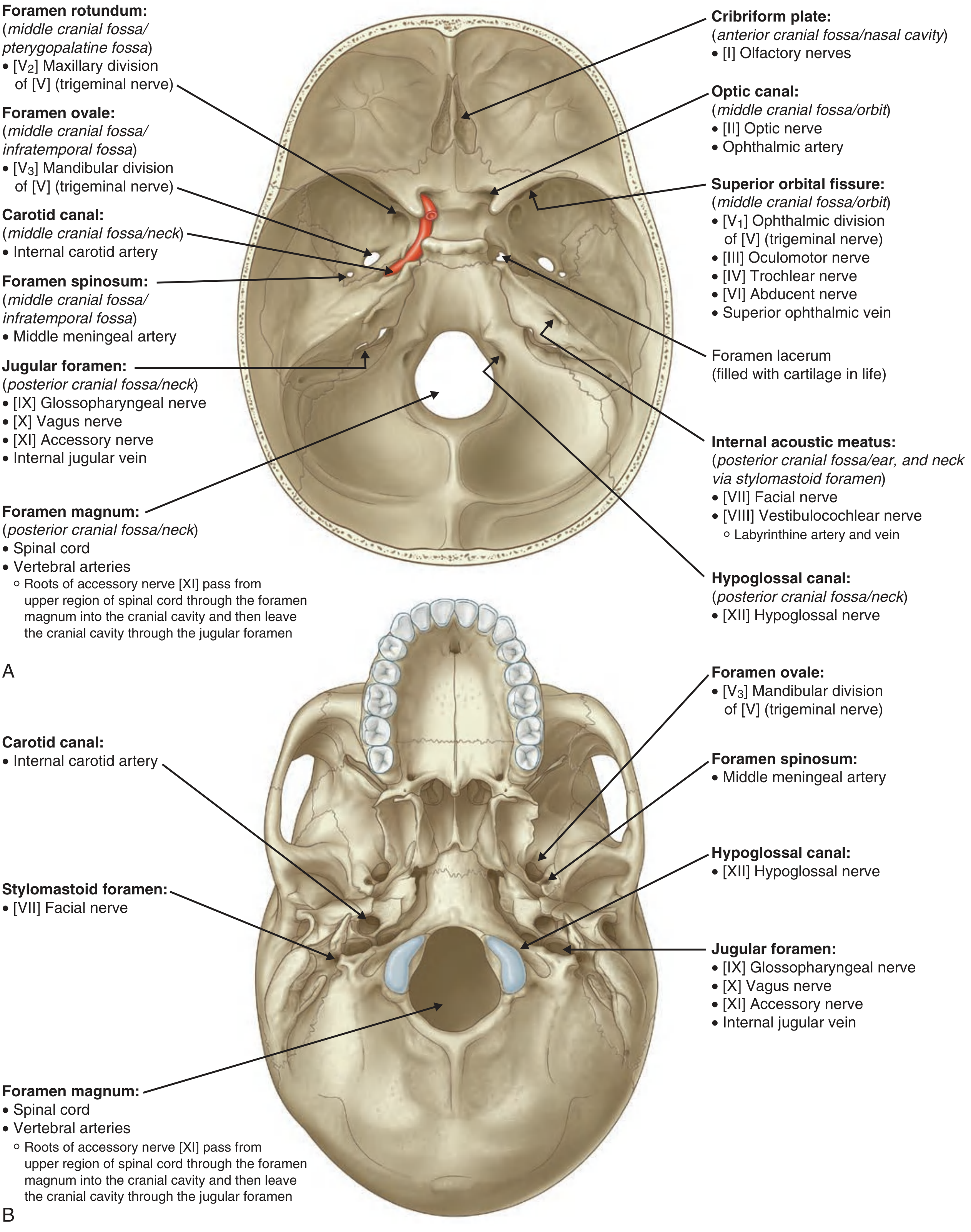
Key foramina and their contents:
| Foramen | Location | Contents |
|---|---|---|
| Cribriform plate | Ant. cranial fossa / nasal cavity | CN I - Olfactory nerves |
| Optic canal | Mid. cranial fossa / orbit | CN II (optic nerve), ophthalmic artery |
| Superior orbital fissure | Mid. cranial fossa / orbit | CN III, IV, V1, VI; superior ophthalmic vein |
| Foramen rotundum | Mid. cranial fossa / pterygopalatine fossa | V2 (maxillary nerve) |
| Foramen ovale | Mid. cranial fossa / infratemporal fossa | V3 (mandibular nerve) |
| Foramen spinosum | Mid. cranial fossa | Middle meningeal artery |
| Carotid canal | Mid. cranial fossa / neck | Internal carotid artery |
| Internal acoustic meatus | Post. cranial fossa | CN VII (facial), CN VIII (vestibulocochlear) |
| Jugular foramen | Post. cranial fossa / neck | CN IX, X, XI; internal jugular vein |
| Hypoglossal canal | Post. cranial fossa / neck | CN XII (hypoglossal nerve) |
| Foramen magnum | Post. cranial fossa / neck | Spinal cord, vertebral arteries |
| Stylomastoid foramen | Skull base | CN VII exits here |
2. Triangles of the Neck
The sternocleidomastoid (SCM) and trapezius muscles divide each side of the neck into two major triangles:
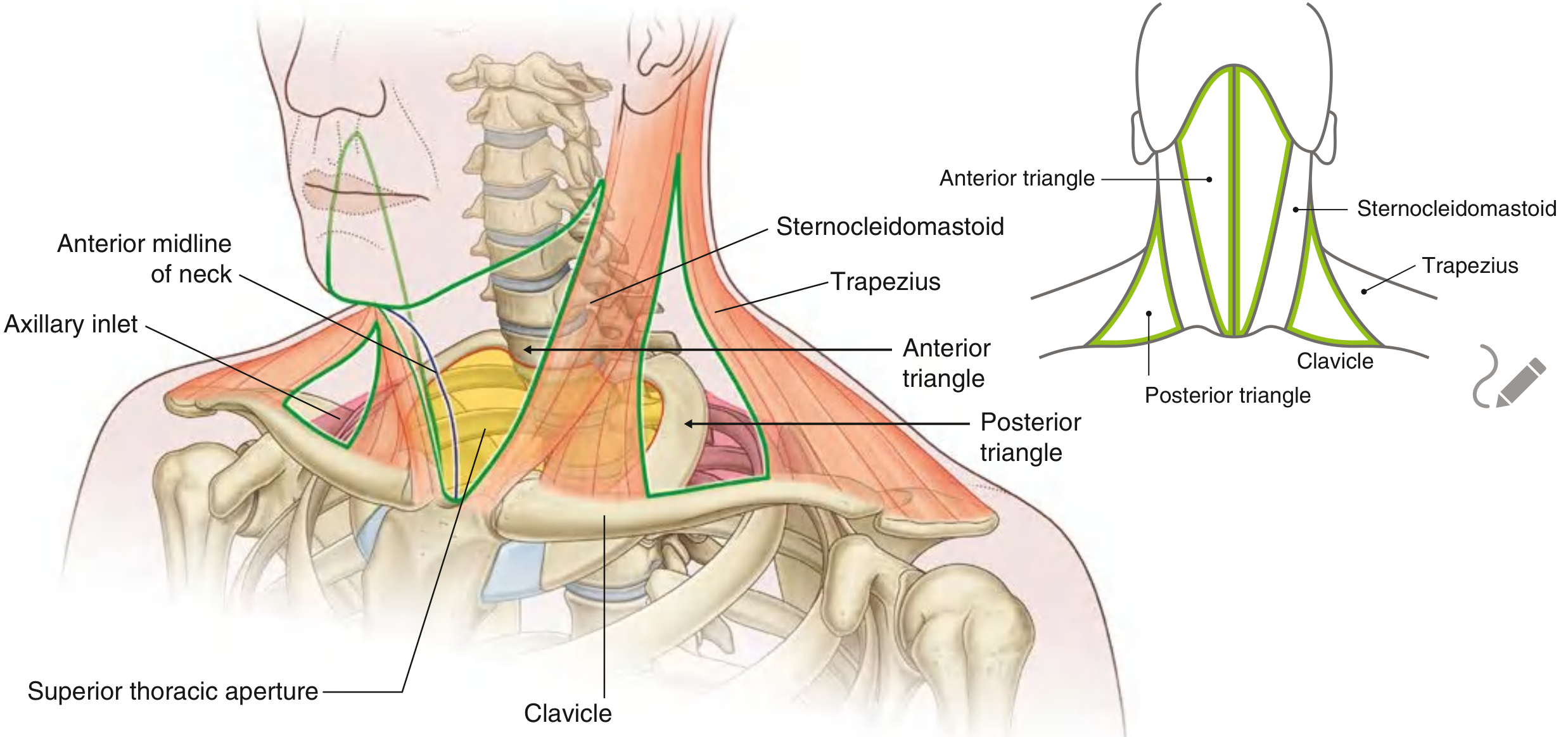
Anterior Triangle
Bounded by:
- The median vertical line of the neck (medially)
- Inferior margin of the mandible (superiorly)
- Anterior margin of the sternocleidomastoid (laterally)
Contains the major structures passing between the head and thorax: carotid arteries, internal jugular vein, vagus nerve, trachea, esophagus, thyroid gland.
Posterior Triangle
Bounded by:
- Middle third of the clavicle (inferiorly)
- Anterior margin of the trapezius (posteriorly)
- Posterior margin of the SCM (anteriorly)
Overlies the axillary inlet and is associated with structures (nerves and vessels) passing into and out of the upper limb - including the brachial plexus and subclavian vessels.
3. Cranial Nerves
There are 12 pairs of cranial nerves, all peripheral nerves that exit through foramina or fissures in the cranial cavity. All except the accessory nerve [XI] originate from the brain.
Functional Component Types
| Component | Abbreviation | Function | Nerves |
|---|---|---|---|
| General somatic afferent | GSA | Touch, pain, temperature | V, VII, IX, X |
| General visceral afferent | GVA | Sensory from viscera | IX, X |
| Special afferent | SA | Smell, taste, vision, hearing, balance | I, II, VII, VIII, IX, X |
| General somatic efferent | GSE | Motor to skeletal muscles | III, IV, VI, XII |
| General visceral efferent | GVE | Motor to smooth muscle and glands | III, VII, IX, X |
| Branchial efferent | BE | Motor to pharyngeal arch muscles | V, VII, IX, X, XI |
The 12 Cranial Nerves - Summary
| # | Nerve | Exit | Key Function |
|---|---|---|---|
| I | Olfactory | Cribriform plate | Smell |
| II | Optic | Optic canal | Vision |
| III | Oculomotor | Superior orbital fissure | Eye movement (4 muscles + levator palpebrae); pupil constriction, lens accommodation |
| IV | Trochlear | Superior orbital fissure | Superior oblique muscle (downward/medial gaze) |
| V | Trigeminal | V1: sup. orb. fissure; V2: foramen rotundum; V3: foramen ovale | Facial sensation + jaw muscles (V3) |
| VI | Abducent | Superior orbital fissure | Lateral rectus (abduction of eye) |
| VII | Facial | Stylomastoid foramen | Facial expression; taste ant. 2/3 tongue; salivary/lacrimal glands |
| VIII | Vestibulocochlear | Internal acoustic meatus | Hearing (cochlear) and balance (vestibular) |
| IX | Glossopharyngeal | Jugular foramen | Taste post. 1/3 tongue; parotid gland; carotid body/sinus |
| X | Vagus | Jugular foramen | Pharynx/larynx/viscera; descends into thorax and abdomen |
| XI | Accessory | Jugular foramen | SCM and trapezius muscles |
| XII | Hypoglossal | Hypoglossal canal | All intrinsic tongue muscles + hyoglossus, genioglossus, styloglossus |
Parasympathetic Distribution in the Head
Four cranial nerves carry parasympathetic fibers in the head - CN III, VII, IX, and X. The fibers in CN III, VII, and IX leave those nerves and hitch a ride on branches of the trigeminal nerve [V] to reach their targets:
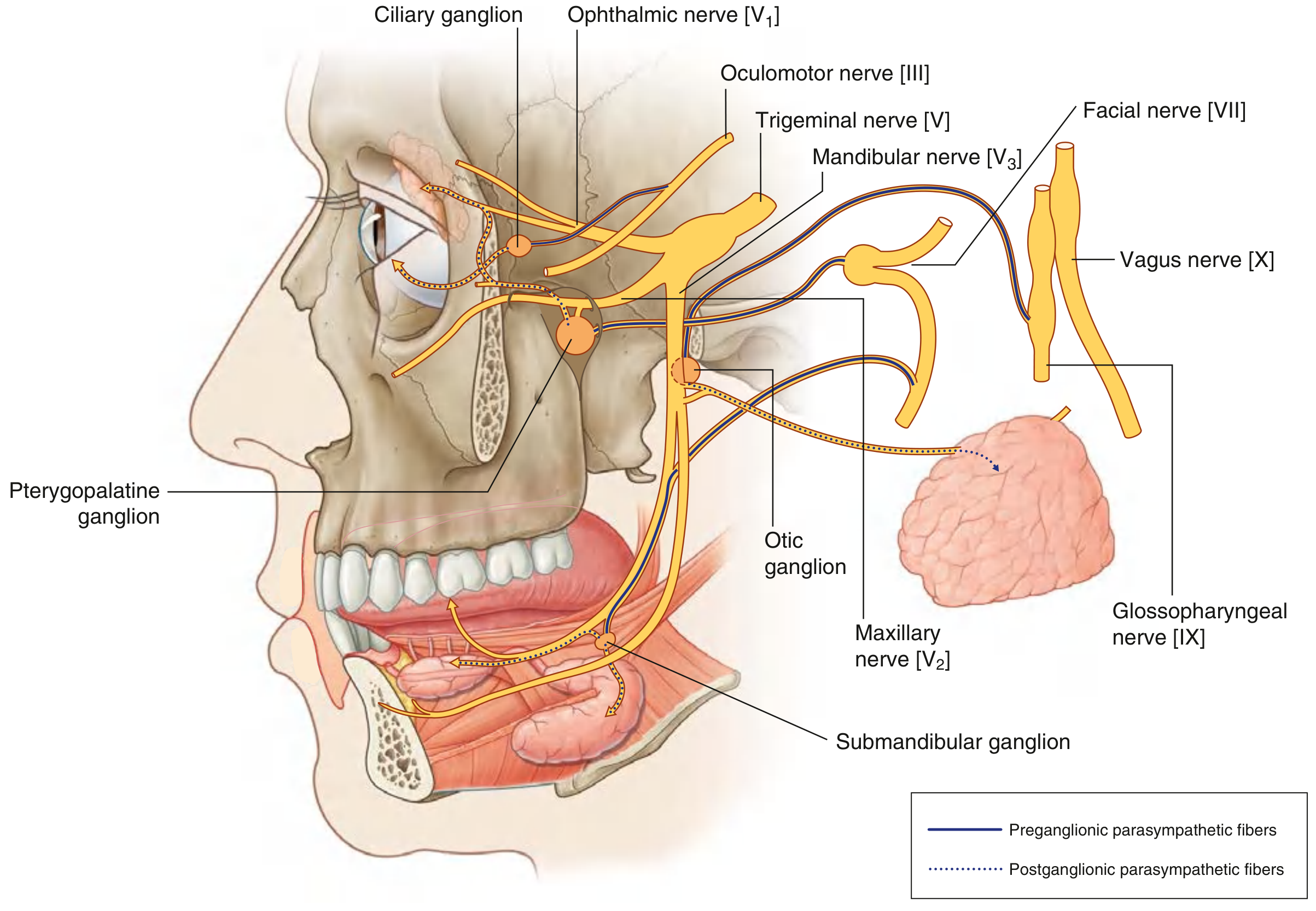
| Ganglion | Pre-ganglionic fiber via | Target |
|---|---|---|
| Ciliary ganglion | CN III | Pupillary sphincter, ciliary muscle (lens) |
| Pterygopalatine ganglion | CN VII | Lacrimal gland, nasal/palatal mucosa |
| Submandibular ganglion | CN VII | Submandibular and sublingual glands |
| Otic ganglion | CN IX | Parotid gland |
4. Cervical Nerves
There are 8 cervical spinal nerves (C1-C8):
- C1-C7 emerge above their respective vertebrae
- C8 emerges between C6 and T1
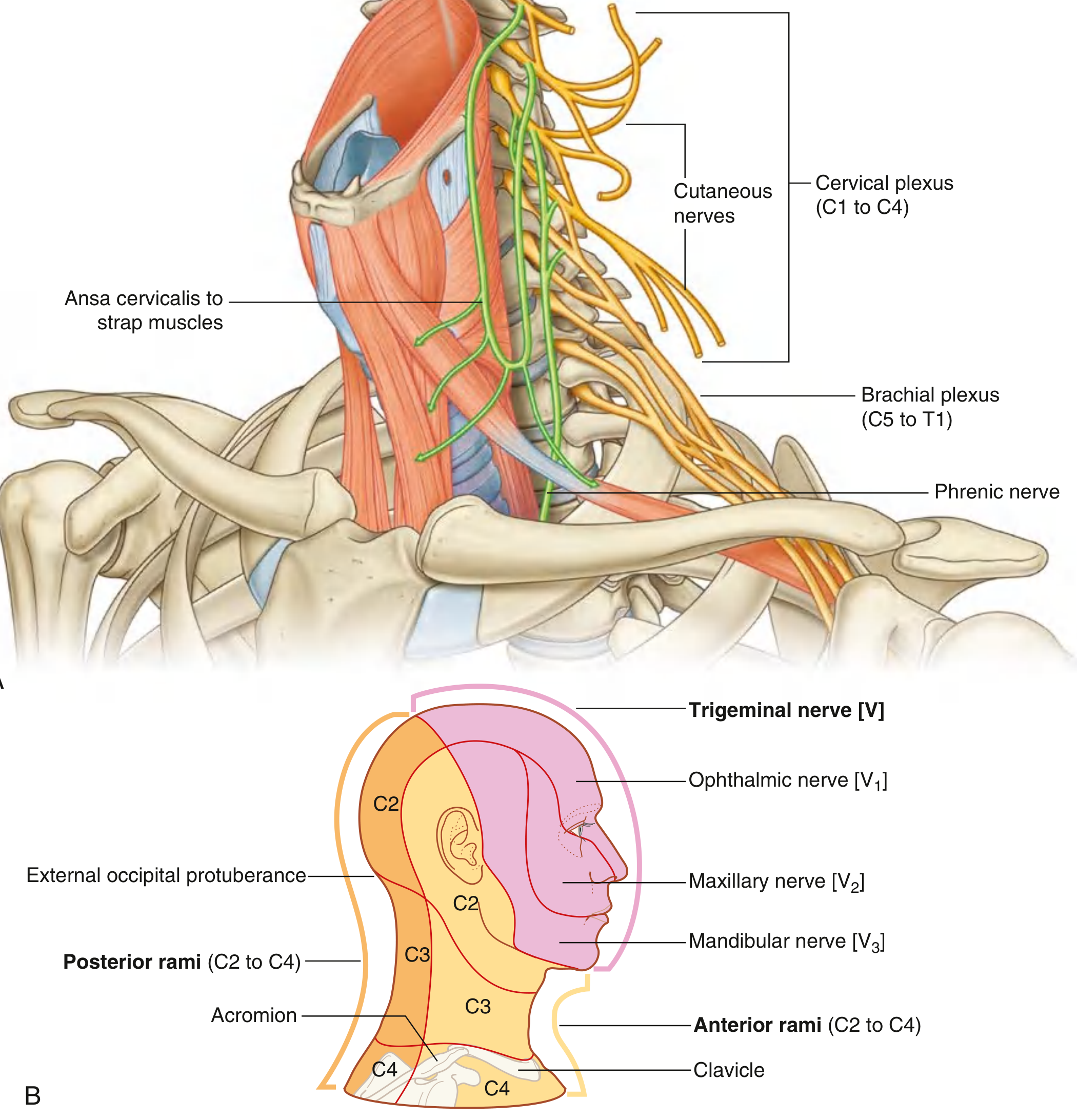
Cervical plexus (C1-C4) - anterior rami supply:
- Strap muscles (via ansa cervicalis: C1-C3)
- Diaphragm (via phrenic nerve: C3, C4, C5)
- Skin on anterior/lateral neck, upper anterior thoracic wall, and inferior head
Brachial plexus (C5-T1) - innervates the upper limb; roots are visible in the posterior triangle of the neck.
5. Vasculature
Arteries
The common carotid artery ascends in the neck and bifurcates at the level of the upper border of the thyroid cartilage (C3/C4) into:
- Internal carotid artery - enters skull via carotid canal; supplies brain (via circle of Willis), eye, and forehead
- External carotid artery - supplies face, scalp, neck structures; main branches include the superior thyroid, lingual, facial, occipital, and maxillary arteries
The vertebral arteries ascend through the transverse foramina of C6-C1, enter the foramen magnum, and unite to form the basilar artery, which supplies the posterior brain.
Venous Drainage
Venous drainage of the brain begins as cerebral veins draining into dural venous sinuses - endothelial-lined spaces within the dura mater.
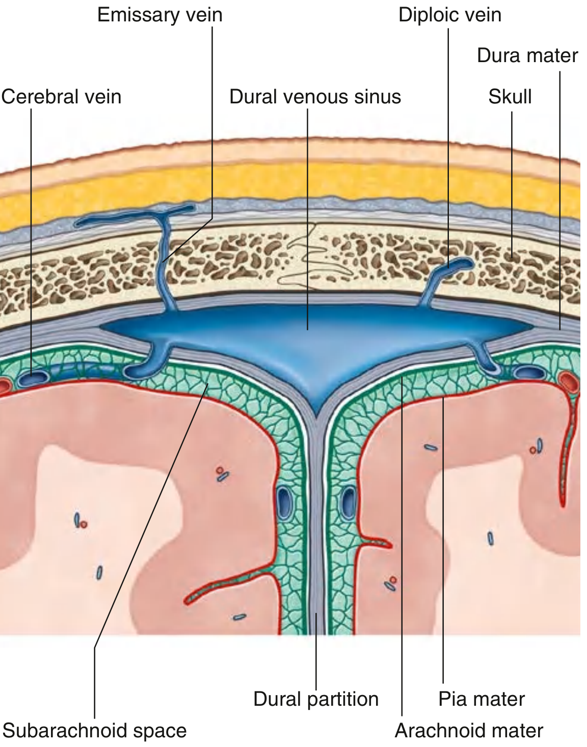
Major dural sinuses and their locations:
| Sinus | Location | Notes |
|---|---|---|
| Superior sagittal | Superior border of falx cerebri | Receives CSF via arachnoid granulations |
| Inferior sagittal | Inferior margin of falx cerebri | - |
| Straight | Junction of falx cerebri and tentorium cerebelli | Receives great cerebral vein |
| Transverse | Posterior tentorium cerebelli | - |
| Sigmoid | Temporal bone | Drains into internal jugular vein |
| Cavernous | Sphenoid bone | Contains CN III, IV, V1, V2, VI, and internal carotid artery |
Emissary veins connect dural sinuses to veins outside the skull. Because they have no valves, they can be a conduit for infections spreading intracranially.
All sinuses ultimately drain into the internal jugular vein, which descends in the neck in the carotid sheath alongside the common/internal carotid artery and vagus nerve.
6. Meninges
Three layers cover the brain and spinal cord:
- Dura mater (outermost) - tough fibrous layer; split into periosteal and meningeal layers in the cranium, forming the dural venous sinuses between them. Folds of dura form partitions: falx cerebri (between cerebral hemispheres), tentorium cerebelli (between cerebrum and cerebellum), and falx cerebelli.
- Arachnoid mater (middle) - thin avascular layer; the subarachnoid space beneath it contains CSF.
- Pia mater (innermost) - thin layer adherent to the brain surface.
7. Key Muscles
Muscles of Facial Expression (CN VII)
Derived from the 2nd pharyngeal arch; include orbicularis oculi, orbicularis oris, buccinator, zygomaticus major/minor, frontalis, and platysma.
Muscles of Mastication (CN V3)
- Masseter - closes jaw; most powerful biting muscle
- Temporalis - closes and retracts jaw
- Medial pterygoid - closes jaw; contraction = protrusion
- Lateral pterygoid - opens jaw; protrudes mandible; side-to-side grinding
Extrinsic Tongue Muscles (CN XII)
Genioglossus (protrudes tongue), hyoglossus (depresses), styloglossus (retracts/elevates).
Strap (Infrahyoid) Muscles (ansa cervicalis C1-C3)
Sternohyoid, sternothyroid, thyrohyoid, omohyoid - depress the hyoid and larynx.
Scalene Muscles (C3-C8)
Anterior, middle, and posterior scalenes; flex the neck laterally; the subclavian artery and brachial plexus pass between the anterior and middle scalene.
8. Pharynx and Larynx (Overview)
The pharynx is a shared chamber for the digestive and respiratory tracts, divided into:
- Nasopharynx (behind nasal cavity)
- Oropharynx (behind oral cavity)
- Laryngopharynx/hypopharynx (behind larynx)
The larynx sits at C3-C6 and contains the vocal folds. Its skeleton includes the thyroid cartilage, cricoid cartilage, epiglottis, and arytenoid cartilages. Intrinsic laryngeal muscles are innervated by the recurrent laryngeal nerve (branch of CN X), except the cricothyroid (external branch of superior laryngeal nerve).
Quick Clinical Correlates
- Craniosynostosis - premature fusion of cranial sutures; alters skull growth direction and shape
- Cavernous sinus thrombosis - dangerous; CN III/IV/V1/V2/VI all run through it, causing ophthalmoplegia
- Horner syndrome - interruption of sympathetic chain in the neck (ptosis, miosis, anhidrosis)
- Subclavian steal - occlusion proximal to vertebral artery origin causes reverse flow in vertebral artery
- Parotid surgery risk - CN VII runs through the parotid gland; damage causes ipsilateral facial palsy
- Neck dissection - accessory nerve [XI] is at risk in posterior triangle; damage causes trapezius weakness (shoulder drop)
Sources: Gray's Anatomy for Students (9780323934237, block 4 & 5), Color Atlas of Human Anatomy Vol. 2 (9783132424487)
This is a shared conversation. Sign in to Orris to start your own chat.