
dermatolog
It looks like your message got cut off! What would you like to know about dermatology? Feel free to ask about:
- Skin conditions or diseases (eczema, psoriasis, acne, melanoma, etc.)
- Dermatological treatments or medications
- Diagnostic criteria or clinical features
- Procedures (biopsies, laser therapy, etc.)
- Anything else skin, hair, or nail related
What's your question?
skin disorder with pathologyor clinical feature with images
There are hundreds of skin disorders. Let me narrow this down for you before diving in.
Asking for Preferences
Which category of skin disorders are you interested in? · What level of detail do you need?
Searching sources...
Reading File
Finding Sources
Finding Sources
Finding Sources
Finding Sources
Finding Sources
Now let me read the key sections from Fitzpatrick's (best dermatology source) and Robbins Pathology for all three disorders simultaneously:
Reading File
Reading File
Reading File
Excellent content found. Now let me read more on psoriasis histology and fetch images for all three conditions:
Reading File
Finding Sources
Finding Sources
Finding Sources
Finding Sources
Reading File
Reading File
Now let me fetch the key images and get the acne histology image URL from Robbins:
Loading Image
Finding Sources
Reading File
Searching the Web
psoriasis histology acanthosis parakeratosis Munro microabscess image dermatology
Loading Image
Loading Image
Searching the Web
atopic dermatitis eczema spongiosis histology image skin pathology
Loading Image
Loading Image
Searching the Web
acne vulgaris clinical photo comedone pustule inflammatory face
Loading Image
Searching the Web
atopic dermatitis eczema clinical photo flexural rash child adult
Loading Image
Loading Image
Here is your in-depth guide to the three major inflammatory skin disorders - with pathology, histology, and clinical images from authoritative dermatology textbooks.
Inflammatory Skin Disorders: In-Depth Pathology & Histology
1. ATOPIC DERMATITIS (ECZEMA)
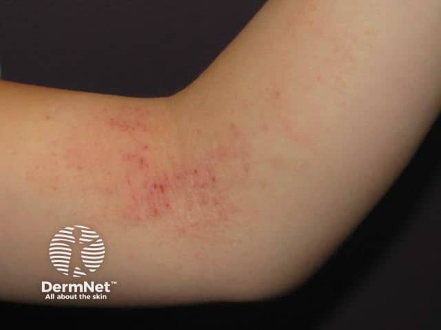
Clinical Features
Atopic dermatitis (AD) is the most common chronic inflammatory skin disease worldwide, affecting ~30% of people in developed nations. It presents with:
- Intense pruritus (the "itch that rashes")
- Xerosis (dry skin) as a background feature
- Eczematous plaques - erythematous, weeping, crusted in acute phases; lichenified (thickened) in chronic phases
- Distribution by age:
- Infants: cheeks, scalp, extensor surfaces
- Children/Adults: flexural areas - antecubital fossae, popliteal fossae, neck
- Atopic triad association: AD + asthma + allergic rhinitis ("atopic march")
- Dennie-Morgan lines, periorbital darkening, Hertoghe sign (lateral eyebrow thinning)
Pathogenesis
AD is a Th2-mediated disease with key elements:
| Component | Role |
|---|---|
| Filaggrin (FLG) gene mutation | Disrupts the epidermal barrier - allows allergen penetration |
| Th2 cytokines (IL-4, IL-5, IL-13) | Promote IgE production, eosinophil recruitment, barrier dysfunction |
| CCR4/TARC (CCL17) | Recruits Th2 cells to atopic skin; serum levels correlate with disease severity |
| CCR10/CTACK (CCL27) | Skin-selective homing of memory T cells |
| Eotaxin (CCL11)/CCR3 | Recruits eosinophils; elevated in lesional skin |
| Staphylococcus aureus | Colonizes 90%+ of AD skin; superantigens activate T cells, worsening inflammation |
| IgE | Elevated in most patients; mediates sensitization to environmental allergens |
- Fitzpatrick's Dermatology, block2, p.212
Histology (3 stages)
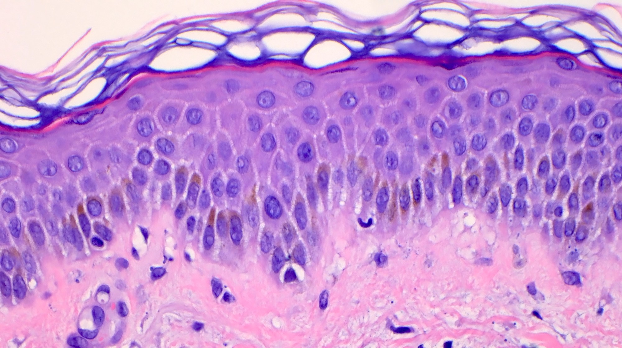
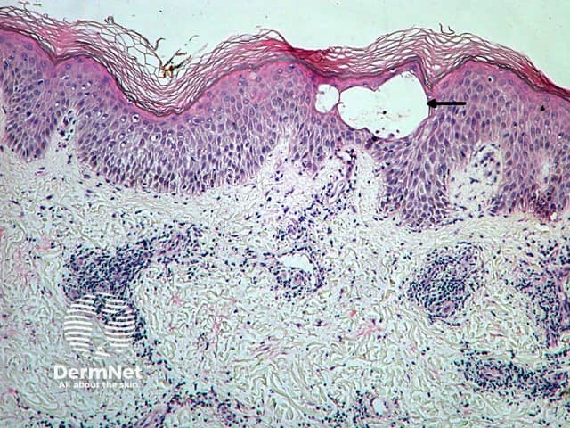
The hallmark is spongiotic dermatitis:
| Stage | Histological Features |
|---|---|
| Acute | Marked intercellular epidermal edema (spongiosis), microvesicle formation, exocytosis of lymphocytes, superficial perivascular lymphocytic infiltrate with eosinophils |
| Subacute | Moderate spongiosis, acanthosis (epidermal thickening), mild parakeratosis, continued eosinophilic infiltrate |
| Chronic | Minimal spongiosis, marked acanthosis, hyperkeratosis, fibrosis, lichenification |
Key distinguishing histological point: Spongiosis (intercellular edema) is the defining feature. Neutrophils are notably absent (unlike psoriasis). Eosinophils are prominent in the dermis.
2. PSORIASIS
Clinical Features
Psoriasis is a chronic, immune-mediated skin disease affecting 2-3% of the population. Classic presentation:
- Well-demarcated erythematous plaques with silvery-white, micaceous (mica-like) scales
- Sites of predilection: scalp, elbows, knees, lumbosacral area, umbilicus, intergluteal fold
- Auspitz sign - removal of scale reveals pinpoint bleeding (dilated dermal capillaries exposed)
- Koebner phenomenon - new lesions appear at sites of skin trauma
- Nail changes: pitting, onycholysis, oil-drop sign, subungual hyperkeratosis
- Psoriatic arthritis in 5-30% of cases
Pathogenesis
Psoriasis is now understood as primarily a Th17/IL-23-driven disease (not Th1 as previously thought):
| Pathway | Details |
|---|---|
| IL-23/Th17 axis | Dendritic cells produce IL-23 (p19+p40), which drives Th17 differentiation |
| IL-17A | Key effector cytokine - acts on keratinocytes to drive hyperproliferation, antimicrobial peptides, and neutrophil recruitment |
| IL-22 | Produced by Th17/Th22 cells; drives acanthosis (epidermal thickening) |
| TNF-alpha | Augments IL-17 effects on keratinocytes; produced by dermal dendritic cells |
| CCR6/CCL20 | Skin homing of Th17 cells; CCR6-deficient mice fail to develop psoriasis |
| Keratinocyte hyperproliferation | Basal keratinocyte transit time reduced from ~28 days to ~3-4 days |
The discovery of IL-17's role is confirmed by the dramatic efficacy of secukinumab and ixekizumab (anti-IL-17A) as well as guselkumab (anti-IL-23 p19) in clinical trials.
- Fitzpatrick's Dermatology, block2, p.228
Histology
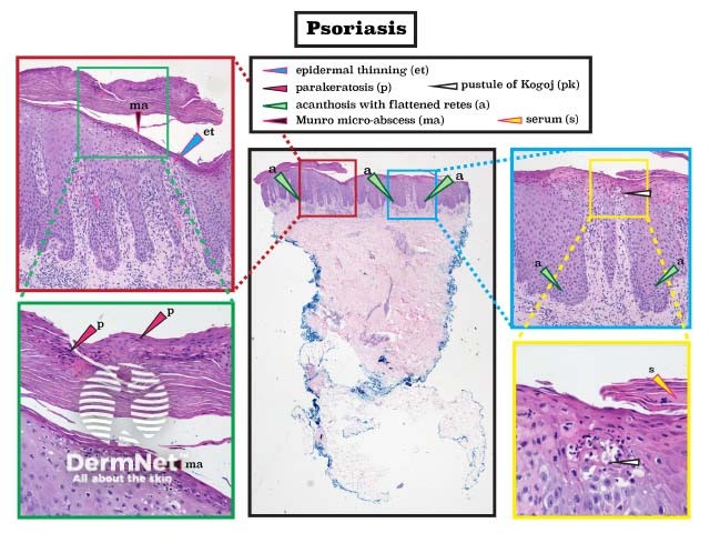
Psoriasis has a highly characteristic histological pattern:
| Feature | Description |
|---|---|
| Acanthosis | Regular epidermal thickening with elongated, club-shaped rete ridges |
| Parakeratosis | Retention of keratinocyte nuclei in the stratum corneum (incomplete keratinization) |
| Hypogranulosis/agranulosis | Loss of the granular layer (focally absent) |
| Munro microabscesses | Neutrophilic collections within the stratum corneum (atop parakeratosis) |
| Kogoj's spongiform pustules | Intraepidermal neutrophilic pustules (more prominent in pustular psoriasis) |
| Suprapapillary plate thinning | Epidermis is thinned directly over dermal papillae |
| Dilated tortuous capillaries | Prominent in papillary dermis - explains Auspitz sign |
| Perivascular lymphocytic infiltrate | With neutrophils migrating upward |
| Minimal spongiosis | (Key differentiator from eczema) |
"Histologically, all psoriasis is pustular. The microscopic pustules include spongiform intraepidermal pustules and Munro microabscesses within the stratum corneum." - Andrews' Diseases of the Skin
3. ACNE VULGARIS
Clinical Features
Acne vulgaris affects virtually all adolescents and many adults. It involves the pilosebaceous unit on areas with the densest sebaceous follicles: face, chest, and back.
Lesion types:
| Type | Description |
|---|---|
| Open comedone (blackhead) | Dilated follicular ostium with oxidized melanin plug (black color = oxidized melanin, NOT dirt) |
| Closed comedone (whitehead) | Plugged follicle with invisible ostium; precursor to inflammatory lesions |
| Papule | Erythematous inflamed elevated lesion |
| Pustule | Visible pus-filled lesion |
| Nodule/Cyst | Deep, painful; risk of scarring |
| Acne conglobata | Severe variant with sinus tract formation and scarring |
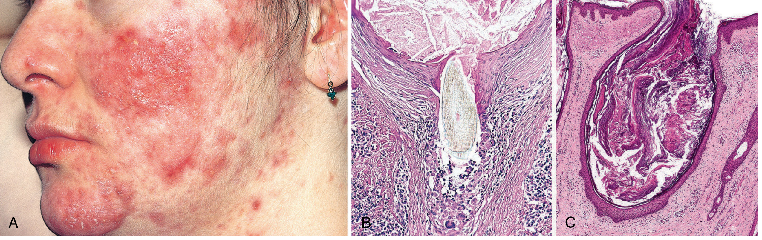
Pathogenesis (4 key factors)
- Follicular keratinization abnormality - keratin plug forms in the lower infundibulum, blocking sebum outflow
- Androgen-driven sebaceous hyperactivity - androgens at puberty cause sebaceous gland hypertrophy and increased sebum production
- Cutibacterium acnes (formerly Propionibacterium acnes) - colonizes the follicle; produces lipases that convert sebum triglycerides into free fatty acids - these are directly proinflammatory and comedogenic
- Secondary inflammation - follicle rupture releases contents into dermis, triggering a foreign body/immune reaction
Key therapeutic implications:
-
Antibiotics (doxycycline, clindamycin) target C. acnes
-
Isotretinoin (13-cis-retinoic acid) - potent antisebaceous action; reduces all 4 factors
-
Benzoyl peroxide - bactericidal, reduces keratinization
-
Androgens link explains why castrated males historically did not develop acne
-
Robbins, Cotran & Kumar Pathologic Basis of Disease, block12, p.1074
Histology
| Feature | Description |
|---|---|
| Open comedone | Dilated follicular lumen with laminated keratin and oxidized melanin; wide patulous orifice |
| Closed comedone | Plugged follicle with microscopically visible orifice only; thin overlying epithelium at risk of rupture |
| Inflammatory papule/pustule | Perifollicular lymphocytic and neutrophilic infiltrate; edema |
| Ruptured follicle | Hair shaft piercing follicular wall; intense surrounding inflammation with lymphocytes, macrophages, and foreign body giant cells |
| Nodule/cyst | Dermal abscess with fibrosis; leads to scarring |
Comparison Table: Key Pathology Features
| Feature | Atopic Dermatitis | Psoriasis | Acne Vulgaris |
|---|---|---|---|
| Main T cell type | Th2 | Th17 | - (innate/adaptive) |
| Key cytokines | IL-4, IL-13, IL-5 | IL-17, IL-22, IL-23, TNF-alpha | IL-1, TNF, IL-8 |
| Epidermal change | Spongiosis (intercellular edema) | Acanthosis + parakeratosis | Follicular plugging |
| Granular layer | Preserved (or increased) | Absent (hypogranulosis) | Normal |
| Neutrophils | Absent | Prominent (Munro, Kogoj) | Present in inflammatory lesions |
| Eosinophils | Prominent | Absent | Absent |
| Spongiosis | Hallmark feature | Minimal | Absent |
| Rete ridges | Normal or mild acanthosis | Elongated, club-shaped | Not applicable |
| Vascular changes | Mild perivascular edema | Dilated tortuous capillaries (Auspitz sign) | Perifollicular inflammation |
| Sebaceous gland | Normal | Normal | Enlarged/hyperactive |
| Scarring | Rare | Rare | Common (nodular/cystic) |
Sources:
- Fitzpatrick's Dermatology (9th Ed.), Volumes 1-2 - Chapters on Atopic Dermatitis and Psoriasis
- Andrews' Diseases of the Skin - Pathology section on Psoriasis
- Robbins, Cotran & Kumar Pathologic Basis of Disease - Chapter 25, Acne Vulgaris
- Dermatology 2-Volume Set (5th Ed.) - Key features on inflammatory dermatosesThis covers the three major inflammatory skin disorders in depth. Here's a quick summary of what was covered:
Atopic Dermatitis - Th2-driven disease centered on a defective epidermal barrier (filaggrin mutations), with spongiosis as the histological hallmark. Three stages (acute/subacute/chronic) each with distinct histology.
Psoriasis - Th17/IL-23-driven disease with dramatic keratinocyte hyperproliferation. Classic histology: regular acanthosis + parakeratosis + Munro microabscesses + dilated papillary capillaries. Minimal spongiosis distinguishes it from eczema.
Acne Vulgaris - Pilosebaceous unit disorder driven by 4 factors: follicular keratinization, androgen excess, Cutibacterium acnes, and secondary inflammation. Histology ranges from simple comedones to ruptured follicles with foreign body granulomas.
Would you like me to go deeper on any specific aspect - such as treatment pathways, grading systems, differential diagnosis, or additional skin disorders?
vedio lecture about skin disorder
I'm not able to create or stream video lectures — I'm a text and image-based AI assistant.
However, here are the best free video lecture resources for skin disorders:
🎓 Top Video Lecture Sources for Dermatology
1. Osmosis (YouTube / osmosis.org)
- Excellent animated pathology videos
- Covers: Psoriasis, Eczema, Acne, Melanoma, Pemphigus, SLE rash, and more
- Osmosis Dermatology playlist
2. Ninja Nerd (YouTube)
- In-depth whiteboard lectures with pathophysiology
- Very popular for med students
- Search: "Ninja Nerd Dermatology"
3. Armando Hasudungan (YouTube)
- Beautiful illustrated hand-drawn explanations
- Great for mechanisms and histology
4. Lecturio / Amboss
- Full dermatology course with clinical cases
- Lecturio has a free tier
- lecturio.com
5. Medscape / UpToDate Videos
- Clinical-level, case-based
- For practicing clinicians
6. Khan Academy Medicine
- Free, beginner-friendly
- Basic skin disorder overview
📌 Specific Topic Searches (YouTube)
| Disorder | Search Term |
|---|---|
| Psoriasis | "Osmosis Psoriasis" or "Ninja Nerd Psoriasis" |
| Eczema/AD | "Atopic dermatitis pathophysiology Osmosis" |
| Acne | "Acne vulgaris pathology Ninja Nerd" |
| Skin cancers | "Melanoma vs BCC vs SCC Lecturio" |
| Blistering diseases | "Pemphigus vs Pemphigoid Osmosis" |
What I can do instead:
- Build a detailed written lecture outline on any skin disorder
- Create visual diagrams or flashcards
- Produce a downloadable PowerPoint presentation
Would you like any of those? Just let me know the topic!
Generate a study guide for dermatology exams
Let me clarify a few things before diving in to make sure the guide fits your needs perfectly.
Asking for Preferences
What is your level? · What format do you want? · Which topics to cover?
This is a shared conversation. Sign in to Orris to start your own chat.