
Milking of subcutaneous emphysema full details how it done , action, indication,contrain6
milking subcutaneous emphysema technique procedure indication contraindication
https://pmc.ncbi.nlm.nih.gov/articles/PMC4692895
"subcutaneous emphysema" milking management chest drain
subcutaneous emphysema milking technique chest tube infraclavicular incision blow hole
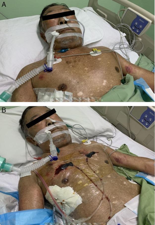
Two-panel clinical photograph illustrating the management of extensive subcutaneous emphysema (SE). Image A shows an elderly male patient with severe swelling and distension of the face, neck, and upper torso, secondary to trapped air in the subcutaneous tissues (subcutaneous emphysema). The patient is intubated with an endotracheal tube secured for airway protection due to compromise from tissue tension. Image B, taken 24 hours later, demonstrates a significant reduction in soft tissue swelling following the placement of subcutaneous negative pressure wound therapy (NPWT). Two incision sites are visible on the anterior chest wall (bilateral infraclavicular region) where subcutaneous drains are inserted and secured under transparent occlusive dressings. The chest also shows an existing surgical dressing on the right lower quadrant and cardiac monitoring leads. This comparison highlights a surgical intervention technique for rapidly decompressing life-threatening subcutaneous emphysema when conventional drainage fails.
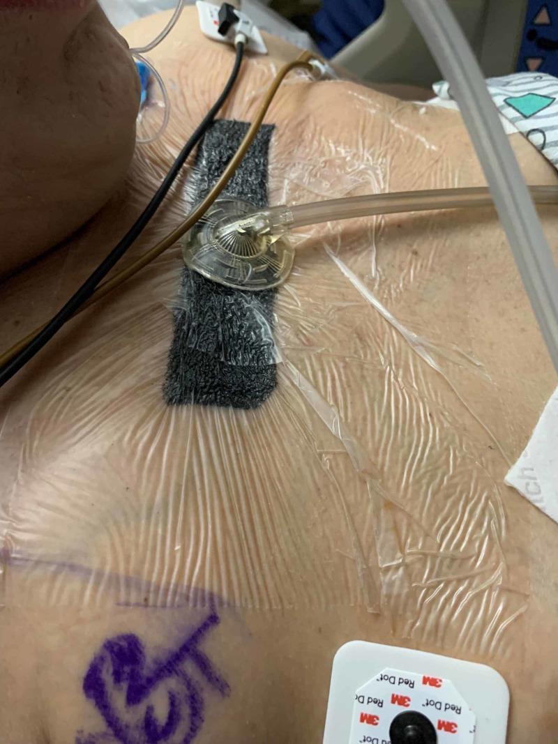
This clinical photograph displays a closed incision negative pressure wound therapy (ciNPWT) system applied to the anterior thoracic region of a patient. Centrally, a rectangular piece of black, reticulated porous foam is visible, secured under a large, wrinkled transparent adhesive drape. A circular suction port with a radial ribbed design is positioned atop the foam, connected to flexible tubing that exits toward the patient's left. The dressing covers loosely approximated 'blow hole' incisions intended to manage massive subcutaneous emphysema. Adjacent to the dressing, several clinical monitoring components are visible, including EKG electrodes (3M Red Dot) and various leads. A purple surgical marking is seen on the lower-left aspect of the chest wall. The image illustrates a specific procedural application for thoracic decompression and wound management in the context of severe chest trauma and air-leak syndromes.
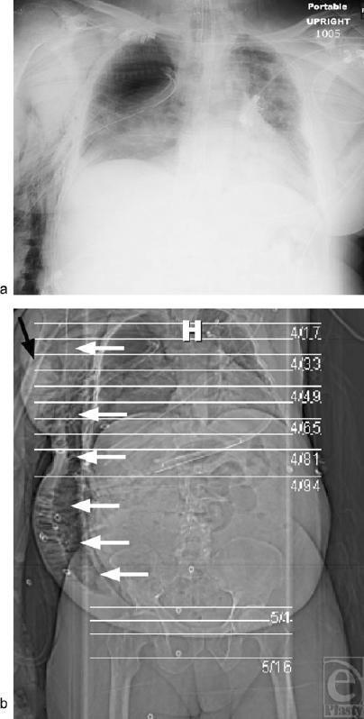
This diagnostic imaging composite displays a case of severe, extensive subcutaneous emphysema. Panel (a) is a portable anteroposterior chest roentgenogram showing radiolucent (dark) air streaks dissecting through the soft tissues of the chest wall and neck, disrupting normal tissue planes. A right-sided chest tube is visible, and the chest wall appears significantly thickened due to air entrapment. Panel (b) is a computed tomography (CT) scout film/topogram of the torso. White arrows highlight the craniocaudal distribution of subcutaneous air extending from the thoracic region down into the abdominal and pelvic subcutaneous tissues. A black arrow indicates a prepectoral 'blowhole' incision on the right chest wall, designed for air decompression. The image illustrates the radiographic manifestations of barotrauma or persistent air leak under positive pressure ventilation, characterized by the 'ginkgo leaf sign' on the chest wall and diffuse mottled low-density areas throughout the trunk's soft tissues.
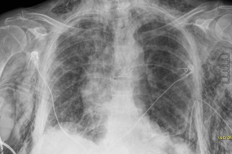
Anteroposterior (AP) chest X-ray demonstrating severe multi-compartmental air leaks. Extensive subcutaneous emphysema is visible as diffuse, streaky radiolucencies (dark areas) dissecting through the soft tissues of the chest wall, axillae, neck, and upper arms, creating a characteristic 'gingko leaf' appearance over the pectoral regions. A prominent pneumomediastinum is indicated by thin lucent lines outlining the mediastinal borders and a 'continuous diaphragm sign,' where air trapped behind the pericardium allows for visualization of the diaphragm across the midline. A left basilar pneumothorax is present, associated with a malpositioned left-sided thoracostomy tube showing a visible sentinel hole near the lateral chest wall, suggesting partial retraction. These findings illustrate critical complications of thoracic trauma or barotrauma, emphasizing the radiological manifestations of extra-alveolar air in pulmonary pathology and emergency medicine.
"subcutaneous emphysema" management decompression blowhole infraclavicular
Milking of Subcutaneous Emphysema (Surgical Emphysema)
What Is Subcutaneous Emphysema (SE)?
Severity Grading (Anatomical Extension)
| Grade | Extent of Air |
|---|---|
| 1 | Base of neck only |
| 2 | Entire neck |
| 3 | Neck + subpectoral area |
| 4 | Chest wall + entire neck |
| 5 | Chest wall + neck + orbit + scalp + abdomen + upper limbs + scrotum |
Pathophysiology
- Parenchymal or tracheobronchial injury → air leaks through the parietal pleura
- Interstitial lung injury → air dissects back along bronchi into the hilum → mediastinum → extrapleural spaces
- Trauma to pharynx, larynx, or esophagus
- Barotrauma from positive-pressure ventilation
- Post-surgical (laparoscopy, thoracic surgery, tracheostomy)
"Milking" - What It Means
Indications for Milking / Decompression of SE
- Rapidly progressive or massive SE (grade 3-5) - when air is spreading to face, neck, orbit, genitals, or limbs
- Airway compromise - SE in the neck causing dysphagia, stridor, or impending airway obstruction
- Respiratory failure - restriction of full lung re-expansion due to air, causing high airway pressures or ventilator failure
- Tension phenomena - SE contributing to tension physiology
- Haemodynamic compromise - in rare cases, SE can cause thoracic outlet compression or restrict cardiac output
- Pacemaker malfunction due to air surrounding the device
- Patient discomfort - severe pain, disfigurement, difficulty swallowing from massive SE
- Worsening SE despite existing chest tube - when the current drain is insufficient to keep up with the air leak
Contraindications
Absolute Contraindications
- Uncontrolled coagulopathy - for incision-based procedures (blowholes)
- Active necrotizing infection at proposed incision site
- No drainage exit - milking without a chest tube or blowhole in place is pointless and should not be done
Relative Contraindications
- Small, asymptomatic SE (grades 1-2) - these self-resolve; no intervention needed
- SE secondary to esophageal perforation where air tracks from the mediastinum - treat the source first; milking may worsen mediastinal contamination
- SE without a functioning pleural drain in place - treat the underlying pneumothorax first; insert a chest tube before any milking manoeuvre
- Severe coagulopathy (relative, for blowhole incisions)
How Milking Is Performed - Step by Step
Prerequisites
- Ensure a chest tube or drainage exit exists - an intercostal drain, blowhole incision, or subcutaneous drain must be in place before milking will be effective
- Confirm the chest tube is patent and connected to an underwater seal or suction
- Assess extent of SE clinically and with CXR/CT
Technique 1: Manual Milking Over an Existing Chest Tube
- Patient positioning: supine or semi-recumbent
- Identify the tract of air by palpating crepitus - map the extent of SE
- Using both hands with flat palms, apply firm but gentle pressure to the subcutaneous tissue distal to the drain (i.e., furthest from the chest tube)
- Using a stroking, rolling movement (like squeezing a tube of toothpaste), work the air proximally toward the drain insertion site
- Work systematically from the periphery inward - neck to chest, or limb to trunk
- The chest drain bubbles vigorously as air escapes into the pleural drain system - this confirms the manoeuvre is working
- Repeat in multiple passes; monitor respiratory status, SpO2, and patient comfort throughout
- Document reduction in crepitus after each session
Technique 2: Milking After Blowhole Incisions
- Mark bilateral infraclavicular incisions in the midclavicular line
- Infiltrate with local anaesthetic (e.g., 1% lignocaine with adrenaline)
- Make 2-4 cm incisions deep to the external thoracic fascia (superficial to the pectoral fascia) - bilaterally
- Blunt dissect to create a subcutaneous channel
- Air escapes spontaneously once the fascia is opened - you will hear/feel air rush out
- After incisions are made, apply systematic manual compression from the periphery toward the blowhole sites
- Compress from the neck downward, from the flanks inward, sweeping air toward the chest wall openings
- The "blowhole" wounds are left open (not sutured) to allow continued egress of air
- Some centres apply negative pressure wound therapy (NPWT/VAC) dressings over the blowhole sites to maintain suction and accelerate air removal
Technique 3: Milking With Subcutaneous Tunnelled Drain
- Under local anaesthesia, create a subcutaneous tract superficial to the pectoral fascia by blunt dissection
- Insert a fenestrated chest drain (14-28 Fr) through the tunnel
- Secure and connect to an underwater seal ± low continuous suction (-5 to -10 cmH₂O)
- Then perform manual milking toward the drain entry site to assist air evacuation
- The angiocatheter variant uses large-bore (14G) IV cannulas inserted at multiple sites; these are more prone to kinking
Mechanism of Action
- Physically displacing air trapped in soft tissue pockets and fascial planes toward the drainage exit
- Reducing tissue tension by evacuating compressed gas, relieving pressure on airways and vascular structures
- Facilitating lung re-expansion by lowering the external pressure exerted by subcutaneous air on the chest wall
- Preventing tension phenomena by ensuring a continuous egress pathway for air
Post-Procedure Monitoring
- Serial SpO2, ABG (watch for CO2 retention / respiratory acidosis)
- CXR after the procedure to confirm reduction in SE and check lung re-expansion
- Monitor chest tube output and bubbling
- Check for pneumothorax recurrence
- Assess neck swelling and airway repeatedly
- Pacemaker function check if a device is present
Important Clinical Points
| Point | Detail |
|---|---|
| Always treat the source | SE is a sign of an underlying air leak - treat the pneumothorax, bronchial tear, or esophageal injury first |
| Insert a chest tube first | Milking without a functional drain achieves nothing |
| Bronchoscopy if massive air leak | Dramatic or increasing SE with massive chest tube air leak suggests major airway injury - investigate with bronchoscopy |
| POCUS > CXR for pneumothorax | Ultrasound is more sensitive than plain film in detecting the underlying pneumothorax |
| Small SE resolves spontaneously | Grades 1-2 (neck base or neck alone) reabsorb on their own; no milking required |
| Positive-pressure ventilation worsens SE | If the patient is on a ventilator, consider reducing tidal volumes and PEEP where possible |
With gauze roll
milking subcutaneous emphysema gauze roll technique how to perform
Milking Subcutaneous Emphysema With a Gauze Roll
What the Gauze Roll Is Used For Here
- Distributes pressure more evenly than fingertips alone
- Allows a longer stroke without skin slippage
- Protects fragile or oedematous skin from direct fingernail pressure
- Can be rolled like a paint roller over large areas of SE efficiently
Equipment Needed
- Gauze roll (crepe bandage or cotton gauze roll, typically 7-10 cm wide)
- Functioning chest tube (or blowhole incision) already in place - mandatory
- Gloves
- Assistant (optional but helpful for large SE)
Step-by-Step Technique
Before You Start
- Confirm chest tube patency - ensure the chest tube is bubbling freely and the underwater seal is intact. Without an exit, milking is futile.
- Position the patient supine or at 30-45° head elevation.
- Palpate the full extent of crepitus - note how far up the neck, down the trunk, and laterally the air has spread.
- Explain the procedure to the patient - it is uncomfortable but not dangerous.
The Milking Manoeuvre
- For neck SE: roll downward from the jaw/neck toward the clavicles and chest
- For chest wall SE: roll from the axilla/lateral chest wall toward the intercostal drain site
- For abdominal/flank SE: roll upward and inward toward the thorax
- Reassess crepitus extent by palpation
- Check SpO2 and respiratory rate
- Observe underwater seal for ongoing bubbling (indicates persistent air leak from source)
- Repeat every 1-4 hours as needed for progressive SE
Why Gauze Roll Is Better Than Bare Hands Alone
| Bare Hands | Gauze Roll |
|---|---|
| Uneven fingertip pressure | Even cylindrical pressure across wide area |
| Skin slippage on sweaty/oedematous tissue | Gauze grips skin surface without slipping |
| Tiring for clinician over large SE | Roll mechanics reduce hand fatigue |
| May cause focal bruising | Distributes force, less trauma |
| Less effective on large body surface area | More efficient for grade 4-5 SE |
Key Rules - Do Not Forget
| Rule | Reason |
|---|---|
| Never milk without a drain/blowhole | Air has nowhere to go - no benefit, potential harm |
| Never milk if chest tube is clamped | Risk of tension pneumothorax |
| Stop if the patient desaturates | May indicate tension phenomena - reassess immediately |
| Never compress the neck forcefully | Risk of airway compression and vascular injury |
| Avoid milking over fresh surgical wounds | Risk of wound dehiscence |
| If air leak is massive and ongoing | Milking is a temporising measure only - the source must be fixed (bronchial repair, chest tube upsizing, or surgical intervention) |
Summary of Action Mechanism
Subcutaneous emphysema o2 treatment why
high flow oxygen subcutaneous emphysema nitrogen washout reabsorption mechanism treatment
Why Oxygen Is Given in Subcutaneous Emphysema
The Core Principle: Nitrogen Washout
- 78% nitrogen (N₂)
- 21% oxygen (O₂)
- ~1% other gases
- Oxygen is continuously consumed by cells (metabolized)
- CO₂ dissolves readily in blood (high solubility)
The Mechanism - Step by Step
Normal State (Room Air Breathing)
Tissue capillaries carry blood with nitrogen
  partially saturated to ~78% of atmospheric
  nitrogen pressure (PN₂ ≈ 593 mmHg at sea level)

Trapped subcutaneous air also has PN₂ ≈ 593 mmHg

→ No pressure gradient → Nitrogen stays in tissues → SE persists
With High-Flow Oxygen
Patient breathes near 100% O₂ (FiO₂ → 1.0)

↓ Alveolar nitrogen drops toward ZERO
↓ Arterial blood nitrogen drops (nitrogen "washed out" of lungs)
↓ Venous and tissue capillary PN₂ falls sharply

NOW a steep pressure gradient exists:
  Subcutaneous air PN₂ (≈593 mmHg)
         >>>>>>
  Capillary PN₂ (≈ 0-10 mmHg)

→ Nitrogen diffuses DOWN the gradient from tissue INTO capillaries
→ Nitrogen carried away in blood → exhaled by lungs
→ Trapped air volume shrinks → SE resolves faster
- Accelerate pneumothorax reabsorption
- Treat decompression sickness in divers
- Pre-oxygenate patients before anaesthesia
How Much Faster Does It Work?
- Room air: SE reabsorbs at approximately 1.25% of total volume per hour spontaneously
- High-flow O₂ (near 100%): reabsorption rate increases 3 to 4 times faster
- Pneumothorax studies show high-flow O₂ increases air reabsorption from ~1.25%/day to ~4-6%/day - the same principle applies to subcutaneous emphysema
Practical Application
| Parameter | Detail |
|---|---|
| Delivery device | Non-rebreather mask (NRM) - delivers FiO₂ ~0.85-0.95 |
| Flow rate | 10-15 L/min via NRM |
| Duration | Continuous until SE is resolving; typically 24-72 hours for mild-moderate cases |
| Alternative | Oxygen concentrator (delivers up to 95% O₂) for less critical cases |
| Goal | SpO₂ >94%; the higher the FiO₂, the steeper the nitrogen gradient, the faster the reabsorption |
Additional Benefits of O₂ in SE
-
Treating hypoxia - SE can compress chest wall mechanics and impair ventilation, causing hypoxaemia; supplemental O₂ corrects this directly
-
Treating respiratory acidosis - especially after laparoscopic surgery where CO₂ is the trapped gas; O₂ therapy + monitoring of ABG for rising CO₂ and respiratory acidosis is standard. As stated in Barash's Clinical Anesthesia: "if concerns for persistent or recurrent hypercarbia from subcutaneous emphysema exist during the postoperative recovery, clinicians should maintain oxygen therapy and monitor for somnolence and acute respiratory acidosis by arterial blood gas"
-
Buffer while treating the source - O₂ is a non-invasive temporising measure while the chest tube drains the pneumothorax or the underlying cause is fixed
-
Reduces total gas partial pressures in blood - when FiO₂ is high, the total gas pressure in capillary blood falls (because N₂ is displaced). This lowers capillary gas pressure below tissue gas pressure across the board, creating a global absorption gradient for all trapped gases
Important Caveats
| Caution | Reason |
|---|---|
| Do not use high-flow O₂ in COPD patients without caution | Risk of hypercapnic respiratory failure (hypoxic drive suppression) |
| O₂ alone is NOT sufficient for massive SE | It is an adjunct - drain placement, milking, and source control are still required |
| Evidence base is limited | Current guidelines note supportive evidence for O₂ in SE reabsorption is "lacking" for definitive recommendation, but the physiological rationale is sound and the practice is widely used |
| For CO₂-based SE (post-laparoscopy) | CO₂ dissolves so readily in blood that it reabsorbs within 24 hours regardless; here O₂ mainly treats hypoxia and monitors for respiratory acidosis |
Summary in One Sentence
- Barash's Clinical Anesthesia, 9e (CO₂ Extravasation / Subcutaneous Emphysema section)
- Management of Subcutaneous Emphysema in Pneumothorax - nitrogen partial pressure gradient mechanism
- High Flow Oxygen and Pneumothorax reabsorption - nitrogen washout evidence
Give a simple explanation to everyone easy to understand
Why We Give Oxygen for Subcutaneous Emphysema - Simple Explanation
First, What Is the Problem?
- 🟦 Nitrogen - 78% of the air
- 🟥 Oxygen - 21% of the air
Why Does the Air Stay Stuck?
- ✅ Oxygen - the body USES it. Cells eat it up. So it gets absorbed quickly.
- ✅ CO₂ - dissolves easily in blood. Gets absorbed and breathed out fast.
- ❌ Nitrogen - the body has NO use for it. It just sits there. The blood cannot carry it away easily.
Now - What Does Giving Oxygen Do?
🫧 Think of the blood vessels as tiny vacuum cleaners running past the trapped air bubble.
- The lungs fill with pure oxygen - almost no nitrogen
- The blood dumps its nitrogen into the lungs to be breathed out
- Now the blood vessels passing by the trapped air bubble are empty of nitrogen - like a vacuum cleaner that just been emptied
- The nitrogen in the trapped air bubble rushes into the empty blood vessels
- Blood carries the nitrogen to the lungs → patient breathes it out
- The trapped air bubble shrinks and disappears 🎉
The Simple Picture
WITHOUT oxygen treatment:
Blood near bubble: already has nitrogen → can't absorb much → bubble stays

WITH high-flow oxygen:
Blood near bubble: nitrogen flushed out → acts like empty sponge →
sucks nitrogen OUT of bubble → bubble shrinks → skin goes back to normal
A Real-Life Analogy
🧃 Imagine a juice carton that still has juice in it. If you try to pour more juice in, it won't go in because it's already full.But if you first empty the carton - now it can absorb a lot more.High-flow oxygen empties the blood of nitrogen first, so the blood can then soak up the nitrogen trapped under the skin.
How Is It Given?
- A tight-fitting oxygen mask (called a non-rebreather mask) is placed on the patient's face
- Oxygen flows at 10-15 litres per minute
- The patient breathes almost pure oxygen (about 85-95%)
- This is kept going for 24 to 72 hours depending on severity
Does It Work Alone?
| What oxygen does | What else is still needed |
|---|---|
| Helps the body absorb the trapped air 3-4x faster | Fix the source of the air leak |
| Treats low oxygen levels | Chest tube to drain the pneumothorax |
| Buys time safely | Milking / blowhole incisions for massive cases |
One-Line Summary
💡 Breathing pure oxygen flushes nitrogen out of the blood, turning the blood vessels into an empty sponge that can then suck the trapped nitrogen out from under the skin - making the subcutaneous emphysema disappear faster.
Create pdf file with protocol and guidelines
ls -la /tmp/workspace/ 2>/dev/null || mkdir -p /tmp/workspace && echo "workspace ready"
mkdir -p /tmp/workspace/subcutaneous-emphysema && cat > /tmp/workspace/subcutaneous-emphysema/generate_pdf.py << 'PYEOF' from reportlab.lib.pagesizes import A4 from reportlab.lib import colors from reportlab.lib.styles import getSampleStyleSheet, ParagraphStyle from reportlab.lib.units import cm, mm from reportlab.platypus import ( SimpleDocTemplate, Paragraph, Spacer, Table, TableStyle, HRFlowable, KeepTogether, PageBreak ) from reportlab.lib.enums import TA_CENTER, TA_LEFT, TA_JUSTIFY from reportlab.pdfgen import canvas from reportlab.platypus import BaseDocTemplate, Frame, PageTemplate import datetime OUTPUT = "/tmp/workspace/subcutaneous-emphysema/Subcutaneous_Emphysema_Protocol.pdf" # ── Colour palette ────────────────────────────────────────────────────────── NAVY = colors.HexColor("#1B3A6B") TEAL = colors.HexColor("#0D7377") LIGHTBLUE = colors.HexColor("#D6EAF8") LIGHTTEAL = colors.HexColor("#D1F2EB") LIGHTYELLOW= colors.HexColor("#FEF9E7") LIGHTRED = colors.HexColor("#FDEDEC") LIGHTGREY = colors.HexColor("#F2F3F4") RED = colors.HexColor("#C0392B") ORANGE = colors.HexColor("#E67E22") GREEN = colors.HexColor("#1E8449") WHITE = colors.white BLACK = colors.black # ── Styles ────────────────────────────────────────────────────────────────── styles = getSampleStyleSheet() def make_style(name, parent='Normal', **kwargs): s = ParagraphStyle(name, parent=styles[parent], **kwargs) return s title_style = make_style('DocTitle', fontSize=22, textColor=WHITE, alignment=TA_CENTER, spaceAfter=4, fontName='Helvetica-Bold') subtitle_style = make_style('DocSubtitle', fontSize=12, textColor=LIGHTBLUE, alignment=TA_CENTER, spaceAfter=2, fontName='Helvetica') section_style = make_style('Section', fontSize=13, textColor=WHITE, spaceAfter=6, spaceBefore=14, fontName='Helvetica-Bold', backColor=NAVY, leftIndent=-6, rightIndent=-6, borderPad=5) subsection_style = make_style('Subsection', fontSize=11, textColor=NAVY, spaceAfter=4, spaceBefore=8, fontName='Helvetica-Bold') body_style = make_style('Body', fontSize=9.5, textColor=BLACK, spaceAfter=4, leading=14, alignment=TA_JUSTIFY) bullet_style = make_style('Bullet', fontSize=9.5, textColor=BLACK, spaceAfter=3, leading=13, leftIndent=14, firstLineIndent=-10) small_style = make_style('Small', fontSize=8.5, textColor=colors.HexColor("#555555"), spaceAfter=2, leading=12, alignment=TA_CENTER) warning_style = make_style('Warning', fontSize=9.5, textColor=RED, spaceAfter=4, leading=13, fontName='Helvetica-Bold', leftIndent=10) note_style = make_style('Note', fontSize=9, textColor=colors.HexColor("#555555"), spaceAfter=4, leading=12, leftIndent=10, fontName='Helvetica-Oblique') # ── Header / Footer ───────────────────────────────────────────────────────── def header_footer(canvas_obj, doc): canvas_obj.saveState() W, H = A4 # Header bar canvas_obj.setFillColor(NAVY) canvas_obj.rect(0, H - 2.2*cm, W, 2.2*cm, fill=1, stroke=0) canvas_obj.setFillColor(TEAL) canvas_obj.rect(0, H - 2.4*cm, W, 0.2*cm, fill=1, stroke=0) canvas_obj.setFillColor(WHITE) canvas_obj.setFont("Helvetica-Bold", 13) canvas_obj.drawCentredString(W/2, H - 1.35*cm, "SUBCUTANEOUS EMPHYSEMA — CLINICAL PROTOCOL & GUIDELINES") canvas_obj.setFont("Helvetica", 8) canvas_obj.setFillColor(LIGHTBLUE) canvas_obj.drawCentredString(W/2, H - 1.85*cm, "Emergency Medicine | Thoracic Surgery | Critical Care | Nursing") # Footer bar canvas_obj.setFillColor(NAVY) canvas_obj.rect(0, 0, W, 1.2*cm, fill=1, stroke=0) canvas_obj.setFillColor(TEAL) canvas_obj.rect(0, 1.2*cm, W, 0.15*cm, fill=1, stroke=0) canvas_obj.setFillColor(WHITE) canvas_obj.setFont("Helvetica", 8) canvas_obj.drawString(1.5*cm, 0.45*cm, f"Protocol Version 1.0 | Date: {datetime.date.today().strftime('%B %Y')}") canvas_obj.drawCentredString(W/2, 0.45*cm, "For clinical use — always treat underlying cause first") canvas_obj.drawRightString(W - 1.5*cm, 0.45*cm, f"Page {doc.page}") canvas_obj.restoreState() # ── Document setup ─────────────────────────────────────────────────────────── doc = SimpleDocTemplate( OUTPUT, pagesize=A4, topMargin=2.8*cm, bottomMargin=1.8*cm, leftMargin=1.8*cm, rightMargin=1.8*cm, title="Subcutaneous Emphysema Protocol", author="Clinical Protocol Unit", subject="SE Management Guidelines" ) story = [] def section(text): story.append(Spacer(1, 6)) story.append(HRFlowable(width="100%", thickness=2, color=TEAL, spaceAfter=0)) p = Paragraph(f" {text}", section_style) story.append(p) story.append(HRFlowable(width="100%", thickness=1, color=TEAL, spaceBefore=0, spaceAfter=4)) def subsection(text): story.append(Paragraph(text, subsection_style)) def body(text): story.append(Paragraph(text, body_style)) def bullet(text, symbol="•"): story.append(Paragraph(f"{symbol} {text}", bullet_style)) def spacer(h=6): story.append(Spacer(1, h)) def warning(text): story.append(Paragraph(f"⚠ {text}", warning_style)) def note(text): story.append(Paragraph(f"ℹ {text}", note_style)) # ── COVER BLOCK ───────────────────────────────────────────────────────────── cover_data = [[ Paragraph("<b>SUBCUTANEOUS EMPHYSEMA</b>", make_style('CT', fontSize=20, textColor=WHITE, fontName='Helvetica-Bold', alignment=TA_CENTER)), "" ],[ Paragraph("Clinical Protocol & Management Guidelines", make_style('CS', fontSize=13, textColor=LIGHTBLUE, fontName='Helvetica', alignment=TA_CENTER)), "" ],[ Paragraph("Emergency Medicine • Thoracic Surgery • ICU • Nursing", make_style('CD', fontSize=10, textColor=colors.HexColor("#A9CCE3"), alignment=TA_CENTER)), "" ]] cover_table = Table(cover_data, colWidths=["100%", 0]) cover_table.setStyle(TableStyle([ ('BACKGROUND', (0,0), (-1,-1), NAVY), ('TOPPADDING', (0,0), (-1,-1), 10), ('BOTTOMPADDING', (0,0), (-1,-1), 10), ('SPAN', (0,0), (1,0)), ('SPAN', (0,1), (1,1)), ('SPAN', (0,2), (1,2)), ('LINEBELOW', (0,2), (-1,2), 3, TEAL), ])) story.append(cover_table) spacer(10) # ── DEFINITION ────────────────────────────────────────────────────────────── section("1. DEFINITION & PATHOPHYSIOLOGY") body("Subcutaneous emphysema (SE) — also called surgical emphysema — is the accumulation of " "free air within the subcutaneous tissue and deeper soft tissue planes. It is identified " "clinically by <b>crepitus</b> on palpation (a crackling sensation, 'like walking on snow') " "and radiographically by radiolucent striations dissecting tissue planes.") spacer(4) subsection("Common Causes") causes = [ ["Traumatic", "Rib fractures, penetrating chest injury, pneumothorax"], ["Iatrogenic", "Chest tube insertion, tracheostomy, laparoscopic surgery (CO₂), intubation"], ["Barotrauma", "Mechanical ventilation (positive pressure), HFNC"], ["Infection", "Gas-forming organisms (necrotising fasciitis — rare)"], ["Spontaneous", "Ruptured alveoli, Valsalva, forceful coughing"], ["Esophageal", "Boerhaave syndrome, endoscopic perforation"], ] t = Table([["Cause", "Examples"]] + causes, colWidths=[4.5*cm, 12*cm]) t.setStyle(TableStyle([ ('BACKGROUND', (0,0), (-1,0), TEAL), ('TEXTCOLOR', (0,0), (-1,0), WHITE), ('FONTNAME', (0,0), (-1,0), 'Helvetica-Bold'), ('FONTSIZE', (0,0), (-1,-1), 9), ('ROWBACKGROUNDS', (0,1), (-1,-1), [WHITE, LIGHTGREY]), ('GRID', (0,0), (-1,-1), 0.5, colors.HexColor("#CCCCCC")), ('TOPPADDING', (0,0), (-1,-1), 5), ('BOTTOMPADDING', (0,0), (-1,-1), 5), ('LEFTPADDING', (0,0), (-1,-1), 8), ('VALIGN', (0,0), (-1,-1), 'MIDDLE'), ])) story.append(t) # ── SEVERITY GRADING ───────────────────────────────────────────────────────── spacer(8) section("2. SEVERITY GRADING (Anatomical Extension)") grades = [ ["Grade", "Extent of Air", "Clinical Urgency", "Action"], ["1", "Base of neck only", "Low", "Observe, O₂, treat cause"], ["2", "Entire neck", "Low–Moderate", "Observe, O₂, treat cause"], ["3", "Neck + subpectoralis area", "Moderate", "Chest tube + O₂ + milking"], ["4", "Chest wall + entire neck", "High", "Bilateral chest tubes + blowhole"], ["5", "Chest wall + neck + face/orbit\n+ abdomen + limbs + scrotum", "CRITICAL", "Immediate decompression\n± surgical intervention"], ] gt = Table(grades, colWidths=[1.5*cm, 5.5*cm, 3.5*cm, 6*cm]) gt.setStyle(TableStyle([ ('BACKGROUND', (0,0), (-1,0), NAVY), ('TEXTCOLOR', (0,0), (-1,0), WHITE), ('FONTNAME', (0,0), (-1,0), 'Helvetica-Bold'), ('FONTSIZE', (0,0), (-1,-1), 9), ('BACKGROUND', (0,1), (-1,2), LIGHTTEAL), ('BACKGROUND', (0,3), (-1,3), LIGHTYELLOW), ('BACKGROUND', (0,4), (-1,4), colors.HexColor("#FAD7A0")), ('BACKGROUND', (0,5), (-1,5), LIGHTRED), ('GRID', (0,0), (-1,-1), 0.5, colors.HexColor("#CCCCCC")), ('TOPPADDING', (0,0), (-1,-1), 6), ('BOTTOMPADDING', (0,0), (-1,-1), 6), ('LEFTPADDING', (0,0), (-1,-1), 7), ('VALIGN', (0,0), (-1,-1), 'MIDDLE'), ('FONTNAME', (0,5), (0,5), 'Helvetica-Bold'), ('TEXTCOLOR', (0,5), (0,5), RED), ])) story.append(gt) note("Grades 1–2 are generally not clinically dangerous and resolve with conservative management. " "Grades 3–5 require active intervention.") # ── ASSESSMENT ────────────────────────────────────────────────────────────── section("3. INITIAL ASSESSMENT") subsection("Clinical Signs") bullet("Palpable crepitus — 'crackling' or 'snow crunching' sensation on touch") bullet("Visible swelling of neck, face, chest wall, or limbs") bullet("Dysphagia, hoarseness, stridor (neck involvement)") bullet("Dyspnoea, tachypnoea, hypoxia (chest wall restriction)") bullet("Pain, tenderness over affected area") spacer(4) subsection("Investigations") inv = [ ["Investigation", "Purpose", "Key Finding"], ["Chest X-ray (CXR)", "First-line imaging", "'Ginkgo leaf sign' — streaky radiolucencies in soft tissue"], ["CT Chest", "Extent mapping, source identification", "Dark pockets of gas; identify pneumothorax or mediastinum"], ["POCUS (Ultrasound)", "Bedside pneumothorax detection", "More sensitive than CXR for pneumothorax"], ["ABG", "Respiratory compromise assessment", "Hypoxia, hypercapnia, respiratory acidosis"], ["SpO₂ monitoring", "Continuous oxygenation", "Target SpO₂ ≥94%"], ] it = Table(inv, colWidths=[4*cm, 5*cm, 7.5*cm]) it.setStyle(TableStyle([ ('BACKGROUND', (0,0), (-1,0), TEAL), ('TEXTCOLOR', (0,0), (-1,0), WHITE), ('FONTNAME', (0,0), (-1,0), 'Helvetica-Bold'), ('FONTSIZE', (0,0), (-1,-1), 9), ('ROWBACKGROUNDS', (0,1), (-1,-1), [WHITE, LIGHTGREY]), ('GRID', (0,0), (-1,-1), 0.5, colors.HexColor("#CCCCCC")), ('TOPPADDING', (0,0), (-1,-1), 5), ('BOTTOMPADDING', (0,0), (-1,-1), 5), ('LEFTPADDING', (0,0), (-1,-1), 7), ('VALIGN', (0,0), (-1,-1), 'MIDDLE'), ])) story.append(it) # ── OXYGEN TREATMENT ──────────────────────────────────────────────────────── story.append(PageBreak()) section("4. OXYGEN THERAPY — PROTOCOL") subsection("Why Oxygen Works (Nitrogen Washout Mechanism)") body("The trapped air under the skin is 78% nitrogen. The body cannot metabolise nitrogen, " "so it persists. When the patient breathes high-flow oxygen:") bullet("Lung alveoli fill with near-pure oxygen — almost no nitrogen present") bullet("Blood nitrogen is washed out into the lungs and exhaled") bullet("Blood vessels near the trapped air bubble become nitrogen-depleted ('empty sponge')") bullet("Nitrogen diffuses FROM the subcutaneous bubble INTO the blood vessels") bullet("Blood carries nitrogen to the lungs → patient exhales it out") bullet("Trapped air bubble shrinks → SE resolves 3–4× faster than breathing room air") spacer(6) subsection("Oxygen Delivery Protocol") o2_data = [ ["Severity", "Device", "Flow Rate", "FiO₂", "Duration"], ["Mild (Grade 1–2)", "Nasal cannula", "2–4 L/min", "~28–36%", "Until SE resolves"], ["Moderate (Grade 3)", "Simple face mask", "5–8 L/min", "~40–60%", "24–48 hours"], ["Severe (Grade 4–5)", "Non-rebreather mask (NRM)", "10–15 L/min", "~85–95%", "Continuous until decompressed"], ["Post-laparoscopy SE", "NRM or nasal cannula", "As needed", "Variable", "Usually resolves in 24h (CO₂ gas)"], ] o2t = Table(o2_data, colWidths=[3.5*cm, 4.5*cm, 2.5*cm, 2.5*cm, 3.5*cm]) o2t.setStyle(TableStyle([ ('BACKGROUND', (0,0), (-1,0), NAVY), ('TEXTCOLOR', (0,0), (-1,0), WHITE), ('FONTNAME', (0,0), (-1,0), 'Helvetica-Bold'), ('FONTSIZE', (0,0), (-1,-1), 9), ('ROWBACKGROUNDS', (0,1), (-1,-1), [LIGHTTEAL, WHITE]), ('GRID', (0,0), (-1,-1), 0.5, colors.HexColor("#CCCCCC")), ('TOPPADDING', (0,0), (-1,-1), 5), ('BOTTOMPADDING', (0,0), (-1,-1), 5), ('LEFTPADDING', (0,0), (-1,-1), 7), ('VALIGN', (0,0), (-1,-1), 'MIDDLE'), ])) story.append(o2t) spacer(4) warning("CAUTION in COPD patients — high-flow oxygen may suppress hypoxic drive and cause " "hypercapnic respiratory failure. Use controlled O₂ (24–28% via Venturi mask) and " "monitor ABG closely.") note("Oxygen alone is NOT sufficient for massive SE. It is an adjunct — " "drain placement and source control remain the priority.") # ── MILKING TECHNIQUE ─────────────────────────────────────────────────────── section("5. MILKING OF SUBCUTANEOUS EMPHYSEMA") subsection("Indications") bullet("Rapidly progressive or massive SE (Grade 3–5)") bullet("Airway compromise — neck SE causing dysphagia, hoarseness, stridor") bullet("Respiratory failure from chest wall restriction") bullet("Worsening SE despite a functioning chest tube") bullet("Haemodynamic compromise or pacemaker malfunction from SE") bullet("Severe patient discomfort or disfigurement") spacer(4) subsection("Contraindications") ctable = [ ["Absolute Contraindications", "Relative Contraindications"], ["No drainage exit in place (chest tube/blowhole)\n— milking is futile without an exit", "Small asymptomatic SE (Grade 1–2)\n— resolves spontaneously"], ["Chest tube is clamped\n— risk of tension pneumothorax", "Uncontrolled coagulopathy (for incision-based methods)"], ["Active necrotising infection at incision site", "SE from oesophageal perforation\n— treat source first"], ] ct = Table(ctable, colWidths=[8.3*cm, 8.3*cm]) ct.setStyle(TableStyle([ ('BACKGROUND', (0,0), (-1,0), NAVY), ('TEXTCOLOR', (0,0), (-1,0), WHITE), ('FONTNAME', (0,0), (-1,0), 'Helvetica-Bold'), ('FONTSIZE', (0,0), (-1,-1), 9), ('BACKGROUND', (0,1), (0,-1), LIGHTRED), ('BACKGROUND', (1,1), (1,-1), LIGHTYELLOW), ('GRID', (0,0), (-1,-1), 0.5, colors.HexColor("#CCCCCC")), ('TOPPADDING', (0,0), (-1,-1), 6), ('BOTTOMPADDING', (0,0), (-1,-1), 6), ('LEFTPADDING', (0,0), (-1,-1), 8), ('VALIGN', (0,0), (-1,-1), 'TOP'), ('ALIGN', (0,0), (-1,-1), 'LEFT'), ])) story.append(ct) # ── MANUAL MILKING STEPS ───────────────────────────────────────────────────── spacer(8) section("6. STEP-BY-STEP MILKING PROCEDURE") subsection("A. Manual Milking With Gauze Roll") body("<b>Equipment:</b> Gauze roll (7–10 cm wide crepe/cotton roll), gloves, " "functioning chest tube or blowhole incision (mandatory prerequisite).") spacer(4) steps = [ ("Step 1 — Confirm drain patency", "Ensure the chest tube is bubbling freely in the underwater seal. " "NEVER milk with a clamped or non-functional drain."), ("Step 2 — Position patient", "Supine or 30–45° head elevation. Explain the procedure — it is uncomfortable but safe."), ("Step 3 — Map the SE extent", "Palpate the full extent of crepitus: note spread to neck, face, axilla, abdomen, limbs."), ("Step 4 — Prepare gauze roll", "Hold the gauze roll in both hands like a rolling pin. Palms on top, fingers around each end."), ("Step 5 — Start distal, work proximal", "Begin at the furthest point from the drain (e.g. neck, face, upper arm). " "Place gauze roll flat on the skin surface."), ("Step 6 — Rolling stroke", "Apply firm, steady, downward-forward pressure. Roll toward the chest tube site — " "like squeezing toothpaste toward the opening. Work in parallel strips, overlapping each stroke."), ("Step 7 — Direction guide", "Neck SE → roll downward toward clavicles. Chest wall SE → roll toward drain site. " "Abdominal SE → roll upward toward thorax."), ("Step 8 — Confirm effectiveness", "Listen for increased bubbling in underwater seal. Palpate — tissue should feel less tense and crepitant."), ("Step 9 — Repeat passes", "Perform 3–5 passes per zone. Crepitus progressively diminishes. " "Repeat every 1–4 hours as needed for progressive SE."), ("Step 10 — Post-milking check", "Reassess crepitus extent. Check SpO₂, RR, HR. Observe drain for ongoing bubbling."), ] for title, desc in steps: row_data = [[Paragraph(f"<b>{title}</b>", make_style('StepT', fontSize=9.5, textColor=NAVY, fontName='Helvetica-Bold')), Paragraph(desc, make_style('StepD', fontSize=9.5, leading=13))]] st = Table(row_data, colWidths=[4.5*cm, 12*cm]) st.setStyle(TableStyle([ ('VALIGN', (0,0), (-1,-1), 'TOP'), ('TOPPADDING', (0,0), (-1,-1), 4), ('BOTTOMPADDING', (0,0), (-1,-1), 4), ('LEFTPADDING', (0,0), (0,0), 8), ('BACKGROUND', (0,0), (0,0), LIGHTBLUE), ('LINEBELOW', (0,0), (-1,0), 0.5, colors.HexColor("#CCCCCC")), ])) story.append(st) # ── BLOWHOLE ──────────────────────────────────────────────────────────────── story.append(PageBreak()) section("7. BLOWHOLE INCISION TECHNIQUE") body("Used when SE is massive, rapidly progressive, and not controlled by chest tube alone. " "Performed under sterile conditions with local anaesthesia.") bh_steps = [ "Mark bilateral infraclavicular positions in the midclavicular line.", "Infiltrate with local anaesthetic (1% lignocaine ± adrenaline).", "Make 2–4 cm incisions DEEP to the external thoracic fascia (superficial to pectoral fascia).", "Blunt dissect to open subcutaneous channel — air escapes spontaneously on fascial opening.", "Leave wounds OPEN (do NOT suture) — this maintains the egress pathway.", "Perform manual milking (gauze roll technique) from periphery toward blowhole sites.", "Optional: Apply negative pressure wound therapy (NPWT/VAC) dressing over blowhole for continuous suction.", "Reassess CXR, SpO₂, and SE extent at 1, 6, and 24 hours.", ] for i, step in enumerate(bh_steps, 1): bullet(f"<b>{i}.</b> {step}") spacer(6) warning("Do NOT suture blowhole wounds — closing them defeats the purpose. " "Air must continue to escape until the source is controlled.") # ── OXYGEN MECHANISM SIMPLE ───────────────────────────────────────────────── section("8. OXYGEN THERAPY — SIMPLE MECHANISM SUMMARY") mech_data = [ ["Without Oxygen (Room Air)", "With High-Flow Oxygen"], ["Blood already carries nitrogen at 78% saturation", "Patient breathes near-pure O₂ — lungs contain almost NO nitrogen"], ["Blood near trapped bubble is 'full' of nitrogen\n→ cannot absorb more from bubble", "Nitrogen washed OUT of blood by the lungs"], ["Bubble stays — nitrogen cannot diffuse into blood\n→ SE persists for days", "Blood vessels near bubble are nitrogen-depleted\n→ act like an empty sponge"], ["Resolution: ~1.25% of bubble volume per day", "Nitrogen rushes from bubble INTO blood → exhaled out\n→ Bubble shrinks 3–4× faster"], ] mt = Table(mech_data, colWidths=[8.3*cm, 8.3*cm]) mt.setStyle(TableStyle([ ('BACKGROUND', (0,0), (-1,0), NAVY), ('TEXTCOLOR', (0,0), (-1,0), WHITE), ('FONTNAME', (0,0), (-1,0), 'Helvetica-Bold'), ('FONTSIZE', (0,0), (-1,-1), 9), ('BACKGROUND', (0,1), (0,-1), LIGHTRED), ('BACKGROUND', (1,1), (1,-1), LIGHTTEAL), ('GRID', (0,0), (-1,-1), 0.5, colors.HexColor("#CCCCCC")), ('TOPPADDING', (0,0), (-1,-1), 6), ('BOTTOMPADDING', (0,0), (-1,-1), 6), ('LEFTPADDING', (0,0), (-1,-1), 8), ('VALIGN', (0,0), (-1,-1), 'TOP'), ])) story.append(mt) # ── MONITORING ────────────────────────────────────────────────────────────── section("9. MONITORING PROTOCOL") mon_data = [ ["Parameter", "Frequency", "Target / Action"], ["SpO₂", "Continuous", "≥94% — increase O₂ if below target"], ["Crepitus extent", "Every 1–2 hours", "Mark extent on skin with pen — document spread or regression"], ["Respiratory rate", "Every 1 hour", ">25/min → reassess airway and drain function"], ["ABG (arterial blood gas)", "On admission + every 4–6h if ventilated", "Watch for hypercapnia (rising pCO₂) and respiratory acidosis"], ["CXR", "On admission, post-procedure, 24h", "Confirm lung re-expansion, check drain position"], ["Chest drain bubbling", "Every 2 hours", "Active bubbling = ongoing air leak → source not controlled"], ["Airway assessment", "Every 1 hour if neck SE", "Stridor or inability to swallow → immediate senior review"], ["Pacemaker function", "If applicable", "Check device function — SE can cause malfunction"], ["Blood pressure & HR", "Every 30 min if severe", "Haemodynamic compromise → tension phenomena"], ] mont = Table(mon_data, colWidths=[4*cm, 4.5*cm, 8.1*cm]) mont.setStyle(TableStyle([ ('BACKGROUND', (0,0), (-1,0), TEAL), ('TEXTCOLOR', (0,0), (-1,0), WHITE), ('FONTNAME', (0,0), (-1,0), 'Helvetica-Bold'), ('FONTSIZE', (0,0), (-1,-1), 9), ('ROWBACKGROUNDS', (0,1), (-1,-1), [WHITE, LIGHTGREY]), ('GRID', (0,0), (-1,-1), 0.5, colors.HexColor("#CCCCCC")), ('TOPPADDING', (0,0), (-1,-1), 5), ('BOTTOMPADDING', (0,0), (-1,-1), 5), ('LEFTPADDING', (0,0), (-1,-1), 7), ('VALIGN', (0,0), (-1,-1), 'MIDDLE'), ])) story.append(mont) # ── MANAGEMENT ALGORITHM ───────────────────────────────────────────────────── story.append(PageBreak()) section("10. MANAGEMENT ALGORITHM") algo = [ ["SUBCUTANEOUS EMPHYSEMA DETECTED"], ["▼"], ["ASSESS SEVERITY (Grade 1–5) + IDENTIFY SOURCE"], ["▼"], ["IMMEDIATE: Airway assessment — Is there stridor, dysphagia, or SpO₂ <94%?"], ["YES → Secure airway (call anaesthetics) | NO → Continue assessment"], ["▼"], ["START HIGH-FLOW OXYGEN (NRM 10–15 L/min)"], ["▼"], ["Is a chest tube/drain already in place?"], ["NO → Insert intercostal chest drain (ICD) | YES → Check patency, apply suction"], ["▼"], ["Grade 1–2: Conservative — O₂ + treat cause + observe"], ["Grade 3: O₂ + unilateral ICD + manual milking every 1–4h"], ["Grade 4: O₂ + bilateral ICD + milking + consider blowholes"], ["Grade 5: O₂ + bilateral ICD + bilateral blowholes + milking + surgical review"], ["▼"], ["MONITOR: SpO₂, RR, crepitus extent, drain output, ABG, CXR at 24h"], ["▼"], ["Is SE improving?"], ["YES → Wean O₂, continue monitoring until full resolution (typically 5–14 days)"], ["NO → Upsize drain / add blowholes / bronchoscopy / surgical source control"], ] at = Table([[Paragraph(row[0], make_style(f'A{i}', fontSize=9 if i not in [0,2,7,17] else 10, fontName='Helvetica-Bold' if i in [0,2,7,17,18] else 'Helvetica', textColor=WHITE if i in [0,2,7,17] else (NAVY if i % 2 == 0 else BLACK), alignment=TA_CENTER, backColor=NAVY if i in [0] else (TEAL if i in [2,7,17] else LIGHTBLUE if i in [4,8,10,13,14,15,19,20] else WHITE)))] for i, row in enumerate(algo)], colWidths=[16.6*cm]) at.setStyle(TableStyle([ ('TOPPADDING', (0,0), (-1,-1), 5), ('BOTTOMPADDING', (0,0), (-1,-1), 5), ('LEFTPADDING', (0,0), (-1,-1), 10), ('GRID', (0,0), (-1,-1), 0.5, colors.HexColor("#CCCCCC")), ('VALIGN', (0,0), (-1,-1), 'MIDDLE'), ])) story.append(at) # ── KEY CLINICAL RULES ─────────────────────────────────────────────────────── section("11. KEY CLINICAL RULES") rules = [ ("ALWAYS treat the underlying cause", "SE is a sign of an air leak — the pneumothorax, bronchial tear, or oesophageal injury must be addressed first."), ("INSERT CHEST TUBE BEFORE milking", "Milking subcutaneous air without a functional drain achieves nothing and may worsen tension."), ("BRONCHOSCOPY for massive air leak", "Dramatic or increasing SE with massive chest tube output suggests major airway injury — investigate with bronchoscopy."), ("POCUS is more sensitive than CXR", "Ultrasound detects pneumothorax more reliably than plain radiograph, especially in early or small pneumothorax."), ("SMALL SE resolves spontaneously", "Grades 1–2 (neck/base of neck only) reabsorb on their own. No milking or invasive procedure required."), ("POSITIVE PRESSURE VENTILATION WORSENS SE", "Consider reducing tidal volumes and PEEP where clinically safe in ventilated patients with SE."), ("CAUTION: O₂ in COPD", "Use controlled low-flow O₂ in Type 2 respiratory failure — monitor for rising CO₂."), ("NEVER clamp the drain while milking", "This creates a closed system and risks tension pneumothorax."), ] for title, desc in rules: rd = [[ Paragraph(f"<b>{title}</b>", make_style('RT', fontSize=9.5, textColor=NAVY, fontName='Helvetica-Bold')), Paragraph(desc, make_style('RD', fontSize=9.5, leading=13)) ]] rt = Table(rd, colWidths=[5.5*cm, 11.1*cm]) rt.setStyle(TableStyle([ ('VALIGN', (0,0), (-1,-1), 'TOP'), ('TOPPADDING', (0,0), (-1,-1), 5), ('BOTTOMPADDING', (0,0), (-1,-1), 5), ('LEFTPADDING', (0,0), (0,0), 8), ('BACKGROUND', (0,0), (0,0), LIGHTYELLOW), ('LINEBELOW', (0,0), (-1,0), 0.5, colors.HexColor("#CCCCCC")), ])) story.append(rt) # ── REFERENCES ─────────────────────────────────────────────────────────────── spacer(10) section("12. REFERENCES & EVIDENCE BASE") refs = [ "Tintinalli JE et al. Tintinalli's Emergency Medicine: A Comprehensive Study Guide, 8th ed. McGraw-Hill.", "Barash PG et al. Barash, Cullen, and Stoelting's Clinical Anesthesia, 9th ed. Wolters Kluwer.", "Nouraei SAR et al. Classification and Management of Subcutaneous Emphysema: a 10-Year Experience. Ann R Coll Surg Engl. 2015; PMC4692895.", "Deakin C. The Management of Subcutaneous Emphysema in Pneumothorax. Current Pulmonology Reports. 2020.", "Cleveland Clinic. Subcutaneous Emphysema: What It Is, Causes & Treatment. my.clevelandclinic.org.", "Herald Open Access. High Flow Oxygen Therapy and Secondary Spontaneous Pneumothorax. 2021.", "ACLSCERTIFICATION.org. Chest Tubes: Indications, Care & Nursing Assessments.", ] for ref in refs: story.append(Paragraph(f"• {ref}", make_style('Ref', fontSize=8.5, leading=12, leftIndent=12, firstLineIndent=-8, spaceAfter=3, textColor=colors.HexColor("#333333")))) # ── DISCLAIMER ─────────────────────────────────────────────────────────────── spacer(8) disc_data = [[Paragraph( "<b>DISCLAIMER:</b> This protocol is for educational and clinical reference purposes. " "Clinical decisions must always be made by qualified healthcare professionals based on " "individual patient assessment, local guidelines, and institutional policies. " "Always follow your institution's current evidence-based protocols.", make_style('Disc', fontSize=8.5, textColor=colors.HexColor("#333333"), alignment=TA_JUSTIFY, leading=12))]] dt = Table(disc_data, colWidths=[16.6*cm]) dt.setStyle(TableStyle([ ('BACKGROUND', (0,0), (-1,-1), LIGHTGREY), ('TOPPADDING', (0,0), (-1,-1), 8), ('BOTTOMPADDING', (0,0), (-1,-1), 8), ('LEFTPADDING', (0,0), (-1,-1), 10), ('RIGHTPADDING', (0,0), (-1,-1), 10), ('BOX', (0,0), (-1,-1), 1, NAVY), ])) story.append(dt) # ── BUILD ──────────────────────────────────────────────────────────────────── doc.build(story, onFirstPage=header_footer, onLaterPages=header_footer) print(f"PDF created: {OUTPUT}") PYEOF echo "Script written successfully"
cd /tmp/workspace/subcutaneous-emphysema && python generate_pdf.py
ls -lh /tmp/workspace/subcutaneous-emphysema/Subcutaneous_Emphysema_Protocol.pdf
Subcutaneous Emphysema Protocol
PDF Document · PDF
| Section | Content |
|---|---|
| 1 | Definition & Pathophysiology — causes table |
| 2 | Severity Grading — colour-coded Grade 1–5 table |
| 3 | Initial Assessment — clinical signs + investigations table |
| 4 | Oxygen Therapy Protocol — delivery devices, flow rates, duration per severity |
| 5 | Milking — Indications & Contraindications (colour-coded table) |
| 6 | Step-by-Step Milking Procedure with Gauze Roll (10 steps) |
| 7 | Blowhole Incision Technique (step-by-step) |
| 8 | Oxygen Mechanism — side-by-side comparison table (simple) |
| 9 | Monitoring Protocol — frequency + targets for all parameters |
| 10 | Management Algorithm — full decision flowchart |
| 11 | 8 Key Clinical Rules (never-miss rules) |
| 12 | References + Disclaimer |