
Cerebral palsy treatment
"cerebral palsy"[MeSH Terms] AND treatment
cerebral palsy spasticity treatment management diagram
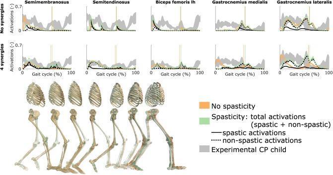
This medical infographic presents a comparative analysis of muscle activation patterns during the human gait cycle in a child with cerebral palsy (CP). The visual features a series of line graphs organized by two motor control conditions: 'No synergies' and '4 synergies.' Five specific muscles are analyzed across a 100% gait cycle: Semimembranosus, Semitendinosus, Biceps femoris long head (lh), Gastrocnemius medialis, and Gastrocnemius lateralis. Each plot compares predictive simulation data against experimental results (shaded gray area, representing mean ± 2 SD). The simulation data differentiates between 'No spasticity' (orange), 'Spasticity: total activations' (green), 'spastic activations' (solid black), and 'non-spastic activations' (dotted black). Vertical lines denote the transition from stance to swing phase. Below the data plots, a sequential musculoskeletal diagram illustrates the right leg's progression from heel strike through the gait cycle. This educational material demonstrates the influence of spasticity and reduced neuromuscular complexity on muscle recruitment and gait mechanics in pediatric CP research.
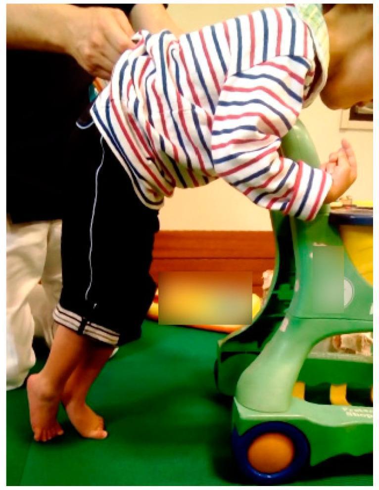
This clinical photograph displays a four-year-old child with spastic cerebral palsy demonstrating a characteristic standing posture while supported by a standing frame and manual assistance. The lower extremities exhibit a classic 'scissors gait' or scissors posture, characterized by hip adduction and internal rotation, causing the legs to cross. The feet are held in a marked equinus position (toe-walking), with the heels significantly elevated off the ground and weight borne on the forefoot. The child shows a forward-leaning trunk posture, utilizing the upper extremities for weight-bearing and balance on a mobility device. This visual illustrates spasticity and motor impairment consistent with Gross Motor Function Classification System (GMFCS) Level IV. The image is an educational example of pediatric neuromotor dysfunction, specifically demonstrating the compensatory postural adaptations and lower limb deformities associated with spastic diplegia.
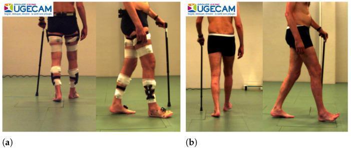
This clinical photograph comparison demonstrates the functional outcome of Botulinum Toxin Type A (BTX-A) treatment for lower limb spasticity. Panel (a) shows the subject 'before treatment' from posterior and lateral views. The subject exhibits significant motor impairment requiring a walking cane and bilateral assistive devices, including wraps and bracing on the thighs and lower legs. The gait is characterized by a forward-leaning trunk, uneven trunk alignment, and abnormal foot placement, with the left foot notably in a position of plantar flexion (equinus deformity). Panel (b) shows the same subject 'after treatment.' The subject is walking without the previous lower limb braces or wraps. The posture is visibly more upright and symmetric, with improved trunk-pelvis alignment. Gait analysis reveals a more natural and fluid stride, characterized by improved knee extension and better ankle dorsiflexion during the swing and initial contact phases. While a cane is still held for balance, the dependency on physical support is reduced, illustrating the efficacy of BTX-A in managing spasticity in conditions like Cerebral Palsy, Multiple Sclerosis, or post-stroke hemiparesis.
Cerebral Palsy: Treatment
CP Classification - Guides Treatment Choice
| Type | Tone | Distribution | Key Concern |
|---|---|---|---|
| Spastic (most common) | High, velocity-dependent | Hemiplegia, Diplegia, Quadriplegia | Contractures, hip dislocation |
| Dyskinetic - Dystonic | Increased, stiff | Generalized | Posture, pain |
| Dyskinetic - Choreoathetoid | Low tone, jerky | Generalized | Coordination, communication |
| Ataxic | Low tone | Generalized | Balance, coordination |
| Mixed | Variable | Variable | Combined spasticity + dystonia |
1. Rehabilitation & Physical Modalities (First-Line)
- Physical therapy (PT): Slow stretching, daily passive range-of-motion (ROM) exercises reduce stretch reflex activity and contracture risk. Weight-bearing in standing frames (for non-ambulatory children) maintains bone density and hip alignment.
- Occupational therapy (OT): Fine motor skills, ADL training, upper limb function.
- Speech and language therapy: Dysphagia management, AAC devices for non-verbal patients.
- Serial casting: Reduces stretch reflex activity and contractures progressively by increasing joint angle over weeks; particularly useful for equinus (ankle) and knee flexion contractures.
- Orthoses (AFO/KAFO): Ankle-foot orthoses maintain joint alignment, support gait, and prevent dynamic deformity from progressing to fixed deformity. Solid-ankle vs. hinged AFOs are selected based on gait analysis findings.
- Gait analysis: Instrumented 3D gait analysis is essential for planning surgical interventions in ambulant children - it guides which muscles/joints to address and in what order.
- Hippotherapy: A 2024 systematic review (PMID 39300490) found hippotherapy significantly improves motor function in children with CP.
2. Pharmacological Management of Spasticity/Hypertonia
Oral Agents
| Drug | Mechanism | Notes |
|---|---|---|
| Oral Baclofen | GABA-B agonist - inhibits motor neuron output at spinal level | Efficacy for CP specifically is not well established; useful adjunct, dose-limited by sedation |
| Diazepam | GABA-A agonist - facilitates spinal motor inhibition | Useful for severe spasticity/spasms; sedation and dependence limit long-term use |
| Tizanidine | Central alpha-2 agonist | Shorter-acting; especially useful for limiting spasms during sleep or brief activities (e.g., wheelchair-to-bed transfers) |
| Dantrolene | Peripherally acting - reduces calcium release from SR | Acts at the muscle fiber level, not centrally; causes muscle weakness |
Botulinum Toxin A (BTX-A) - Major Non-Surgical Spasticity Treatment
- Mechanism: Inhibits acetylcholine release at the neuromuscular junction, causing temporary chemodenervation.
- Indications: Dynamic (not fixed) deformity - equinus, hip adductor spasticity, hamstring tightness, upper limb flexor spasticity.
- Effect: Lasts 3-6 months; allows a "window" for stretching, casting, and functional rehabilitation.
- Evidence: A Cochrane review confirmed benefit for lower limb spasticity in CP. A 2025 network meta-analysis (PMID 40494559) across non-surgical therapies found BTX-A among the most effective for spastic CP.
3. Neurosurgical Treatments
Intrathecal Baclofen (ITB) Pump
- A pump is implanted subcutaneously and delivers baclofen directly into the intrathecal space, achieving 100x higher CSF concentrations than oral dosing with minimal systemic effects.
- Best for: Severe, generalized spasticity (GMFCS III-V); whole-body involvement; patients for whom oral agents are insufficient.
- Not useful for dystonia (unlike spasticity, dystonia does not respond to baclofen reliably).
- Requires ongoing pump maintenance and refills; risk of acute withdrawal (potentially fatal).
Selective Dorsal Rhizotomy (SDR)
- Neurosurgical procedure cutting 25-50% of sensory nerve rootlets (L1-S1) that show abnormal EMG responses on intraoperative stimulation, permanently reducing afferent input driving spasticity.
- Ideal candidate: Child with spastic diplegia, GMFCS II-III, age 3-8 years, adequate underlying muscle strength, motivated family able to commit to intensive post-op rehabilitation.
- Outcomes: Gross motor function improves ~1 GMFCS level; improvements in lower extremity function, gait, bladder function, and pain. Reduces subsequent need for BTX-A and orthopedic surgery.
- A 10-year follow-up shows peak improvement at 3 years post-procedure, then gradual decline; 84% of patients still required a mean of 3 orthopedic procedures, showing contracture is not purely spasticity-mediated.
- Complications: Scoliosis, hyperlordosis, spondylolysis, sensory changes, urinary incontinence, hip subluxation, and postoperative weakness.
- Less effective for spastic quadriplegia or hemiplegia; not useful for dystonia.
Deep Brain Stimulation (DBS)
- Reserved for dyskinetic CP (dystonic/choreoathetoid), where SDR and baclofen are ineffective.
- Targets the globus pallidus internus (GPi).
4. Orthopedic Surgical Treatment
| Goal | Procedures |
|---|---|
| Correct static/dynamic deformity | Musculotendinous lengthening (recession, Z-plasty, tenotomy), serial casting |
| Balance muscle forces | Tendon transfer (e.g., split anterior tibial tendon transfer for foot varus) |
| Stabilize joints / correct bony malalignment | Osteotomies (femoral, tibial, pelvic), joint fusion |
| Reduce spasticity (neurological) | SDR, ITB pump |
Hip Management
- Hip surveillance is mandatory in all CP children - especially non-ambulatory (GMFCS IV-V) who have up to 90% risk of subluxation/dislocation.
- Adductor/psoas release: For early hip subluxation to reduce deforming forces.
- Bony reconstruction (femoral/pelvic osteotomy): For established subluxation with Reimer's migration percentage >50%.
- Hip pain affects up to 47% of young adults with CP and is a major driver of surgical intervention.
Foot/Ankle
- Equinus (most common): Achilles tendon lengthening/recession, BTX-A injections first.
- Crouch gait (overlengthened Achilles): Distal femoral extension osteotomy, patellar tendon shortening.
- Spastic equinovarus: Split anterior tibial tendon transfer (SPLATT) to third cuneiform.
Spine
- Neuromuscular scoliosis: Common in GMFCS IV-V; posterior spinal fusion when curves progress beyond 40-50°; must rule out diastematomyelia before correction.
Knee
- Crouch gait (knee flexion): Hamstring lengthening, serial stretch-casting for resistant flexion contractures, distal femoral extension osteotomy.
- Stiff knee gait: Rectus femoris transfer (distal rectus femoris to gracilis/semitendinosus).
5. Management of Associated Comorbidities
| Comorbidity | Prevalence | Management |
|---|---|---|
| Cognitive impairment | ~50% | Special education, individualized learning plans |
| Epilepsy | 25-40% | Antiepileptic drugs |
| Pain | Common in adults | Multimodal analgesia; address musculoskeletal causes |
| Feeding/dysphagia | Very common in GMFCS IV-V | NG tube, gastrostomy (G-tube) |
| Visual/hearing impairment | ~20% | Corrective lenses, hearing aids |
| Bladder dysfunction | Common | Oxybutynin, clean intermittent catheterization |
| Respiratory complications | GMFCS IV-V | Chest PT, secretion management |
6. Prevention
Summary: Treatment by GMFCS Level
| GMFCS | Ambulation | Priority Treatments |
|---|---|---|
| I | Independent | PT, orthotics if needed, BTX-A for focal spasticity |
| II | Independent with aids | PT, AFO, BTX-A, gait analysis to guide surgery |
| III | With walking aids | BTX-A, serial casting, SDR (selected), SEMLS |
| IV | Self-mobility limited | ITB pump, hip surveillance, G-tube assessment, SEMLS |
| V | No self-mobility | ITB pump, hip/spine surgery, G-tube, comfort care, pain management |
- Campbell's Operative Orthopaedics 15th Ed 2026, Ch. 35
- Bailey and Love's Short Practice of Surgery, Ch. 44 (Table 44.15-16)
- Bradley and Daroff's Neurology in Clinical Practice, Ch. 55
- Goldman-Cecil Medicine, Ch. 385
- Katzung's Basic and Clinical Pharmacology, 16th Ed