
What is standardised loop technique and pour plate technique
Both of these are microbiology techniques used to isolate or enumerate bacteria. Here's a quick overview before I pull up the textbook details:
- Standardised loop technique: Uses a calibrated inoculation loop of fixed volume (usually 0.001 mL or 0.01 mL) to streak a defined quantity of specimen onto culture media, allowing semi-quantitative colony counts.
- Pour plate technique: A sample is mixed with molten agar (cooled to ~45-50°C), poured into a petri dish, and allowed to solidify - colonies grow both on the surface and embedded within the agar.
Searching sources...
Reading File
Now let me search the library for both techniques simultaneously.
Finding Sources
Finding Sources
Good - I found pour plate content. Let me now search more broadly for the standardised loop technique and read the full plating section.
Finding Sources
Reading File
Excellent - I have the pour plate content and the calibrated loop info. Let me read the full plating section and the calibrated loop section from Henry's.
Reading File
Reading File
Now let me get the calibrated loop details from Henry's and fetch the pour plate diagram image.
Reading File
Loading Image
I now have all the information needed to give a comprehensive answer. Let me also fetch the inoculation technique diagram.
Loading Image
I now have everything needed for a comprehensive answer.
Standardised (Calibrated) Loop Technique
What it is
The standardised loop technique (also called the calibrated loop method) is a semi-quantitative to quantitative culture method. It uses a plastic or wire inoculating loop that is precisely manufactured to hold a fixed, known volume of specimen - typically 0.001 mL (1 µL) or 0.01 mL (10 µL).
Purpose
Its primary goal is not just to isolate organisms, but to estimate bacterial counts in a specimen. This is especially important for urine cultures, where distinguishing infection from contamination depends on knowing how many colony-forming units (CFU) per mL are present.
How it works (urine culture example)
-
The appropriate calibrated loop is selected:
- 0.001 mL loop - used for most routine urine specimens
- 0.01 mL loop - used for women with suspected acute urethral syndrome and for suprapubic aspirate specimens (which may have lower bacterial counts but still indicate infection)
-
The loop is inserted vertically into the well-mixed urine sample and withdrawn with a loopful of urine.
-
The specimen is spread over the entire surface of the agar plate in a standardised pattern - typically a series of parallel horizontal lines across the plate (as seen in the diagram below).
-
Without re-flaming, the loop is reinserted into the urine and spread onto a second plate (e.g., blood agar and MacConkey agar).
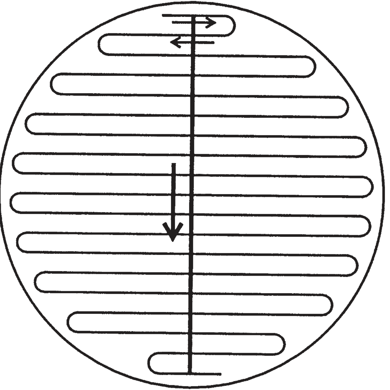
Standardised spreading pattern for calibrated loop inoculation
Interpreting results
Because the volume deposited is known, colony counts can be back-calculated:
-
0.001 mL loop: Number of colonies × 1,000 = CFU/mL
-
0.01 mL loop: Number of colonies × 100 = CFU/mL
-
≥ 10⁵ CFU/mL - traditionally considered significant bacteriuria indicating infection
-
< 10³ CFU/mL - usually considered contamination
-
Intermediate counts require clinical correlation
Source: Henry's Clinical Diagnosis and Management by Laboratory Methods, p. 1566-1567
Pour Plate Technique
What it is
The pour plate method is a classic technique for isolating microorganisms in pure culture and for counting viable organisms in a sample. It was pioneered by Robert Koch in 1881.
Principle
Unlike cells in liquid medium, cells immobilised in solidified agar cannot move - so each bacterium grows into an isolated, countable colony. Because the original sample is serially diluted before plating, the number of colonies directly reflects the number of viable organisms.
Step-by-step procedure
-
Serial dilution - The original sample is diluted stepwise (e.g., 10⁻¹, 10⁻², 10⁻³, 10⁻⁴) by transferring 1.0 mL into tubes containing 9 mL of water or buffer.
-
Mixing with molten agar - 1.0 mL of a diluted sample is pipetted into an empty Petri dish, then molten agar cooled to ~50°C is poured in and mixed gently. The agar must be cooled enough not to kill the cells but still fluid enough to pour.
-
Solidification - The agar is allowed to harden. Cells become trapped and immobilised within the gel.
-
Incubation - Plates are incubated at the appropriate temperature.
-
Colony counting and picking - Colonies that are well separated (ideally 30-300 per plate) are counted. Each colony theoretically arose from a single cell, so it can be used to establish a pure culture.
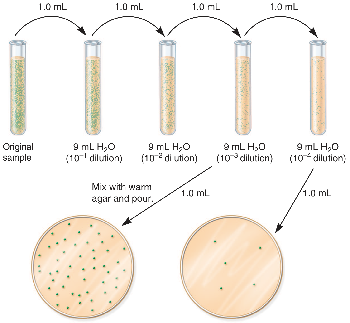
Pour plate technique: serial dilutions are mixed with warm agar and poured into Petri dishes. The more dilute sample (right) gives fewer, well-separated colonies.
Important features
- Surface colonies appear circular; subsurface colonies (embedded in agar) appear lenticular (lens-shaped)
- The cell suspension must be sufficiently dilute so colonies are well separated and each has a high probability of originating from a single cell
- For counting purposes, a plate with 30-300 colonies gives the most reliable count
Source: Jawetz, Melnick & Adelberg's Medical Microbiology 28th Ed, p. 82-83
Comparison Table
| Feature | Standardised Loop | Pour Plate |
|---|---|---|
| Primary use | Quantitative / semi-quantitative culture (especially urine) | Isolation of pure cultures; viable count |
| Volume deposited | Fixed known volume (0.001 or 0.01 mL) | Variable (usually 1.0 mL of a diluted suspension) |
| How bacteria are distributed | Spread on agar surface | Mixed throughout agar depth |
| Colony location | Surface only | Surface (round) + subsurface (lenticular) |
| Serial dilution needed? | No - loop volume standardises this | Yes - essential before plating |
| Speed | Fast, simple | More steps (dilution + pouring) |
| Main clinical application | Urine, BAL, wound cultures | Microbial enumeration, pure culture isolation |
This is a shared conversation. Sign in to Orris to start your own chat.