
Area supplied by each branch of circle of Willis...
Reading File
Reading File
Finding Sources
Searching PubMed
"Circle of Willis"[MeSH Terms] AND "anatomy"
Reading File
Reading File
Finding Sources
Reading File
Reading File
Territories supplied by the circle of Willis and its branches
The circle joins the anterior circulation (internal carotids) to the posterior circulation (vertebrobasilar). The three main cerebral arteries arise from it. Each gives cortical branches to the surface and small penetrating branches to deep structures (Neuroanatomy through Clinical Cases, 3rd ed., p. 416-420).
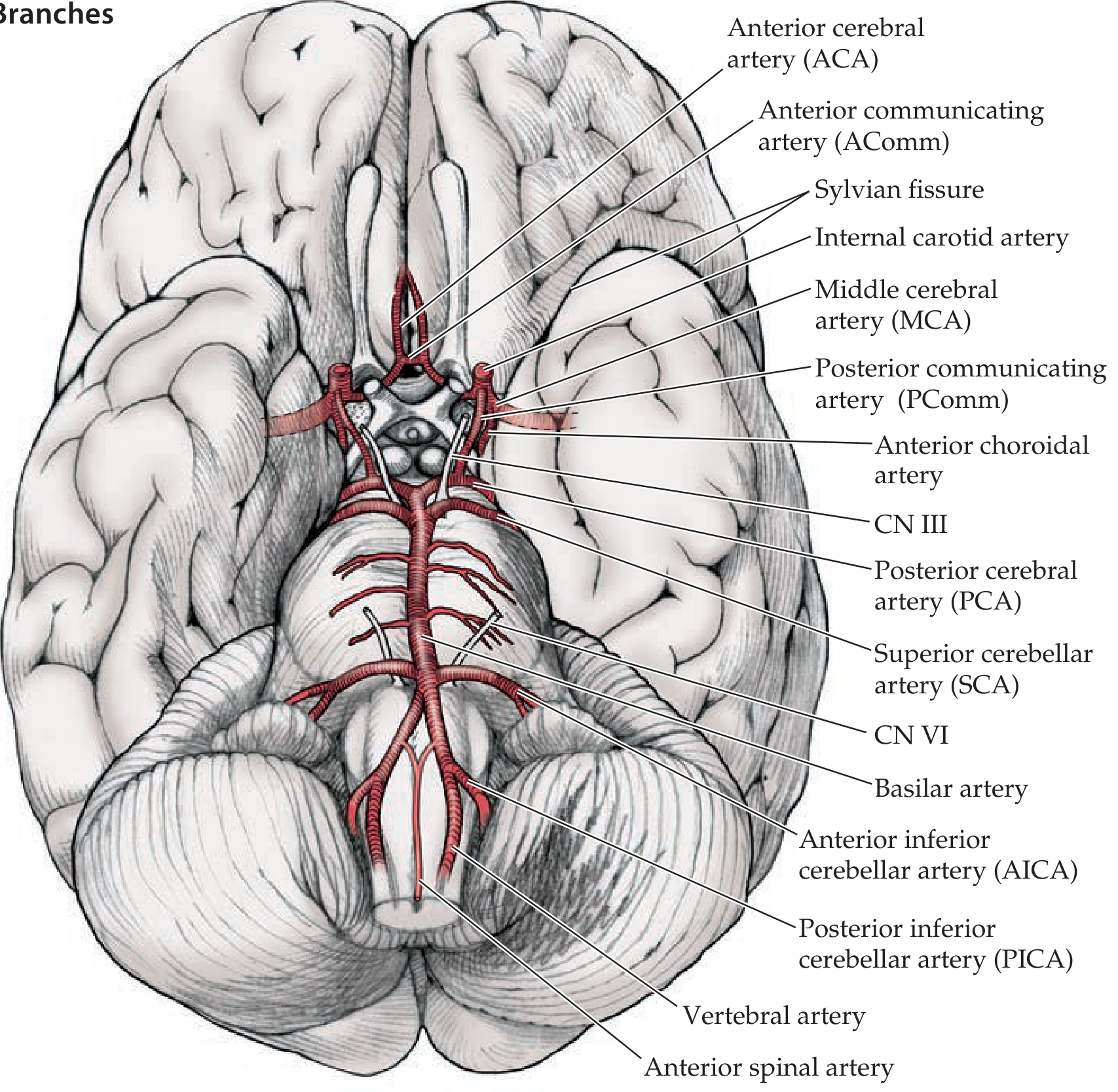
Components of the ring
| Vessel | Origin and role | Area supplied |
|---|---|---|
| Internal carotid artery (ICA) | Gives rise to the anterior circulation. Its terminal branches are the ACA and MCA. | Supplies the ACA and MCA territories through those branches. It also gives off the ophthalmic artery (eye), anterior choroidal artery and posterior communicating artery. |
| Anterior communicating artery (AComm) | Links the two ACAs. | Small perforators to the anterior hypothalamus, optic chiasm and fornix. This is a common aneurysm site. |
| Posterior communicating artery (PComm) | Links the ICA to the PCA. | Perforators to the thalamus, hypothalamus and posterior limb of the internal capsule. |
| Basilar artery | Formed by the two vertebral arteries. It ends by dividing into the two PCAs. | Pons, cerebellum and midbrain, through its pontine, labyrinthine, AICA and SCA branches. |
Cortical (superficial) territories
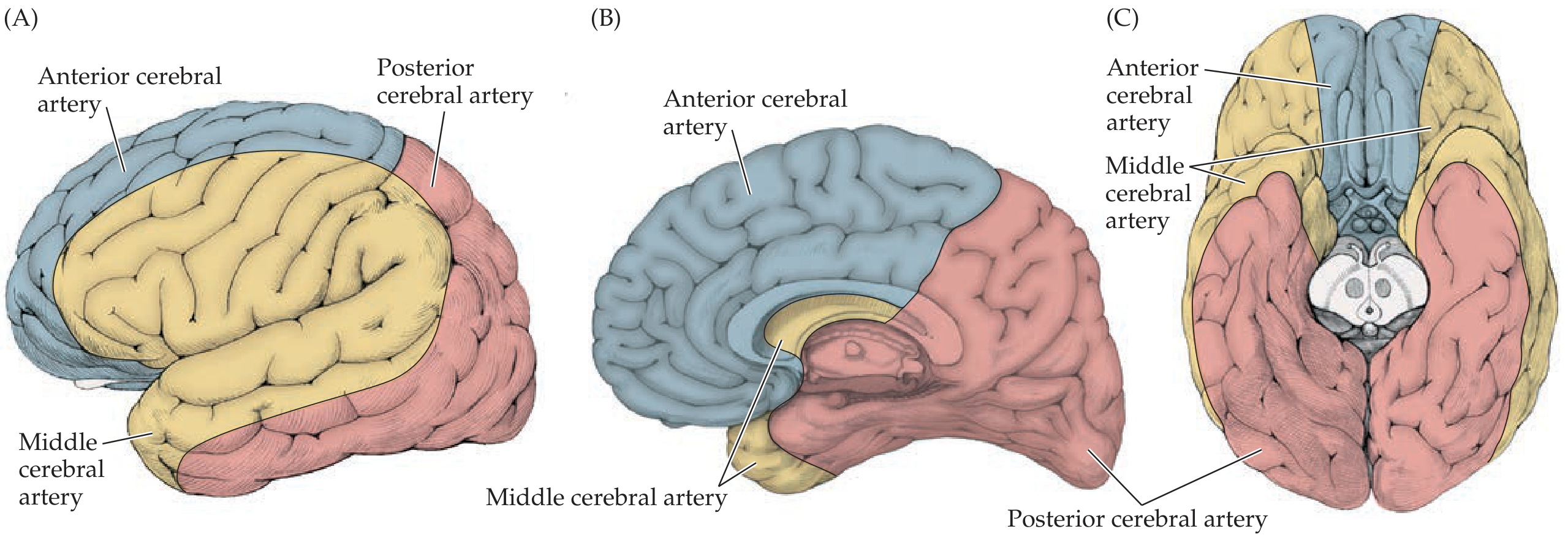
Anterior cerebral artery (ACA)
- It runs in the interhemispheric fissure and over the corpus callosum. Its main branches are the pericallosal and callosomarginal arteries.
- It supplies most of the anteromedial cortex, from the frontal lobe to the anterior parietal lobe. This usually includes the medial sensorimotor cortex, which serves the leg and foot.
- From general knowledge, it also supplies the anterior part of the corpus callosum and the superior medial frontal gyrus.
- Occlusion causes contralateral leg weakness and sensory loss, with abulia or frontal signs.
Middle cerebral artery (MCA)
- It enters the Sylvian fissure and divides into a superior and an inferior division.
- The superior division supplies the lateral frontal and parietal lobes. The inferior division supplies the lateral temporal lobe (Goldman-Cecil Medicine, Middle Cerebral Artery).
- Together they supply most of the lateral convexity and the insula. From general knowledge, this includes the face and arm motor and sensory areas, Broca and Wernicke areas, and the optic radiations (partly).
- Occlusion causes contralateral face and arm weakness, aphasia (dominant hemisphere) or neglect (non-dominant hemisphere), and a contralateral homonymous hemianopia or quadrantanopia.
Posterior cerebral artery (PCA)
- It arises from the top of the basilar artery.
- It supplies the occipital lobe (visual cortex) and the inferomedial temporal lobe. From general knowledge, it also supplies the splenium of the corpus callosum.
- Occlusion causes a contralateral homonymous hemianopia, often with macular sparing.
Deep (penetrating) territories
| Artery | Origin | Deep structures supplied |
|---|---|---|
| Lenticulostriate arteries | MCA, before it enters the Sylvian fissure | Large parts of the basal ganglia and internal capsule |
| Recurrent artery of Heubner | Proximal ACA | Head of the caudate, anterior putamen, globus pallidus and the anterior limb of the internal capsule |
| Anterior choroidal artery | ICA | Parts of the globus pallidus, putamen and thalamus (sometimes the lateral geniculate nucleus), and the posterior limb of the internal capsule up to the lateral ventricle |
| Thalamoperforators, thalamogeniculate and posterior choroidal arteries | Proximal PCA | Thalamus, sometimes part of the posterior limb of the internal capsule. Perforators from the top of the basilar artery also supply the midbrain. |
(Neuroanatomy through Clinical Cases, 3rd ed., p. 419-420)
Lacunar infarcts or hemorrhages in the lenticulostriate or anterior choroidal territories often cause contralateral hemiparesis. Hypertension makes the lenticulostriate vessels particularly prone to this.
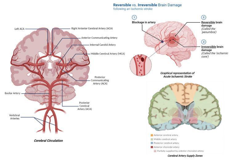
Summary diagram
Points to remember
- The ACA and MCA are the terminal branches of the ICA. The PCAs come from the basilar artery.
- A complete, full-caliber circle is present in only about 34% of people. Variants such as a fetal-type PCA are common (Neuroanatomy through Clinical Cases, p. 416).
- A mnemonic for cortical territories: ACA supplies the leg, MCA supplies the face and arm plus language, and PCA supplies vision.
A quick PubMed check of recent systematic reviews and meta-analyses found only anatomical studies (for example, sex differences in intracranial arteries, PMID 37209774, and the recurrent artery of Heubner, PMID 39472321). Nothing contradicts the textbook territories above.
In hemiplegia involving internal capsule, which brach of circle of Willis is relevant?
Reading File
Finding Sources
Searching Images
internal capsule blood supply lenticulostriate anterior choroidal artery axial section
Hemiplegia from an internal capsule lesion: which arteries are relevant
The main arteries are the lenticulostriate arteries, which come from the MCA. The anterior choroidal artery (from the ICA) and the recurrent artery of Heubner (from the ACA) are also involved. The internal capsule gets its blood from several small penetrating arteries, not one vessel.
| Part of internal capsule | Artery | Source |
|---|---|---|
| Anterior limb | Medial lenticulostriate branches and the recurrent artery of Heubner | MCA, and proximal ACA |
| Genu and posterior limb (corticospinal and corticobulbar fibres) | Lateral lenticulostriate arteries (most common), and the anterior choroidal artery | M1 segment of the MCA, and the ICA |
| Posterior limb, small part | Perforating branches of the PCA (thalamoperforators and thalamogeniculate arteries) | Proximal PCA |
Why these arteries cause hemiplegia
The posterior limb carries the corticospinal and corticobulbar motor pathways. A lacunar infarct in the lenticulostriate or anterior choroidal territory often causes contralateral hemiparesis (Neuroanatomy through Clinical Cases, 3rd ed., p. 420). The lenticulostriate arteries are the most common culprit. They arise from the proximal MCA before it enters the Sylvian fissure, and they are prone to narrowing and to rupture in hypertension.
Typical presentations
- Lenticulostriate lacune (MCA): Pure motor hemiparesis, with weakness of the face, arm and leg together, usually with dysarthria. There is little or no sensory loss (Bradley and Daroff's Neurology in Clinical Practice, Subcortical Lesions). The posterior limb of the internal capsule is the commonly affected site, and lenticulostriate arteries are the common vessels (Neuroanatomy through Clinical Cases, Mechanisms of Ischemic Stroke).
- Anterior choroidal artery (ICA) infarct: The classic triad is contralateral hemiparesis, hemisensory loss and homonymous hemianopia. The hemiparesis comes from the corticospinal tract in the posterior limb. The sensory loss comes from the thalamic radiations. The hemianopia comes from the optic tract or lateral geniculate nucleus (Localization in Clinical Neurology, 8e; the hemianopia part is from general knowledge).
- Recurrent artery of Heubner (ACA): It supplies the anterior limb and the head of the caudate. A lesion here can cause a faciobrachial-predominant weakness (from general knowledge).
Exam points
- The commonest answer for "hemiplegia due to an internal capsule lesion" is the lenticulostriate artery, a branch of the MCA. Charcot's "artery of cerebral hemorrhage" is the same vessel.
- If the question mentions hemisensory loss and hemianopia as well, think of the anterior choroidal artery.
- A capsular lesion produces a dense, contralateral face, arm and leg weakness, because the fibres are packed tightly in the posterior limb.
The image search returned no matching figure for this question. Figures 10.7 to 10.9 in Neuroanatomy through Clinical Cases, p. 420, show the deep territories.
Types of speech see in different CNS cases
Reading File
Finding Sources
Finding Sources
Searching Images
dysarthria types table spastic flaccid ataxic hypokinetic speech
Reading File
Reading File
Reading File
Reading File
Types of speech disturbance in CNS disease
Speech disorders fall into two groups. Motor speech disorders affect articulation only. Language is normal, so the patient can write and comprehend normally. Aphasias affect language itself (Bradley and Daroff's Neurology in Clinical Practice, Motor Speech Disorders, p. 149).
1. Dysarthria (abnormal articulation)
The Mayo classification (Duffy) has six types. Details not found in the retrieved text are marked as from general knowledge.
| Type | Speech quality | Lesion site | Typical causes |
|---|---|---|---|
| Flaccid | Breathy, nasal, indistinct consonants. In myasthenia, speech fatigues as the patient keeps talking. | Lower motor neuron (cranial nerve nuclei, nerves, NMJ, bulbar muscle) | Bulbar palsy, myasthenia gravis, polymyositis, bulbar poliomyelitis |
| Spastic | Harsh, "strain-strangle" voice, slow rate, low pitch, consonant errors. Often with pseudobulbar palsy: dysphagia, brisk jaw jerk and gag, emotional incontinence. | Bilateral corticobulbar tracts or motor cortex | Bilateral strokes, ALS (upper motor neuron part), multiple sclerosis |
| Unilateral upper motor neuron | A milder spastic pattern. One of the commonest dysarthrias. | Unilateral UMN lesion | Unilateral stroke |
| Ataxic ("scanning speech") | Either irregular breakdowns with bursts of syllables and pauses, or a slow cadence with equal stress on every syllable. | Cerebellum, especially the superior vermis. Adams and Victor also link scanning speech to mesencephalic lesions of the brachium conjunctivum. | Cerebellar stroke, tumour, multiple sclerosis, cerebellar degenerations |
| Hypokinetic | Soft, monotonous in loudness and pitch, rapid rate, occasional consonant errors | Basal ganglia (nigrostriatal system) | Parkinson disease |
| Hyperkinetic (general knowledge) | Irregular, variable articulation, with sudden changes in loudness and pitch. In dystonia the voice is strained. | Basal ganglia | Huntington chorea, dystonia, Tourette syndrome, tremor |
| Mixed | Combined features | Multiple systems | ALS (spastic and flaccid), multiple sclerosis (spastic and ataxic, giving scanning dysarthria), Wilson disease |
(Bradley and Daroff's, Dysarthrias; Adams and Victor's Principles of Neurology, 12th ed., Ataxic Dysarthria; Scott-Brown's, Cerebellar Disorders)
Other motor speech disorders (Bradley and Daroff's, p. 149 to 151):
- Apraxia of speech: a motor programming disorder. Speech is effortful and halting, with groping and inconsistent errors.
- Aphemia, foreign accent syndrome, acquired stuttering, and the opercular syndrome, which are rarer.
- Anarthria is total loss of the ability to articulate.
2. Aphasia (language disorder)
Aphasias are classified by fluency, comprehension and repetition. Naming difficulty and paraphasias occur in almost all types and have little localising value (Neuroanatomy through Clinical Cases, 3rd ed., KCC 19.6, Figure 19.4).
| Aphasia | Fluency | Comprehension | Repetition | Lesion |
|---|---|---|---|---|
| Broca | Impaired | Normal | Impaired | Left MCA superior division, inferior frontal gyrus |
| Wernicke | Normal | Impaired | Impaired | Left MCA inferior division, posterior superior temporal gyrus |
| Global | Impaired | Impaired | Impaired | Large left MCA infarct (both divisions), or large subcortical lesions |
| Conduction | Normal | Normal | Impaired | Peri-Sylvian lesion interrupting the arcuate fasciculus and supramarginal gyrus |
| Transcortical motor | Impaired | Normal | Normal | ACA to MCA watershed infarct, or dominant-hemisphere subcortical lesion |
| Transcortical sensory | Normal | Impaired | Normal | MCA to PCA watershed infarct |
| Mixed transcortical (isolation of the language areas) | Impaired | Impaired | Normal | Combined watershed infarcts, or subcortical lesions |
| Anomic | Normal | Normal | Normal | Many sites (the least localising) |
Speech in conduction aphasia is fluent and paraphasic, which can be mistaken for Wernicke aphasia. The difference is that comprehension is spared.
3. Other patterns (general knowledge)
- Mutism: akinetic mutism (bilateral medial frontal or ACA lesions, or reticular lesions), and the locked-in state (ventral pontine lesion).
- Dysphonia: abnormal voice production, for example from vocal cord palsy in a vagal or recurrent laryngeal nerve lesion, or the spasmodic dysphonia of laryngeal dystonia.
- Right hemisphere lesions: aprosodia, meaning loss of the emotional tone and melody of speech, with normal language.
- Palilalia (repeating one's own words) and echolalia: seen in parkinsonism and frontal or dementing disorders.
Quick bedside clues
- Fluent but meaningless speech with poor comprehension points to Wernicke aphasia.
- Effortful, telegraphic, non-fluent speech with good comprehension and right face and arm weakness points to Broca aphasia.
- Slow, equal-stress, "scanning" speech points to the cerebellum.
- Soft, fast, monotone speech points to Parkinson disease.
- Strained, harsh speech with emotional lability points to bilateral upper motor neuron (pseudobulbar) disease.
- Nasal, breathy speech that worsens with prolonged talking points to myasthenia gravis.
I did not run a PubMed check for this answer. The searches I did run returned no suitable figure.
This is a shared conversation. Sign in to Orris to start your own chat.