
What is meant by culture report negative vs culture negative in infective endocarditis
- Culture-negative IE (CNIE): Blood cultures never grow an organism - the causative pathogen is unidentified. This can be due to prior antibiotic use, fastidious/slow-growing organisms (Brucella, HACEK, Coxiella, Bartonella, fungi), or intracellular organisms that don't grow on standard media.
- Culture report negative: A specific blood culture bottle/set came back with no growth reported - but this is a lab result, not a final diagnosis. It doesn't mean the patient has CNIE; other cultures may still be pending or positive.
"culture-negative endocarditis" diagnosis management
PMID: 40094211
Culture Report Negative vs Culture-Negative IE - A Clear Distinction
1. "Culture Report Negative" (Lab Result)
- The patient doesn't have IE
- All cultures are negative
- A causative organism won't be found
2. Culture-Negative Infective Endocarditis (CNIE) - Clinical Diagnosis
"An endocarditis illness with no known etiology 7 days following inoculation and subculturing of three independent blood samples in a standard blood culture system." (Fuster and Hurst's The Heart, 15th Ed., p. 1029)
Why Do Cultures Come Back Negative in True CNIE?
A. Prior Antibiotic Administration (Most Common - 33-50% of CNIE)
- The patient received antibiotics before cultures were drawn
- Suppresses growth of fastidious organisms like viridans streptococci and HACEK more readily than staphylococci
- Cultures may still be positive for staph/enterococcus even after antibiotics - so a "culture-negative" result in that setting still suggests streptococci or HACEK as the culprit (Goldman-Cecil, p. 1823)
B. Fastidious, Slow-Growing, or Intracellular Organisms (when no prior antibiotics)
| Exposure History | Organism |
|---|---|
| Farm animals, abattoirs, unpasteurized milk | Coxiella burnetii (Q fever), Brucella spp. |
| Cats | Bartonella henselae |
| Body lice, homelessness, alcoholism | Bartonella quintana |
| Immunosuppression/HIV | Fungi (Candida, Aspergillus) |
| Soil exposure | Tropheryma whipplei |
| Travel to endemic areas | Brucella, B. quintana |
| Pet birds | Chlamydophila psittaci |
- Bartonella spp.: Blood cultures positive in only ~25% of cases even with special techniques; prolonged incubation (up to 6 weeks) required. Serology (IgG ≥1:800) is the mainstay of diagnosis. (Harrison's 22nd Ed.)
- Coxiella burnetii: Obligate intracellular - will never grow in standard cultures. Diagnosed by phase I/II IgG serology (Duke criterion). Best identified by serology. (Fuster & Hurst)
- HACEK organisms: Slow-growing; in labs without extended incubation protocols, these patients will appear culture-negative. (Comprehensive Clinical Nephrology, 7th Ed.)
- Tropheryma whipplei: Increasingly recognized; in one German cohort, it was the most common cause of CNIE in patients without classical Whipple disease (Goldman-Cecil)
How CNIE Is Diagnosed (the diagnostic pathway beyond blood cultures)
- Serology: Coxiella burnetii (phase I IgG ≥1:800 = major Duke criterion), Bartonella spp., Brucella, Chlamydophila psittaci
- PCR on blood or explanted valve tissue: Identifies streptococci, enterococci, staphylococci, Bartonella, T. whipplei
- Extended/special culture techniques: Notify the microbiology lab; prolonged incubation (>5 days) increases yield for nutritional variant streptococci (Abiotrophia, Granulicatella), Brucella, Legionella, Cutibacterium acnes
- Histopathology of resected tissue: Can provide etiologic clues
- PET-CT: Helps identify periprosthetic abscesses, mycotic aneurysms, and infected devices - useful for fastidious organisms like T. whipplei and C. burnetii
- Consider non-infectious causes: Marantic endocarditis, Libman-Sacks (SLE), atrial myxoma (Goldman-Cecil)
Empirical Treatment of CNIE
- Fastidious streptococci (Abiotrophia, Gemella, Granulicatella)
- HACEK organisms
- Bartonella spp. (gentamicin + doxycycline + ceftriaxone)
Clinical Significance: Why This Distinction Matters
Summary Table
| Feature | "Culture Report Negative" | "Culture-Negative IE" (CNIE) |
|---|---|---|
| Type | Single lab result | Clinical diagnosis |
| Scope | One bottle/set, one time point | 3 independent sets over ≥7 days |
| Meaning | No growth detected yet | No pathogen identified after full workup |
| Action required | Wait, repeat, extend incubation | Serology, PCR, special cultures |
| Frequency | Very common (routine) | 2-7% of IE cases |
| Organism | Unknown | Often Coxiella, Bartonella, HACEK, fungi |
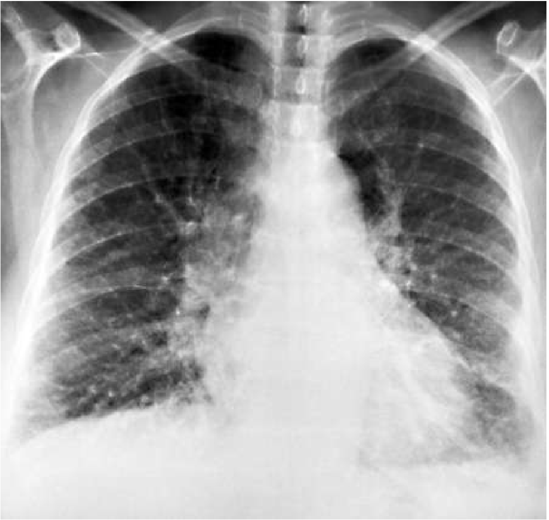
Cephalisation of pulmonary vessels
cephalisation cephalization upper lobe diversion pulmonary vessels chest X-ray
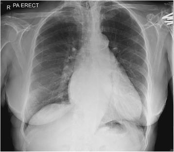
This diagnostic image is a posteroanterior (PA) erect chest X-ray demonstrating signs of pulmonary venous hypertension and early interstitial edema. Key visual findings include cephalization of the pulmonary vasculature, or 'vascular diversion,' characterized by prominent, dilated pulmonary vessels in the upper lung fields relative to the lower lung fields. In the right lower lung zone, there is evidence of interstitial thickening and increased lung markings, consistent with septal edema (Kerley lines). The cardiac silhouette appears slightly enlarged, though the primary focus is the pulmonary vascular congestion. These findings are typical of heart failure or cardiac-related fluid overload. The image serves as an educational tool for identifying the radiological progression from pulmonary venous congestion to interstitial edema, highlighting the redistribution of blood flow and the accumulation of fluid in the lung parenchyma and septal spaces.
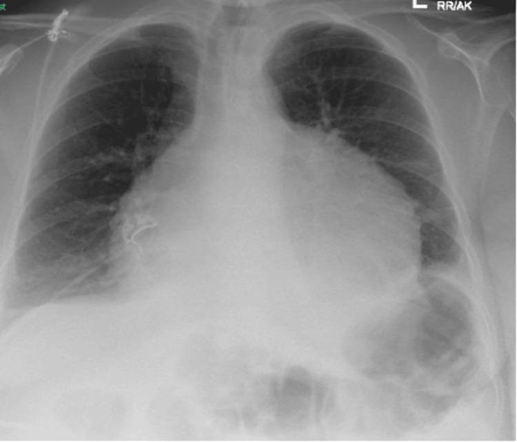
This diagnostic image is an anteroposterior (AP) chest X-ray demonstrating significant cardiomegaly. The cardiac silhouette is markedly enlarged with a global, rounded appearance, commonly referred to as a 'water bottle' heart, which is characteristic of a large pericardial effusion. Lung field analysis reveals cephalization of the pulmonary vessels (upper lobe blood diversion), suggesting pulmonary venous congestion. Bilateral blunting of the costophrenic angles is present, consistent with small pleural effusions. Subtle opacities are noted at the right lung base and in the region of the lingula, possibly representing atelectasis or early pulmonary edema. These radiographic findings are highly suggestive of congestive heart failure or a large pericardial effusion progressing toward cardiac tamponade. The image serves as a clinical teaching tool for identifying markers of left heart failure and pathological enlargement of the mediastinal silhouette in an emergency or critical care context.
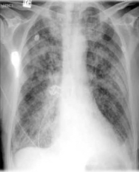
**Imaging Modality:** Anteroposterior (AP) Chest Radiograph (X-ray). **Anatomical Region:** Thoracic cavity, including the lungs, mediastinum, and cardiophrenic angles. **Observed Pathology:** The image demonstrates diffuse, bilateral interstitial and alveolar opacities consistent with severe pulmonary congestion and edema. There is evidence of cephalization of the pulmonary vessels (upper lobe venous diversion). **Characteristic Visual Features:** * **Parenchymal Findings:** Extensive "bat-wing" or perihilar distribution of opacities. Presence of Kerley B lines is suggested by peripheral linear densities at the lung bases. * **Cardiac/Mediastinal Findings:** Marked cardiomegaly with an increased cardiothoracic ratio. The cardiac silhouette is widened, indicating ventricular enlargement. * **Pleural Findings:** Blunting of the costophrenic angles, particularly on the left, indicative of bilateral pleural effusions. **Differentiating Features:** The combination of cardiomegaly, cephalization, perihilar opacification, and pleural effusions strongly characterizes congestive heart failure (CHF) leading to pulmonary edema, distinguishing it from primary pneumonia or focal lung masses. **Clinical Context:** Radiologic features are diagnostic of acute decompensated heart failure with associated pulmonary venous hypertension.
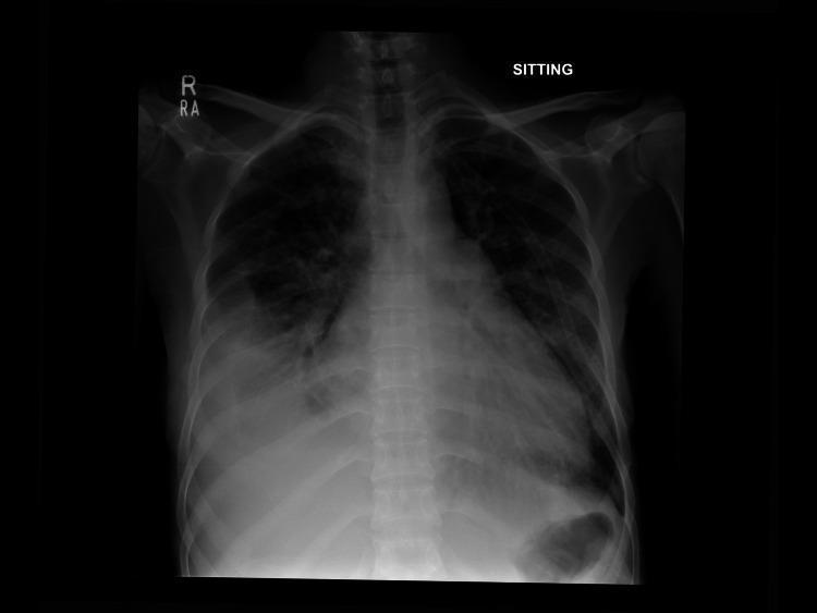
Anteroposterior (AP) chest X-ray in a sitting position demonstrating several findings consistent with pulmonary venous hypertension and fluid overload. The cardiac silhouette is enlarged, indicating cardiomegaly. Notable pulmonary vascular changes include cephalization, where upper lobe vessels appear prominent and distended. Perihilar congestion is evident with increased opacification and blurring of the hilar structures. A moderate right-sided pleural effusion is present, characterized by the blunting and obscuration of the right costophrenic angle, likely associated with underlying compressive atelectasis in the adjacent lung base. The combined findings of cardiomegaly, vascular redistribution, and pleural effusion are highly suggestive of congestive heart failure. The mediastinal structures remain midline, and the bones and soft tissues appear unremarkable within the field of view.
Cephalisation of Pulmonary Vessels
What Is It?
Normal Pattern First: Caudalisation
- Basal pulmonary vessels are 2-3 times wider than upper lobe vessels
- Very little vascularity is visible above the hilum
- Vessels taper gradually from the hilum outward in a tree-like pattern
- The right descending pulmonary artery measures 10-15 mm in males, 9-14 mm in females
- The arterial-bronchial ratio (ABR) is 1.34 ± 0.25 in lower zones (artery > bronchus)
Mechanism of Cephalisation
- Elevated postcapillary pulmonary venous pressure (e.g., left heart failure, mitral stenosis)
- When total intravascular pressure exceeds oncotic pressure, fluid leaks from the capillaries into the interstitium - then into alveoli
- Alveolar flooding causes local hypoxia - predominantly at the bases (where edema accumulates earliest due to gravity)
- Hypoxia triggers hypoxic pulmonary vasoconstriction of the lower lobe arterioles and venules
- Blood is diverted preferentially to the upper lobes, which are less edematous and not vasoconstricted
- Upper lobe vessels dilate; lower lobe vessels constrict
- The ABR ratio between upper and lower lungs equalises, then reverses
"Despite the terms redistribution and cephalization, the diagnosis should be based on constriction of the lower-lobe vessels. Dilatation of the upper lobe vessels can be found without narrowing of the basilar vessels in a number of entities, most noticeably left-to-right shunts." - Fuster & Hurst's The Heart, 15th Ed.
The CXR Progression: PCWP Correlation
| Stage | PCWP | CXR Finding |
|---|---|---|
| Stage 1 | 13-18 mmHg | Cephalisation (upper > lower lobe vessels), widened vascular pedicle, cardiomegaly |
| Stage 2 | 18-25 mmHg | + Interstitial edema: Kerley B lines, peribronchial cuffing, thickened fissures, indistinct vessel contours |
| Stage 3 | >25 mmHg | + Alveolar edema: "bat-wing" / "cotton-wool" confluent airspace opacification, often perihilar |
Chest X-Ray: Patient with Left Ventricular Failure Showing Cephalisation
Causes
- Left ventricular failure (most common) - systolic or diastolic dysfunction
- Mitral stenosis - back pressure from obstructed mitral valve
- Mitral regurgitation (severe)
- Elevated LV diastolic pressure from any cause (e.g., hypertension, aortic stenosis, hypertrophic cardiomyopathy)
How to Distinguish Cephalisation from Other Abnormal Vascular Patterns
| Pattern | Appearance | Cause |
|---|---|---|
| Cephalisation | Upper > lower lobe vessels | Postcapillary PH (LV failure, mitral stenosis) |
| Centralisation | Dilated central PAs, pruned peripheral vessels | Precapillary PH (PAH, Eisenmenger, CTEPH) |
| Equalisation (balanced) | Upper = lower lobe vessels | Left-to-right shunt, anemia, pregnancy, obesity, hyperthyroidism |
| Lateralisation | One lung > other | Massive PE, unilateral emphysema, Swyer-James syndrome |
| Cephalisation + decreased flow | Upper > lower, but all vessels small | Severe mitral stenosis (late) |
Key Points to Remember
- The key diagnostic sign is constriction of lower lobe vessels, not just dilatation of upper lobe vessels
- The upper-lower ABR ratio reverses (upper ABR > lower ABR) in decompensated heart failure
- It is the earliest radiographic sign of elevated pulmonary venous pressure on a PA erect CXR
- On a portable/AP/supine film, cephalisation is not reliable as a sign of heart failure - it may be normal
- Cephalisation is a postcapillary phenomenon; do not confuse with pulmonary arterial hypertension which gives centralisation
- Combined patterns are common (e.g., cephalisation + decreased flow in critical mitral stenosis)