

d77a4da5-328f-4034-af01-2315a3a37572.jpeg
Give me answer to this and explain in appropriate way
analyse_media
Reading File
Now let me search the medical library for detailed content on peptic ulcer pathology.
Finding Sources
Reading File
Finding Sources
Reading File
Finding Sources
Finding Sources
Reading File
I now have all the information needed. Let me also fetch the Robbins textbook image of the peptic ulcer for the response.
Loading Image
Here is a complete, structured answer to all three questions.
Clinical Case: 40-year-old Female with Dyspepsia, Epigastric Pain, and Tarry Stools
Q1. Diagnosis
Chronic Peptic Ulcer Disease (most likely Duodenal Ulcer)
Clinical reasoning:
- Pain occurring 2-3 hours after meals and worse at night is the hallmark of duodenal ulcer (pain occurs when stomach empties and acid is unbuffered, relieved by food/antacids)
- Dyspepsia and epigastric pain fit PUD perfectly
- Tarry stools (melena) indicate upper GI bleeding - a classic complication of peptic ulcers
- A 40-year-old female is in the typical age group for PUD
Q2. Microscopic Features Seen in the Image
The histological slide shows the 4-layer zonal architecture of a chronic peptic ulcer base (from surface/lumen down to deeper tissue):
| Zone | What You See |
|---|---|
| Zone 1 - Superficial Necrosis | Thin layer of eosinophilic amorphous debris + fibrinous exudate at the luminal surface |
| Zone 2 - Active Inflammation | Dense band of neutrophils (PMNs) indicating active tissue destruction |
| Zone 3 - Granulation Tissue | Proliferating capillaries (neovascularization), fibroblasts, and chronic inflammatory cells (lymphocytes, plasma cells, macrophages) |
| Zone 4 - Fibrosis/Cicatrization | Deep dense collagenous scar replacing the normal muscularis - the hallmark of chronicity |
Additional microscopic features:
- Complete breach of the mucosal epithelium extending through the muscularis mucosae
- Destruction of normal glandular architecture in the ulcer bed
- Endarteritis obliterans (thickened vessel walls) may be seen in deeper tissue
- Marginal regenerative epithelial changes at ulcer edges (hyperchromatic nuclei, mucin depletion - should not be confused with dysplasia)
As Robbins & Kumar describe: "The classic peptic ulcer is a round to oval, sharply punched-out defect. The ulcer base is smooth and clean as a result of peptic digestion of exudate and on histologic examination is composed of highly vascular granulation tissue." - Robbins & Kumar Basic Pathology, p. 556
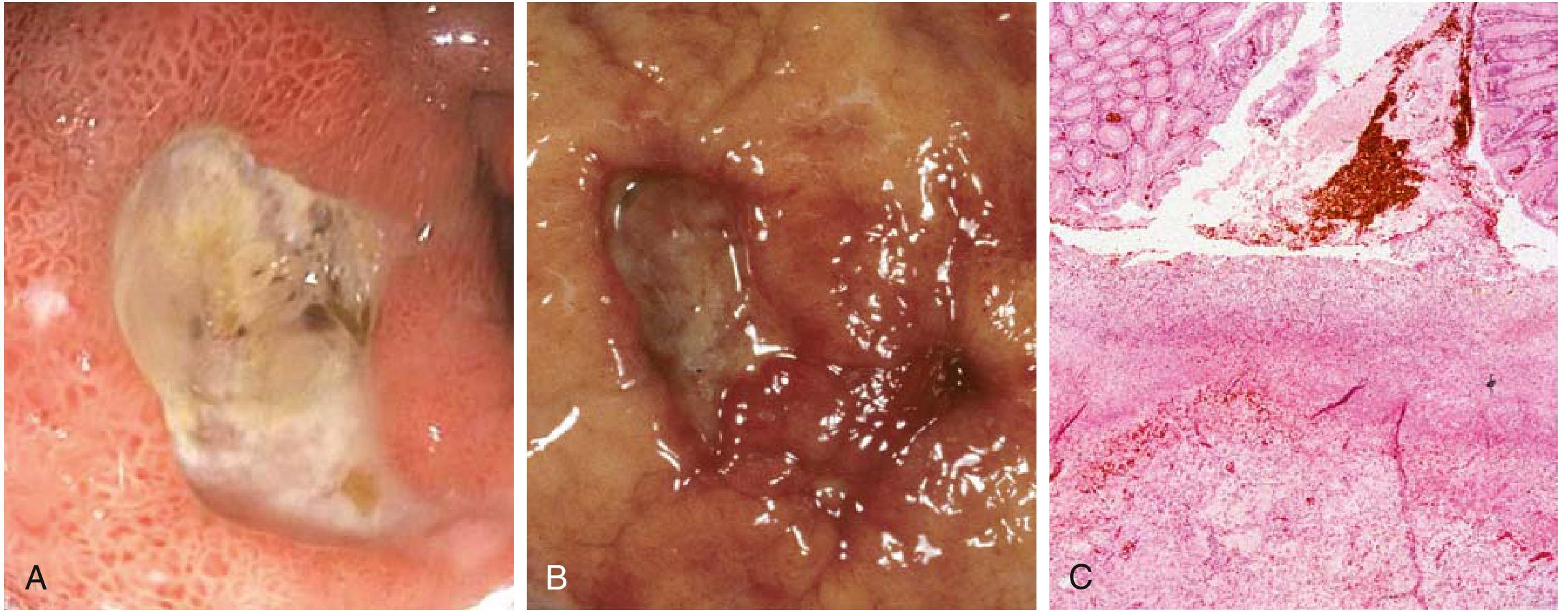
Q3. Complications of Peptic Ulcer Disease
The four major complications are:
1. Hemorrhage (most common)
- Erosion into a submucosal blood vessel leads to bleeding
- Presents as melena (tarry stools) as seen in this patient - from upper GI bleeding
- Massive hemorrhage if a large vessel (e.g., gastroduodenal artery) is eroded
2. Perforation
- Ulcer erodes completely through the bowel wall
- Releases gastric contents into the peritoneal cavity
- Causes acute peritonitis - sudden severe diffuse abdominal pain ("board-like rigidity")
- X-ray shows free air under the diaphragm
3. Pyloric Obstruction / Gastric Outlet Obstruction
- Repeated ulceration and scarring of the pylorus/duodenum causes fibrosis and narrowing
- Presents with projectile vomiting of undigested food, succussion splash, weight loss
4. Penetration
- Ulcer erodes into an adjacent organ (pancreas, liver, biliary tract)
- Posterior duodenal ulcer penetrating the pancreas causes back pain and raised serum amylase/lipase
5. Malignant Transformation
- Gastric ulcers (NOT duodenal) carry a small risk of transformation into gastric adenocarcinoma
- Duodenal ulcers almost never become malignant
Key Teaching Points:
- This patient's melena = hemorrhage complication already present
- H. pylori infection and NSAID use are the two biggest causes of PUD
- Treatment: H. pylori eradication (triple therapy) + proton pump inhibitors (PPIs)
- Robbins notes: "Pain tends to occur 1 to 3 hours after meals during the day, is worse at night, and is relieved by alkali or food" - matching this patient exactly
This is a shared conversation. Sign in to Orris to start your own chat.