
HELP WITH THE CHAPER OF DISTURBANCE OF RESPIRATOY FUNCTIONS FROM HARRISON BOOK CH -285
Reading File
Searching sources...
Finding Sources
Finding Sources
Finding Sources
Finding Sources
Finding Sources
I can see this is in block32. Let me find the exact chapter 285 content on disturbances of respiratory function.
Finding Sources
Finding Sources
Finding Sources
Let me search for the chapter heading directly with the chapter number and look around line 1500-1700 area where HYPOVENTILATION/HYPERVENTILATION was seen, and also look in block31.
Finding Sources
I can see chapter 296 covers Ventilation, Gas Exchange, Pathophysiology and Functional Measurements in block31. Now let me read the actual content starting from "VENTILATION" at line 1654 through "GAS EXCHANGE" in block31, which is the core disturbances of respiratory function chapter.
Reading File
Excellent! I found the chapter. Now let me read more of this content to get the full chapter coverage including Gas Exchange, Pathophysiology, and Functional Measurements.
Reading File
I have enough content. Let me now read the Gas Exchange and Pathophysiology sections from block31 (lines 1739-1840):
Reading File
I now have comprehensive content from Harrison's Chapter on Disturbances of Respiratory Function (Chapter 296 in the 22nd edition). Let me compile this into a complete, well-organized study guide.
Harrison's - Disturbances of Respiratory Function (Ch. 296)
Harrison's Principles of Internal Medicine, 22nd Edition (2025)
Overview
The primary functions of the respiratory system are to oxygenate blood and eliminate CO2. This requires intimate contact between blood and fresh alveolar air across an enormous surface area (~70 m²) packed within a 7 L thoracic cavity. The alveolar membrane is extremely thin (flattened endothelial + epithelial cells), across which gases diffuse and equilibrate.
For normal respiratory function, the system must:
- Ventilate the lung tidally to freshen alveolar gas
- Perfuse alveoli in proportion to their ventilation
- Allow rapid diffusion of gases between alveolar air and capillary blood
- Accommodate several-fold increases in O2 demand during exercise or metabolic derangement
PART 1: VENTILATION
The Three Components of the Respiratory System
Conceptualize the system as three independently functioning parts:
| Component | Description |
|---|---|
| The Lung | Airways and lung parenchyma |
| Neuromuscular system | Respiratory muscles + neural drive |
| Chest wall | Everything else (ribs, diaphragm mechanically, pleura) |
Lung Volumes
- Total Lung Capacity (TLC): Volume at maximal inspiration - when inspiratory muscles exert maximum force
- Residual Volume (RV): Volume remaining after maximal expiration - when expiratory muscles exert maximum force
- Functional Residual Capacity (FRC): The resting volume - where outward chest wall recoil balances inward lung recoil (passive equilibrium, no muscle activity). This is the natural resting position of the respiratory system.
Statics: Volume-Related Mechanical Properties
The lung and chest wall have elastic recoil properties:
- Lung: Always tends to collapse (inward recoil)
- Chest wall: Tends to spring outward at volumes below ~75% TLC
- At FRC, these opposing forces are equal and opposite
- The pressure-volume (P-V) relationship of the lung reflects its compliance (ΔV/ΔP). A stiffer lung (e.g., fibrosis) has reduced compliance; an emphysematous lung has increased compliance.
Dynamic Mechanics: Airflow
During breathing, pressure is needed not just to overcome elastic recoil but also to overcome airflow resistance in the airways.
Key concept: Maximum expiratory flow is effort-independent once effort reaches a threshold. This is because:
- Increased expiratory muscle effort compresses airways (dynamic airway compression)
- Beyond a "choke point," flow is limited by airway properties (caliber, recoil), not effort
- This is why the MEFV (Maximum Expiratory Flow-Volume) curve is reproducible and diagnostically useful
Obstructive disease (COPD, asthma): Expiratory flow is reduced due to airways narrowing and loss of lung recoil. The flow-volume curve shows a "scooped" concave pattern.
Restrictive disease (fibrosis, neuromuscular disease, obesity): All lung volumes are reduced. Flow may be proportionally preserved, but total volume is small.
PART 2: GAS EXCHANGE
A. Diffusion
- The alveolar membrane has a huge surface area and minimal thickness - highly optimized for gas diffusion
- Oxygen uptake is normally perfusion-limited (not diffusion-limited): O2 fully equilibrates between alveolar gas and capillary blood before the RBC completes 1/3 of its journey through the alveolar capillary
- Diffusion limitation can occur in:
- High altitude (low alveolar PO2 gradient)
- Maximal exercise in elite athletes
- Interstitial lung disease (thickened alveolar walls)
B. Ventilation/Perfusion (V/Q) Matching
For maximum efficiency, ventilation (V) to each alveolus must match its perfusion (Q).
Even in health, minor V/Q heterogeneity exists due to gravity and variations in airway/vascular architecture.
Two Extreme V/Q Abnormalities:
| Abnormality | V/Q Ratio | Mechanism | Example |
|---|---|---|---|
| Dead space | Very high (V >> Q) | Ventilated but unperfused alveoli | Pulmonary embolism |
| Shunt | Zero (Q with no V) | Perfused but unventilated alveoli | Consolidation, AVM |
Key Features of Shunt:
- Venous blood passes through lung unaltered (not reoxygenated)
- Mixes with oxygenated blood → lowers mixed arterial PaO2 disproportionately
- Refractory to supplemental O2 (raising FiO2 cannot oxygenate blood through non-ventilated alveoli)
V/Q Mismatch (more common in disease):
- Widening of the distribution of V/Q ratios
- Perfusion of low V/Q units (underventilated) → partial oxygenation → lowers PaO2
- Responds to supplemental O2 (unlike pure shunt), because these alveoli are still ventilated
C. Causes of Arterial Hypoxemia - Summary
| Cause | Mechanism | O2 Response |
|---|---|---|
| Low inspired O2 | Reduced PiO2 (altitude) | Responds to O2 |
| Alveolar hypoventilation | ↑ PaCO2 displaces O2 in alveolar gas | Responds to O2 |
| V/Q mismatch | Low V/Q units → incomplete oxygenation | Responds to O2 |
| Shunt (intrapulmonary or cardiac) | Blood bypasses ventilated lung | Refractory to O2 |
| Diffusion limitation | Thickened membrane (rare) | Usually responds to O2 |
PART 3: PATHOPHYSIOLOGY OF COMMON DISEASES
Alveolar Hypoventilation (Hypoventilation)
- PaCO2 is elevated (>45 mmHg), which displaces O2 in alveolar gas (Alveolar gas equation: PAO2 = PiO2 - PaCO2/R)
- Alveolar-arterial O2 difference (A-a gradient) is normal - hypoxemia is purely due to ↑ CO2
- Causes: CNS depression (drugs, brainstem lesions), neuromuscular disease, obesity (OHS), chest wall deformity
Causes of Elevated PaCO2 (Hypercapnia)
PaCO2 = CO2 production / Alveolar ventilation
Hypercapnia occurs when:
- CO2 production increases AND ventilation fails to compensate
- Alveolar ventilation decreases (dead space ↑ or minute ventilation ↓)
Obesity Hypoventilation Syndrome (OHS)
- BMI ≥30 + PaCO2 >45 mmHg during wakefulness + sleep-disordered breathing
- Mechanism: Increased load on respiratory muscles (excess weight), reduced respiratory drive
- Often coexists with obstructive sleep apnea
Central Hypoventilation Syndrome
- Failure of automatic respiratory drive from brainstem
- Normal voluntary breathing; fails during sleep
- Congenital (PHOX2B mutation) or acquired (brainstem injury)
Hyperventilation
- PaCO2 falls below normal (<35 mmHg)
- Causes: Anxiety/panic, pain, fever, pulmonary embolism, hypoxia stimulating peripheral chemoreceptors, salicylate toxicity, liver failure, early pregnancy
- Symptoms: Perioral and digital tingling (hypocalcemia due to alkalosis), carpopedal spasm, lightheadedness
PART 4: PULMONARY FUNCTION TESTS (PFTs)
Spirometry
| Parameter | Definition | Clinical Use |
|---|---|---|
| FVC | Forced Vital Capacity - total volume exhaled with maximum effort after full inspiration | Reduced in restriction AND obstruction |
| FEV1 | Volume exhaled in first second of FVC maneuver | Reduced in obstruction |
| FEV1/FVC ratio | Proportion exhaled in 1 second | <0.7 = obstruction; normal/↑ = restriction |
| FEF25-75% | Flow rate in middle 50% of FVC | Sensitive for small airways disease |
Lung Volume Measurement
- Body plethysmography or gas dilution techniques
- TLC reduced in restriction; RV and TLC elevated in air trapping/obstruction
Pattern Recognition:
| Pattern | FEV1 | FVC | FEV1/FVC | TLC | RV |
|---|---|---|---|---|---|
| Obstruction | ↓ | Normal or ↓ | ↓ (<0.7) | Normal or ↑ | ↑ |
| Restriction | ↓ | ↓ | Normal or ↑ | ↓ | ↓ or Normal |
| Mixed | ↓ | ↓ | ↓ | ↓ or Normal | Variable |
DLCO (Diffusing Capacity for CO)
- Measures the ability of the lung to transfer gas across the alveolar membrane
- Reduced DLCO with obstruction: emphysema (destroyed alveolar walls)
- Reduced DLCO with restriction: ILD (thickened alveolar walls), pulmonary vascular disease
- Normal or increased DLCO: Asthma, obesity
- Increased DLCO: Polycythemia, pulmonary hemorrhage (extra Hb in alveoli binds CO)
Bronchodilator Response
- Significant if FEV1 or FVC improves by ≥12% AND ≥200 mL after bronchodilator
- Suggests reversible obstruction (asthma > COPD)
PART 5: ASSESSMENT OF VENTILATORY FUNCTION IN DISEASE
Work of Breathing
- Normal breathing requires minimal effort. In disease, the work is increased by:
- ↑ Airway resistance (obstruction)
- ↓ Lung compliance (restriction/fibrosis)
- Dynamic hyperinflation (air trapping in COPD - puts respiratory muscles at mechanical disadvantage)
Dynamic Hyperinflation (COPD)
- Expiratory flow limitation prevents complete exhalation before the next breath
- Auto-PEEP develops: positive alveolar pressure at end-expiration
- Diaphragm becomes flattened (less efficient)
- Sensation of air hunger and increased dyspnea
Hypoxemia vs. Hypercapnia in COPD
- Early COPD: V/Q mismatch → hypoxemia without hypercapnia
- Advanced COPD: Alveolar hypoventilation + V/Q mismatch → both hypoxemia AND hypercapnia
PART 6: KEY EQUATIONS TO KNOW
Alveolar Gas Equation:
PAO2 = FiO2 × (Patm - PH2O) - PaCO2/R where R (respiratory quotient) ≈ 0.8
Alveolar-Arterial (A-a) Gradient:
A-a gradient = PAO2 - PaO2
- Normal: ~10 mmHg in young adults (increases with age: ~4 + age/4)
- Elevated A-a gradient: V/Q mismatch, shunt, diffusion limitation
- Normal A-a gradient with hypoxemia: hypoventilation or low inspired O2
Dead Space Fraction (Bohr equation):
VD/VT = (PaCO2 - PECO2) / PaCO2
- Normal VD/VT ~0.30
- Elevated in pulmonary embolism, COPD
Clinical Pearls for Exams
- Shunt hypoxemia is refractory to 100% O2 - the hallmark distinguishing it from V/Q mismatch
- Pure hypoventilation gives normal A-a gradient; supplemental O2 fully corrects hypoxemia
- DLCO is the key test for emphysema vs. asthma - both obstructive, but DLCO low in emphysema (destroyed alveolar walls) and normal/elevated in asthma
- FRC = passive equilibrium point (no muscle activity) - set by lung vs. chest wall recoil balance
- Dynamic compression makes expiratory flow effort-independent (the basis of the MEFV curve's diagnostic value)
- In V/Q mismatch, high V/Q regions cannot compensate for low V/Q regions' contribution to hypoxemia due to hemoglobin's sigmoidal saturation curve - blood leaving high V/Q alveoli is already near-fully saturated, so extra O2 there is not helpful
- The CO2 dissociation curve is more linear than the oxyhemoglobin curve, so high V/Q regions CAN blow off extra CO2 and compensate for low V/Q hypoventilation - this is why hypercapnia is a late finding in V/Q mismatch (the body compensates with increased ventilation to high V/Q units)
Source: Harrison's Principles of Internal Medicine, 22nd Edition (2025), Chapter 296 - Disturbances of Respiratory Function
ELASIC RECOIL PRESSURE
Elastic recoil pressure is the pressure generated by the elastic properties of the lung (and chest wall) that tends to return them to their resting shape. The lung always wants to collapse inward; the chest wall wants to spring outward. At FRC, these forces balance.
Searching sources...
Reading File
Loading Image
Reading File
Here is a complete, Harrison's-based breakdown of elastic recoil pressure:
Elastic Recoil Pressure - Harrison's Ch. 296
Harrison's Principles of Internal Medicine, 22nd Edition
What Is Elastic Recoil Pressure?
Elastic recoil pressure is the pressure generated by a structure's tendency to return to its natural (unstretched) shape after being deformed. In the respiratory system, two structures produce elastic recoil that work in opposite directions:
| Structure | Direction of Recoil | Tendency |
|---|---|---|
| Lung | Inward (collapses) | Always wants to collapse at all lung volumes |
| Chest wall | Outward (expands) | Wants to spring open at volumes below ~75% TLC |
Why Does the Lung Have Elastic Recoil?
The inward recoil of the lung arises from two sources:
- Surface tension at the air-liquid interface lining the alveolar wall (the dominant factor)
- Elastic tissue within the lung parenchyma itself (collagen and elastin fibers)
Surfactant (produced by type II pneumocytes) dramatically reduces surface tension and prevents alveolar collapse - it directly opposes one component of lung elastic recoil.
Transpulmonary Pressure (Key Formula)
To keep the lung inflated against its own inward recoil, alveolar pressure must exceed pleural pressure:
P_transpulmonary = P_alveolus - P_pleura
- This pressure difference is what holds the lung open
- At rest (FRC), P_pleura is negative (~-5 cmH₂O), which keeps the lung inflated
- The transpulmonary pressure equals the elastic recoil pressure of the lung at any static volume
The Pressure-Volume (P-V) Curve of the Lung
The relationship between elastic recoil pressure and lung volume is not linear - the lung behaves differently at different volumes:
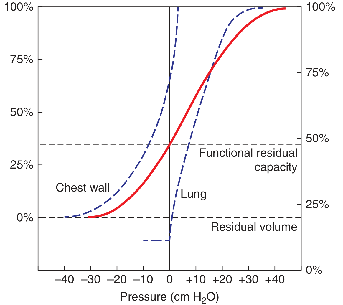
FIGURE 296-1: Pressure-volume curves of the lung (blue dashed) and chest wall (red solid). The intersection at FRC (~35% TLC) is where their opposing recoil pressures are equal.
Reading the Lung Curve (Blue dashed line):
- The lung always requires positive transpulmonary pressure to stay inflated (curve stays to the right of zero)
- At low volumes: the lung is compliant (flat, small pressure change = large volume change) - this is the range of normal tidal breathing
- At high volumes (near TLC): the lung becomes stiff - large pressure changes yield small volume changes
- At zero pressure: some air remains in alveoli (small airways close, trapping gas = the basis of Residual Volume)
Reading the Chest Wall Curve (Red solid line):
- At volumes below ~75% TLC: chest wall recoil is outward (curve is to the left of zero = negative pressure) - it wants to expand
- At volumes above ~75% TLC: chest wall recoil becomes inward - it resists further expansion
- At FRC (~35% TLC): the chest wall recoil is outward (+), lung recoil is inward (-) and they are exactly equal and opposite
FRC: The Passive Equilibrium Point
FRC (Functional Residual Capacity) is the volume at which the system rests with no muscle activity:
FRC is where: Outward chest wall recoil = Inward lung recoil
No muscle force is needed to maintain FRC. The moment you relax completely, you come to rest at FRC automatically.
What Shifts FRC?
| Condition | Effect on FRC | Reason |
|---|---|---|
| Emphysema | ↑ FRC | Lung recoil ↓ (destroyed elastic tissue) → less inward pull, equilibrium shifts outward |
| Pulmonary fibrosis | ↓ FRC | Lung recoil ↑ (stiff fibrotic lung) → more inward pull, equilibrium shifts inward |
| Obesity | ↓ FRC | Weight of chest wall adipose blunts outward chest wall recoil → lung's inward recoil dominates |
| Supine position | ↓ FRC | Abdominal contents push diaphragm up, reducing outward chest wall recoil |
Compliance: The Inverse of Stiffness
Compliance (C) = ΔVolume / ΔPressure (units: L/cmH₂O)
- High compliance (emphysema): lung distends easily, low recoil, flat P-V curve
- Low compliance (fibrosis): stiff lung, high recoil, steep P-V curve
The slope of the P-V curve at any point = compliance at that volume. The P-V curve is steepest (most compliant) in the mid-volume range and flattens at extremes.
Elastic Recoil in Disease - High-Yield Comparisons
Emphysema
- Destruction of alveolar walls → loss of elastic tissue AND reduced surface area → ↓ lung elastic recoil
- P-V curve shifts left (more volume for same pressure)
- FRC ↑, RV ↑, TLC ↑ (barrel chest)
- Small airways lose radial tethering from surrounding parenchyma → collapse during expiration → airflow obstruction even without intrinsic airway disease
Pulmonary Fibrosis (ILD)
- Collagen deposition stiffens lung → ↑ lung elastic recoil
- P-V curve shifts right (less volume for same pressure; requires higher transpulmonary pressure to inflate)
- FRC ↓, TLC ↓, FVC ↓ - restrictive pattern
- Interestingly, MEFV (flow-volume) loops show flows that are actually increased relative to volume because the high recoil drives rapid expiration
Neuromuscular Disease (e.g., Myasthenia Gravis)
- Lung and chest wall recoil are both normal → FRC is normal
- Respiratory muscles too weak to fully inflate (↓ TLC) or deflate (↑ RV) the system
- FVC and FEV1 are low as "innocent bystanders" - airways and parenchyma are unaffected
- DLCO and airway resistance are normal
Key Pearl: Radial Tethering of Airways
Small peripheral airways have no cartilage to hold them open. They stay open because the surrounding inflated lung parenchyma pulls on their adventitia - this is called radial tethering.
- In emphysema: destruction of alveolar walls removes this tethering support → airways collapse during expiration → gas trapping at abnormally high volumes
- In fibrosis: high recoil increases radial tethering → airways are pulled open → flow rates are preserved or increased relative to the reduced lung volumes
Summary Table
| Concept | Definition |
|---|---|
| Elastic recoil pressure | Pressure from a structure's tendency to return to its resting shape |
| Transpulmonary pressure | P_alveolus - P_pleura; holds lung open against its recoil |
| FRC | Passive equilibrium: inward lung recoil = outward chest wall recoil |
| Compliance | ΔV/ΔP; inversely related to stiffness |
| Emphysema | ↓ recoil, ↑ compliance, ↑ FRC, ↑ TLC, airway collapse |
| Fibrosis | ↑ recoil, ↓ compliance, ↓ FRC, ↓ TLC, preserved/increased flows |
| Radial tethering | Inflated parenchyma pulls small airways open; lost in emphysema |
Source: Harrison's Principles of Internal Medicine, 22nd Ed., Ch. 296
This is a shared conversation. Sign in to Orris to start your own chat.