
Fracture, types of fracture phase of healing and complications of fracture
Reading File
This is a broad orthopedic topic. Let me search the library for fracture classification, healing phases, and complications.
Finding Sources
Finding Sources
Finding Sources
Finding Sources
Reading File
Reading File
Reading File
Reading File
Finding Sources
Reading File
Finding Sources
Reading File
Reading File
Reading File
Searching Images
types of bone fracture diagram transverse oblique spiral comminuted greenstick
Searching Images
stages of fracture healing hematoma callus remodeling diagram
query_images_db
query_images_db
Excellent - found a great fracture healing stages diagram. I have enough content now to compose a comprehensive answer.
Searching PubMed
fracture healing complications
These are specific to particular fracture sites, not directly relevant to the general topic. I have enough textbook material to give a thorough answer now.
Fracture: Types, Healing, and Complications
What is a Fracture
A fracture is a break in the structural continuity of bone, caused by trauma exceeding the bone's mechanical tolerance, or (in pathological bone) by forces too small to break normal bone (Bailey and Love's Short Practice of Surgery, p. 444).
Types of Fractures
By completeness:
- Complete - full discontinuity across the bone
- Incomplete - part of the cortex remains intact (greenstick, buckle/torus, plastic bowing - typically in children, whose more elastic bone tolerates greater plastic deformation before breaking) (Grainger & Allison's Diagnostic Radiology)
By fracture line pattern:
- Transverse - line runs perpendicular to the long axis
- Oblique - line runs at an angle
- Spiral - line spirals around the shaft (torsional force)
- Comminuted - more than one fracture line, multiple fragments (includes segmental and butterfly-fragment patterns)
- Greenstick - incomplete break, one side of cortex bends
- Compression/crush - typically in cancellous bone (e.g. vertebral bodies)
- Avulsion - a fragment pulled off by tendon/ligament tension
By skin integrity:
- Closed (simple) - skin intact
- Open (compound) - wound communicates with the fracture site (graded by the Gustilo-Anderson classification)
By cause:
- Traumatic - normal bone, excess force
- Pathological - abnormal bone (tumor, osteoporosis, infection) breaking under normal/minimal load
- Stress fracture - repetitive submaximal loading
By displacement/alignment: described further as displaced or undisplaced, and by angulation, shortening, and rotation.
Formal classification systems (e.g. AO/OTA alphanumeric system, merged with ICD-10 coding) are used clinically to standardize description, guide treatment, and compare outcomes across fracture morphology and location (Campbell's Operative Orthopaedics, p. 58).
Phases of Fracture Healing
Bone heals by two mechanisms (Bailey and Love's Short Practice of Surgery, p. 444-445):
1. Direct (primary) bone healing - occurs only with absolute stability and cortical apposition (e.g. rigid internal fixation with compression), with no callus. Osteoclastic "cutting cones" tunnel across the fracture line followed by osteoblasts laying down lamellar bone directly - essentially normal bone remodeling redirected across the fracture.
2. Indirect (secondary) bone healing - the more common pathway, proceeding through four sequential stages:
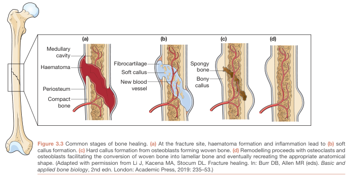
- Stage 1 - Haematoma formation/inflammation: Bleeding from the fracture ends and surrounding soft tissue fills the gap with a haematoma; an inflammatory response begins, bringing in cytokines and cells that initiate repair.
- Stage 2 - Soft callus formation: The haematoma organizes into fibrous tissue/fibrocartilage; new blood vessels invade, and a soft, pliable callus bridges the fragments.
- Stage 3 - Hard callus formation: The soft callus mineralizes into woven (immature) bone, forming a rigid bony callus that bridges the fracture - this is radiographic "union."
- Stage 4 - Remodelling: The bony callus is progressively remodeled by osteoclasts/osteoblasts into mature lamellar bone, restoring the normal cortical shape and medullary canal, guided by the mechanical forces the bone experiences (Wolff's law).
The strain at the fracture site determines the tissue that forms (Perren's strain theory): >100% strain -> fibrous tissue only; <10% -> soft callus; <2% -> hard callus with mineralization. Some interfragmentary movement promotes healing, but too much prevents it. Adequate blood supply (periosteal and nutrient artery) and biological environment are just as important as mechanics - conditions impairing perfusion (vascular disease, diabetes, extensive soft-tissue injury, infection) impair healing.
Key terminology of healing outcomes:
- Union - clinically and radiologically healed enough to bear physiological load
- Delayed union - healing slower than the expected time frame
- Non-union - no healing progress over 3 months (formally labeled at 6 months); subtypes are atrophic (biological problem - poor blood supply/stimulus), hypertrophic (mechanical problem - excess movement), and infected
- Consolidation - bone has returned to full strength, cortex normalized on imaging
- Malunion - healed in a non-anatomical (deformed) position
Complications of Fractures
Classically divided into immediate, early, and late (Pye's Surgical Handicraft, p. 152-154; Bailey and Love's Short Practice of Surgery):
Immediate (at time of injury):
- Haemorrhage (external or internal - can be severe and concealed in pelvic fractures)
- Injury to internal organs (e.g. bladder/urethra with pelvic fracture, lung/spleen/liver with rib fractures)
- Injury to local nerves or vessels (vascular injury risking ischaemia; nerve stretch/contusion)
- Skin loss or damage, and fracture into a joint
Early (during initial treatment period) - local:
- Skin and soft-tissue necrosis
- Ischaemia from vascular damage or external pressure (compartment syndrome, Volkmann's ischaemic contracture - classically after supracondylar humeral fractures in children)
- Pressure sores and nerve palsies from splints/casts/traction (e.g. common peroneal nerve palsy from a Thomas splint)
- Infection and wound breakdown (particularly with open fractures or internal fixation)
Early - general (systemic):
- Fat embolism syndrome
- Deep vein thrombosis and pulmonary embolism
- Crush syndrome/rhabdomyolysis
- Adult respiratory distress syndrome
- Tetanus (in contaminated open wounds)
Late (after initial treatment, may appear years later):
- Delayed union / non-union (atrophic, hypertrophic, or infected)
- Malunion
- Avascular necrosis (e.g. femoral head after neck of femur fracture, scaphoid, talus)
- Myositis ossificans
- Joint stiffness and contractures
- Post-traumatic osteoarthritis
- Growth disturbance (in children, if the growth plate is involved - Salter-Harris injuries)
- Complex regional pain syndrome (Sudeck's atrophy)
- Chronic osteomyelitis (following infected open fractures)
Note on Pediatric Fractures
Children's more elastic, cartilage-rich bones produce distinct injury patterns (greenstick, buckle/torus, plastic bowing) and growth plate (physeal) injuries classified by the Salter-Harris system, where higher grades carry a greater risk of growth disturbance (Grainger & Allison's Diagnostic Radiology).
My quick pubmed check for recent evidence returned only site-specific systematic reviews (olecranon, patella, metacarpal fracture complications) rather than anything that revises this general framework, so the classic teaching above remains current.
This is a shared conversation. Sign in to Orris to start your own chat.