
Acid base disorder not able to understand
Reading File
Now let me search the medical library for comprehensive content on acid-base disorders.
Finding Sources
Finding Sources
Good results. Let me read the most comprehensive sections - Rosen's Emergency Medicine acid-base content and the compensation formulas section.
Reading File
Reading File
Finding Sources
Now let me read the core Rosen's acid-base content and the Barash anesthesia ABG section for a clear clinical framework.
Reading File
Reading File
Reading File
Finding Sources
I have rich content from the textbooks. Let me also fetch the key diagram image and read the Rosen's detailed differential section.
Loading Image
Reading File
I now have more than enough content from authoritative textbooks to write a thorough, clear explanation. Let me compose the complete answer.
Acid-Base Disorders - A Complete Breakdown
The Big Picture First
The body keeps blood pH tightly between 7.35 - 7.45. Two systems control this:
- Kidneys - regulate bicarbonate (HCO₃⁻) - slower, takes 2-5 days
- Lungs - regulate CO₂ - faster, acts within 12-24 hours
The Henderson-Hasselbalch equation ties it all together:
pH = pK + log ([HCO₃⁻] / [0.03 × PaCO₂])
When one system fails, the other compensates to pull pH back toward normal - but never completely to normal, and never past normal.
The 4 Primary Disorders
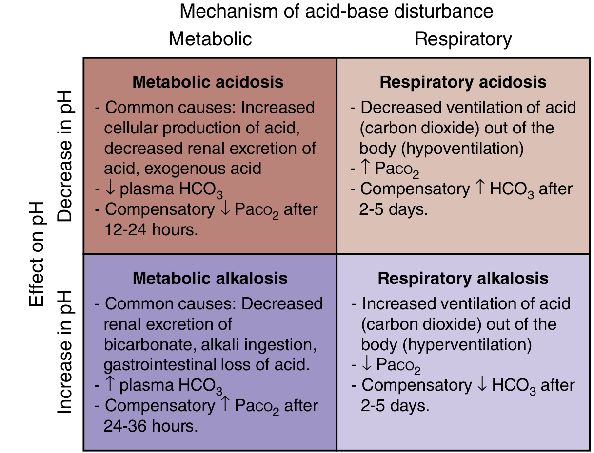
1. Metabolic Acidosis
Definition: Low pH + Low HCO₃⁻ (kidneys losing or not retaining base)
Compensation: Lungs blow off CO₂ (hyperventilation) - kicks in within 12-24 hours
Formula: Expected PaCO₂ = (1.5 × HCO₃⁻) + 8 ± 2 (Winters' formula), or simply: ΔPaCO₂ = ΔHCO₃⁻ × 1.2
Key Step - Calculate the Anion Gap:
AG = Na⁺ - (Cl⁻ + HCO₃⁻) | Normal = ~12 mEq/L
| Type | Anion Gap | Pathology |
|---|---|---|
| High AG (>15) | Elevated | Unmeasured anions accumulate |
| Normal AG (hyperchloremic) | Normal | HCO₃⁻ lost, Cl⁻ rises to compensate |
Causes of High AG Metabolic Acidosis - Mnemonic "MUDPILES":
- M - Methanol
- U - Uremia
- D - DKA (Diabetic ketoacidosis)
- P - Paraldehyde / Paracetamol (acetaminophen)
- I - Iron / Isoniazid
- L - Lactic acidosis
- E - Ethylene glycol
- S - Salicylates
Causes of Normal AG Metabolic Acidosis - Mnemonic "HARDUP":
- H - Hyperalimentation / Hospital saline
- A - Acid infusion / Addison's disease / Acetazolamide
- R - Renal tubular acidosis (RTA)
- D - Diarrhea (loses HCO₃⁻)
- U - Ureterosigmoidostomy
- P - Pancreatic fistula/drainage
2. Metabolic Alkalosis
Definition: High pH + High HCO₃⁻ (acid lost or base gained)
Compensation: Lungs retain CO₂ (hypoventilation) - takes 24-36 hours
Formula: ΔPaCO₂ = ΔHCO₃⁻ × 0.7 (PaCO₂ rarely rises above 60 mmHg even with very high HCO₃⁻)
Common Causes:
- Vomiting / NG tube suction (loss of HCl)
- Diuretic use (loop/thiazide - lose Cl⁻, K⁺)
- Hyperaldosteronism
- Alkali ingestion (antacid overuse)
- Post-hypercapnic state
Important: Metabolic alkalosis is associated with volume depletion and hypokalemia. GI-based chloride losses respond to IV normal saline. Mineralocorticoid-driven cases do not respond to saline (called "chloride-resistant").
3. Respiratory Acidosis
Definition: Low pH + High PaCO₂ (CO₂ not cleared = hypoventilation)
Compensation: Kidneys retain HCO₃⁻
| Timing | Formula |
|---|---|
| Acute | ΔHCO₃⁻ = ΔPaCO₂ × 0.1 (rises ~1 mEq/L per 10 mmHg CO₂ rise) |
| Chronic | ΔHCO₃⁻ = ΔPaCO₂ × 0.4 (rises ~3.5 mEq/L per 10 mmHg CO₂ rise) |
Common Causes:
- COPD / severe asthma (lung disease)
- Opioid/sedative overdose (reduced respiratory drive)
- Neuromuscular disease (ALS, Guillain-Barre, myasthenia)
- Chest wall deformity (obesity hypoventilation, kyphoscoliosis)
- Central sleep apnea
4. Respiratory Alkalosis
Definition: High pH + Low PaCO₂ (hyperventilation = too much CO₂ blown off)
Compensation: Kidneys excrete HCO₃⁻ (most effective compensator - pH can sometimes return to normal)
| Timing | Formula |
|---|---|
| Acute | ΔHCO₃⁻ = ΔPaCO₂ × 0.2 |
| Chronic | ΔHCO₃⁻ = ΔPaCO₂ × 0.5 |
Common Causes:
- Anxiety / panic attack (most common)
- Pain
- Pregnancy (progesterone stimulates respiration)
- High altitude
- Pulmonary embolism
- Salicylate toxicity (early - directly stimulates respiratory center)
- Sepsis (early)
- Liver failure
Step-by-Step ABG Interpretation (5-Step Approach)
Given: pH, PaCO₂, HCO₃⁻ (from basic metabolic panel + blood gas)
Step 1: Check the pH
- pH < 7.35 → Acidemia
- pH > 7.45 → Alkalemia
- pH 7.35-7.45 → Normal (but a mixed disorder may still exist!)
Step 2: Identify the primary disorder
| pH | Primary Change | Disorder |
|---|---|---|
| Low pH | Low HCO₃⁻ | Metabolic acidosis |
| Low pH | High PaCO₂ | Respiratory acidosis |
| High pH | High HCO₃⁻ | Metabolic alkalosis |
| High pH | Low PaCO₂ | Respiratory alkalosis |
Step 3: Check for appropriate compensation
Use the formulas above. If compensation is MORE or LESS than expected, a second primary disorder is present (mixed disorder).
Step 4: Calculate Anion Gap (if metabolic acidosis)
AG = Na⁺ - (Cl⁻ + HCO₃⁻). Normal = ~12.
Step 5: If high AG - calculate the Delta-Delta (Delta Gap)
Used to detect a hidden concurrent metabolic alkalosis or normal-AG acidosis:
Delta Gap = (Measured AG - 12) - (24 - Measured HCO₃⁻)
- Delta gap > +6 → Concurrent metabolic alkalosis hiding behind the AG acidosis
- Delta gap < -6 → Concurrent normal-AG acidosis also present
Compensation Formulas - Quick Reference Table
| Primary Disorder | Expected Compensation |
|---|---|
| Metabolic acidosis | PaCO₂ = ΔHCO₃⁻ × 1.2 |
| Metabolic alkalosis | PaCO₂ = ΔHCO₃⁻ × 0.7 (max ~60 mmHg) |
| Acute resp. acidosis | HCO₃⁻ = ΔPaCO₂ × 0.1 |
| Chronic resp. acidosis | HCO₃⁻ = ΔPaCO₂ × 0.4 |
| Acute resp. alkalosis | HCO₃⁻ = ΔPaCO₂ × 0.2 |
| Chronic resp. alkalosis | HCO₃⁻ = ΔPaCO₂ × 0.5 |
Worked Example
ABG: pH = 7.28, PaCO₂ = 20, HCO₃⁻ = 9, Na = 140, Cl = 105
Step 1: pH 7.28 → Acidemia
Step 2: Low HCO₃⁻ → Primary metabolic acidosis
Step 3: Expected PaCO₂ = 1.5 × 9 + 8 = 21.5 ± 2. Measured = 20 ✓ Appropriate compensation (simple metabolic acidosis)
Step 4: AG = 140 - (105 + 9) = 26 → High AG metabolic acidosis
Step 5: Delta gap = (26 - 12) - (24 - 9) = 14 - 15 = -1 → No additional hidden disorder. Pure high-AG metabolic acidosis.
Think: MUDPILES. Lactic acidosis? DKA? Renal failure?
Simple Memory Framework
| disorder | pH | Primary change | Compensation |
|---|---|---|---|
| Metabolic acidosis | ↓ | ↓ HCO₃⁻ | ↓ PaCO₂ (hyperventilate) |
| Metabolic alkalosis | ↑ | ↑ HCO₃⁻ | ↑ PaCO₂ (hypoventilate) |
| Respiratory acidosis | ↓ | ↑ PaCO₂ | ↑ HCO₃⁻ (kidneys retain) |
| Respiratory alkalosis | ↑ | ↓ PaCO₂ | ↓ HCO₃⁻ (kidneys excrete) |
Key rule: In compensation, PaCO₂ and HCO₃⁻ always move in the same direction. If they move in opposite directions, you are looking at a mixed disorder - guaranteed.
Sources: Rosen's Emergency Medicine (9th ed.), Henry's Clinical Diagnosis and Management by Laboratory Methods, Barash Clinical Anesthesia (9th ed.), Comprehensive Clinical Nephrology (7th ed.)
This is a shared conversation. Sign in to Orris to start your own chat.