
External ear anatomy make it easier
Reading File
Searching sources...
Finding Sources
Searching the Web
external ear anatomy diagram auricle pinna parts labeled
Reading File
Loading Image
Loading Image
Also read the textbook section on gross anatomy of the auricle/temporal bone for fuller coverage:
Reading File
Here is a clear, easy-to-follow breakdown of external ear anatomy:
External Ear Anatomy - Made Easy
The external ear has 2 main parts: the Auricle (Pinna) and the External Auditory Canal (EAC), ending at the Tympanic Membrane.
Visual Reference
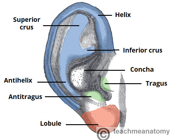
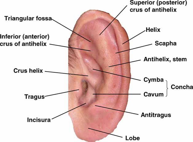
1. The Auricle (Pinna)
The visible part of the ear projecting from the side of the head. It is made of elastic cartilage covered by skin (except the lobule, which is just fat and connective tissue - no cartilage).
Key Landmarks (outer to inner):
| Structure | What it is | Easy Memory Tip |
|---|---|---|
| Helix | The curved outer rim of the ear | "H for Hat brim" - the outermost edge |
| Antihelix | Curved ridge inside/parallel to helix | The Y-shaped ridge running inward |
| Superior crus | Upper branch of antihelix | Splits upward at the top |
| Inferior crus | Lower branch of antihelix | Splits downward |
| Triangular fossa | Depression between the two crura | Triangle-shaped pit at top |
| Scapha | Groove between helix and antihelix | "Scapha = scoop/groove" |
| Concha | Deep bowl-shaped hollow in the center | Leads directly into the ear canal |
| Cymba conchae | Upper part of concha (above crus helix) | |
| Cavum conchae | Lower part of concha (below crus helix) | Opens into the EAC |
| Crus helix | Ridge of helix crossing the concha | Divides the concha in two |
| Tragus | Small cartilage bump in front of ear canal | Flap you push to block sound |
| Antitragus | Small bump opposite the tragus | Across from the tragus |
| Incisura (intertragic notch) | Notch between tragus and antitragus | The gap between the two bumps |
| Lobule (earlobe) | Soft lower tip | Only part with NO cartilage |
2. External Auditory Canal (EAC)
The tube leading from the auricle to the eardrum. About 2.5 cm long in adults.
It has 2 portions:
| Portion | Location | Contents |
|---|---|---|
| Outer 1/3 - Cartilaginous | Lateral | Hair follicles, sebaceous glands, ceruminous (wax) glands |
| Inner 2/3 - Bony | Medial | Thin squamous epithelium, no glands or hair |
Key features:
- Isthmus = narrowest point, at the bony-cartilaginous junction
- Cerumen (earwax) = produced in the outer 1/3; slightly acidic (pH 6.0-6.5), hydrophobic - acts as a protective barrier
- Fissures of Santorini = small slits in the cartilaginous canal; can allow infection/tumor to spread to surrounding tissues
- Foramen of Huschke = a small defect in the anterior bony canal; can allow disease to spread to the parotid gland
Self-cleaning mechanism:
Shed epithelium from the eardrum migrates outward toward the opening, carrying debris with it as cerumen. This is called epithelial migration - nature's built-in cleaning system.
3. Tympanic Membrane (the endpoint)
The eardrum sits at the end of the EAC and marks the boundary between the external and middle ear. The EAC's squamous epithelium is continuous with its lateral (outer) surface.
Innervation - Quick Summary
| Nerve | Area supplied |
|---|---|
| Auriculotemporal nerve (CN V3) | Anterior auricle, anterior EAC, anterior TM |
| Greater auricular nerve (C2, C3) | Posterior/inferior auricle |
| Lesser occipital nerve (C2) | Superior/posterior auricle |
| Auricular branch of vagus (Arnold's nerve) (CN X) | Posterior EAC and TM |
| Facial nerve (CN VII) | Small contribution to posterior EAC |
Clinical pearl: Stimulation of the Arnold's nerve (CN X branch in the EAC) can trigger a cough reflex - this is why cleaning ears with a cotton swab sometimes causes coughing!
Blood Supply - Quick Summary
- Anterior auricle: Superficial temporal artery (branch of ECA)
- Posterior auricle: Posterior auricular artery (branch of ECA)
- Deep auricular artery: Supplies the EAC and TM
Quick Recall Summary
EXTERNAL EAR
├── Auricle (Pinna)
│   ├── Helix (outer rim)
│   ├── Scapha (groove between helix & antihelix)
│   ├── Antihelix (Y-shaped inner ridge)
│   │   ├── Superior crus
│   │   └── Inferior crus
│   ├── Triangular fossa (between crura)
│   ├── Concha (central bowl → EAC)
│   │   ├── Cymba (above crus helix)
│   │   └── Cavum (below crus helix)
│   ├── Tragus (front flap)
│   ├── Antitragus (opposite to tragus)
│   └── Lobule (no cartilage!)
│
├── External Auditory Canal (2.5 cm)
│   ├── Outer 1/3: Cartilaginous (wax here)
│   └── Inner 2/3: Bony (no glands)
│
└── Tympanic Membrane (boundary with middle ear)
This video tutorial is a great visual companion:
Source: Cummings Otolaryngology Head and Neck Surgery; TeachMeAnatomy; NIH Elements of Morphology
This is a shared conversation. Sign in to Orris to start your own chat.