
Relevant history of skin tightening in systemic Sclerosis
"scleroderma, systemic"[MeSH Terms] AND "skin"[MeSH Terms]
systemic sclerosis sclerodactyly skin tightening hands
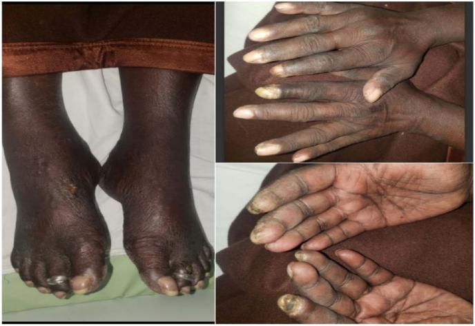
Clinical photograph series of the hands and feet demonstrating dermatologic manifestations of systemic sclerosis (scleroderma). The images show diffuse hyperpigmentation of the extremities with associated sclerodactyly, characterized by taut, thickened skin over the digits. In the dorsal view of the hands, there is notable skin tightening and loss of normal skin creases. The palmar and dorsal views reveal impending digital gangrene, specifically visible as dark, necrotic-appearing lesions at the distal tips of the index fingers bilaterally, indicative of severe Raynaud phenomenon and digital ischemia. The lower extremity photograph shows bilateral diffuse pedal hyperpigmentation and a distinct cutaneous ulcer on the dorsal surface of the right foot. The toes exhibit similar skin tightening and trophic changes. These findings are hallmarks of vascular and fibrotic complications in systemic sclerosis, emphasizing the risk of digital infarction and impaired wound healing in this patient population.
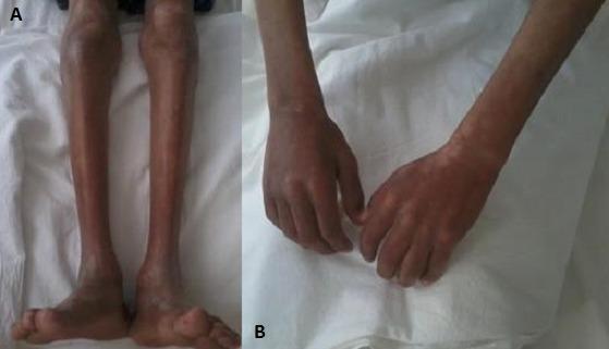
Two-panel clinical photograph demonstrating cutaneous and musculoskeletal manifestations of systemic sclerosis (scleroderma). Image (A) shows the lower limbs with significant cutaneous sclerosis, characterized by skin tightening and thickening. Notable bilateral amyotrophy (muscle wasting) is present, evidenced by a marked reduction in calf circumference and prominence of bony landmarks. Image (B) displays the upper limbs and hands, exhibiting similar skin sclerosis and severe forearm muscle atrophy. The skin in this region also demonstrates salt-and-pepper dyspigmentation, with areas of patchy hyperpigmentation and hypopigmentation. The hands show fixed, slightly flexed posturing consistent with sclerodactyly. These findings are clinically relevant for diagnosing connective tissue diseases and evaluating the extent of skin involvement (modified Rodnan skin score assessment) and secondary muscular complications.
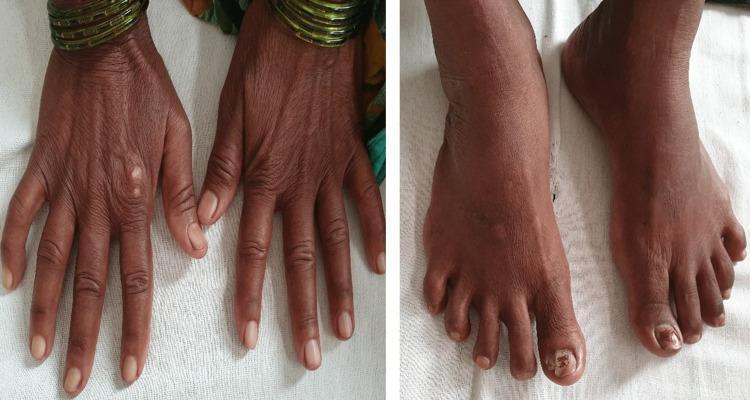
Two clinical photographs demonstrate dermatological manifestations of a systemic connective tissue disease, likely systemic sclerosis. The left panel shows the dorsal aspect of both hands, characterized by significant skin thickening and a taut, bound-down texture. The fingers exhibit tapering and fixed flexion contractures consistent with sclerodactyly. Subtle 'salt and pepper' pigmentary changes (hypopigmentation and hyperpigmentation) are visible on the dorsal surface. The right panel displays the dorsal aspect of the feet, which show similar skin tightening and thickening extending to the toes. Notable findings include xerosis (dryness) and onychodystrophy of the great toenails, suggestive of secondary fungal infection. These visual signs are classic hallmarks of the fibrotic phase of scleroderma, highlighting the loss of skin elasticity and characteristic digital changes used in the clinical diagnosis of CREST syndrome or systemic sclerosis.
Natural history of the skin disease
- Raynaud phenomenon comes first. It is not specific to SSc, but it often precedes skin change, sometimes by many years. It is the first manifestation in more than half of patients. - GOLDMAN-CECIL MEDICINE, Cutaneous Manifestations; Andrews' Diseases of the Skin, Progressive Systemic Sclerosis, p. 201
- Early phase. Patients typically present with swollen digits, then tight hands, then tight skin. The skin is initially erythematous and swollen ("woody" edema). - GOLDMAN-CECIL MEDICINE; Andrews' Diseases of the Skin
- Carpal tunnel misdiagnosis. Early swelling is often misdiagnosed as carpal tunnel syndrome, and EMG may even be positive. Raynaud phenomenon is the clue to the right diagnosis. - Andrews' Diseases of the Skin
- Sclerotic phase. The skin becomes smooth, yellowish and firm, and is bound down to underlying structures. Sclerodactyly develops, with a pinched nose, thin radially furrowed lips and a small mouth.
- Late or atrophic phase. Features include pigment change ("salt and pepper"), hair loss, telangiectasia, calcinosis and digital ulcers. - Andrews' Diseases of the Skin; Firestein & Kelley's Rheumatology, Ch. 111, which describes an edematous phase, then sclerodactyly with increased thickness, then an atrophic phase
Questions to ask in the history
- Where did the tightness start, and where is it now?
- Did the fingers swell or puff first?
- Is it distal only (hands, feet, face, forearms) or proximal to the MCP/MTP joints? Proximal involvement is the key classification feature.
- Has it spread to the trunk or legs? This points to diffuse disease.
- How fast is the change? Rapid progression in the first few years predicts internal organ risk.
- Can the patient make a fist, open the mouth fully, or flex the fingers?
- Is there itch, pigment change, or hair loss?
- Raynaud phenomenon: when did it start, and does it show the three color phases (pallor, cyanosis, hyperemia)?
- Digital pits, fingertip ulcers or gangrene, painful fingertips?
- Telangiectasia?
- Esophagus and gut: reflux, dysphagia, early satiety, bloating, diarrhea or constipation. Esophageal involvement occurs in more than 90% of patients.
- Lung: exertional dyspnea, dry cough. Interstitial lung disease and pulmonary hypertension are the main concerns.
- Cardiac: palpitations, edema.
- Renal: new headache, visual change, high blood pressure. Scleroderma renal crisis is the concern.
- Musculoskeletal: joint pain, weakness, tendon friction rubs.
- Sicca symptoms.
- Patchy, localized hardening suggests morphea.
- Other causes of skin thickening should be considered:
- Scleromyxedema.
- Nephrogenic systemic fibrosis, if there is renal failure and gadolinium exposure.
- Eosinophilic fasciitis, if onset followed exertion.
- Diabetic cheiroarthropathy.
- Scleredema.
- Chronic graft-versus-host disease.
- Ask about exposures such as silica, solvents, vinyl chloride and bleomycin.
- Ask about family history of autoimmune disease.
- Ask about age and sex. Women are affected about 3 times more often than men, with peak onset in the 3rd to 5th decades.
How the history guides subtype
| Feature | Limited cutaneous (CREST) | Diffuse cutaneous |
|---|---|---|
| Skin involvement | Hands, feet, face, forearms | Also proximal limbs and trunk |
| Raynaud phenomenon | Long-standing, precedes skin change by years | Short interval before skin change |
| Antibody | Anticentromere | Anti-topoisomerase I (Scl-70), anti-RNA polymerase |
| Organ risk | Pulmonary hypertension | Interstitial lung disease, renal crisis |
Examination and classification
- Skin thickness is scored with the modified Rodnan skin score, which correlates with severity. Its rate of progression can predict survival.
- Look for the following signs:
- Round finger-pad sign.
- Digital pitting.
- Neck ridging.
- Dilated nailfold capillaries (about 75% of patients).
- Nailfold hemorrhages.
- Pterygium inversum unguis.
- Skin thickening proximal to the MCP joints is the major ACR criterion.
- The 2013 ACR/EULAR criteria also score telangiectasia, abnormal nailfold capillaries, pulmonary involvement, Raynaud phenomenon and SSc-specific antibodies.
- Some patients have "sine scleroderma", with internal organ disease and no skin thickening.
Recent evidence flag
- A 2025 review on whether SSc and morphea coexist (PMID 41043651).
- A 2024 review of regenerative treatments for facial skin involvement (PMID 38976297).