
Give me a detailed 10 marker for shoulder impingement with types of acromian with diagrams
Reading File
Now let me search the medical library for shoulder impingement and acromial types simultaneously.
Finding Sources
Finding Sources
Searching the Web
shoulder impingement syndrome types of acromion Neer classification diagram
Good findings. Now let me read the textbook content on shoulder impingement and fetch the acromion types diagram simultaneously.
Reading File
Reading File
Loading Image
Loading Image
Excellent! I have the acromion X-ray images. Now let me get more detailed content from Campbell's and also fetch more textbook images.
Reading File
Reading File
Loading Image
I have the key diagram URL from Campbell's. Let me fetch the acromion morphology diagram and get the clinical features/management sections.
Loading Image
Reading File
Finding Sources
I now have all the core material needed. Let me get the clinical tests table and Neer stages from Campbell's.
Reading File
Finding Sources
I now have enough comprehensive material. Let me compile the full detailed 10-marker answer.
Shoulder Impingement Syndrome - Detailed 10 Marker
Definition
Shoulder impingement syndrome (SIS) is defined as the encroachment of the acromion, coracoacromial ligament, coracoid process, or acromioclavicular (AC) joint on the rotator cuff tendons as they pass beneath the coracoacromial arch during glenohumeral motion. The supraspinatus tendon is most commonly affected, compressed between the greater tuberosity of the humerus and the anteroinferior surface of the acromion.
Originally described by Neer in 1972 after 100 anatomic shoulder dissections, it remains one of the most common causes of non-traumatic shoulder pain.
Anatomy of the Subacromial Space
The subacromial space is bounded:
- Superiorly: Acromion, AC joint, coracoacromial ligament (forming the coracoacromial arch)
- Inferiorly: Humeral head and greater tuberosity
- Contents: Supraspinatus tendon, subacromial bursa, long head of biceps tendon
Normal subacromial space = 9-10 mm. Narrowing below 7 mm is considered pathological.
Types of Acromion (Bigliani/Morrison Classification)
The shape of the acromion plays a major role in impingement. Bigliani and Morrison (1986) classified acromion morphology into three types based on lateral shoulder X-ray (outlet view):
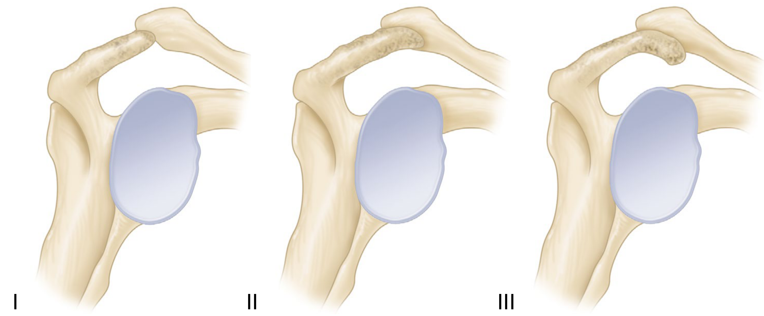
Figure: Three types of acromion morphology. Type I has the least compromise of the supraspinatus outlet; Type III has the highest rate of rotator cuff pathology. (Campbell's Operative Orthopaedics 15th Ed, Fig. 51.24)
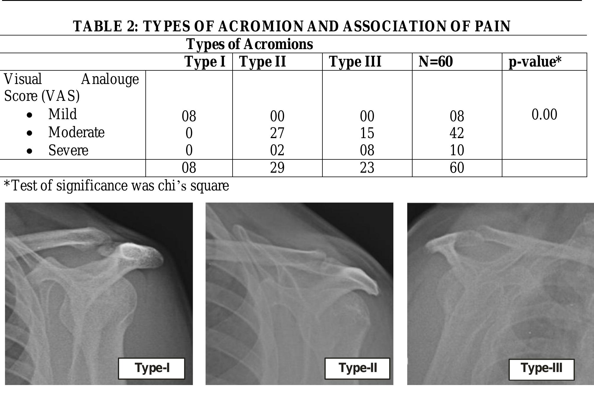
| Type | Shape | Description | Impingement Risk |
|---|---|---|---|
| Type I | Flat | Flat undersurface, normal slope | Lowest - rarely associated with impingement |
| Type II | Curved | Gentle concave curve on undersurface | Moderate - most common type (45%), moderately associated with tears |
| Type III | Hooked | Anterior hook (slope 43°, acromial angle <70°) | Highest - strongly associated with rotator cuff tears and severe impingement |
A Type IV (convex) acromion has also been described but is less widely used clinically.
Key notes:
- Genetics play a role in subacromial space shape
- Type III hooked acromions have a reduced supraspinatus outlet and highest rate of rotator cuff pathology
- Acromions with less slope have a propensity toward impingement
- Anterior acromial spurs (traction osteophytes from coracoacromial ligament) can convert a Type I/II into an effectively Type III morphology
Types of Impingement
1. Primary (External) Impingement
Classic Neer-type impingement. Subdivided into:
- Intrinsic primary: Structures passing beneath the arch enlarge (e.g., rotator cuff thickening, calcium deposits, subacromial bursal thickening) - they abut the arch from below
- Extrinsic primary: Space above the cuff is diminished (subacromial spurring, acromial fracture, os acromiale, AC joint osteophytes, greater tuberosity exostoses)
2. Secondary Impingement
Caused by glenohumeral instability allowing abnormal translation of the humeral head (typically anteriorly), bringing the rotator cuff into contact with the coracoacromial arch. Common in younger patients with lax capsules or labral tears.
3. Internal Impingement
Contact of the articular surface of the rotator cuff with the posterosuperior glenoid rim occurs when the arm is in abduction, extension, and external rotation (the "cocked" throwing position). This is a normal phenomenon that becomes pathological in overhead athletes. Associated with glenohumeral internal rotation deficit (GIRD).
4. Subcoracoid Impingement
Impingement between the subscapularis tendon/lesser tuberosity and the coracoid process. Less common; associated with coracoid morphology or post-surgical changes.
Neer's Stages of Impingement (Pathological Progression)
| Stage | Pathology | Typical Age | Reversibility |
|---|---|---|---|
| Stage I | Oedema and haemorrhage in supraspinatus tendon/bursa | <25 years | Fully reversible with conservative treatment |
| Stage II | Fibrosis and tendinitis; irreversible changes begin | 25-40 years | Partial - may need surgery |
| Stage III | Tendon rupture (partial or full thickness), bone changes, anterior acromial spur | >40 years | Irreversible - surgery often required |
Aetiology / Risk Factors
Extrinsic (structural) factors:
- Type III (hooked) acromion
- Acromial osteophytes/spurs
- AC joint osteophytes impinging from above
- Os acromiale
- Subacromial narrowing
Intrinsic (tendon) factors:
- Age-related tendon degeneration: decreased cellularity, fascicular thinning, hypovacularity, dystrophic calcification - likely irreversible
- Degenerative tears associated with narrower subacromial space, larger lateral extension, steeper acromion angulation
- Shear stress from repetitive overhead activities
Dynamic/functional factors:
- Rotator cuff weakness leading to loss of force couples → superior humeral head translation
- Scapular dyskinesia (reduced scapular upward rotation narrows the outlet)
- Kyphosis reducing shoulder elevation
- Overhead sports (swimmers, throwers, racquet sports)
Clinical Features
Symptoms:
- Anterolateral shoulder pain, worsened by overhead activity
- Pain at night, difficulty lying on the affected shoulder
- "Painful arc" - pain between 60° and 120° of abduction
Signs:
- Tenderness over anterolateral acromion and supraspinatus insertion on greater tuberosity
- Painful arc of abduction (60°-120°)
- Weakness in abduction and external rotation (if cuff involved)
- Muscle wasting (supraspinatus/infraspinatus fossa) in chronic cases
Clinical Tests
| Test | Technique | Sensitivity | Specificity | Notes |
|---|---|---|---|---|
| Neer's sign | Passive forced forward flexion of arm (scapula stabilised) - pain = positive | 68% | 68.7% | Positive in all stages |
| Hawkins-Kennedy sign | Arm at 90° forward flexion, elbow flexed 90°, internal rotation - pain = positive | 71.5% | 66.3% | Most sensitive |
| Painful arc sign | Active abduction 60°-120° causes pain | 73.5% | 81.1% | High overall accuracy |
| Jobe (Supraspinatus) test | Arm at 90° abduction, 30° forward flexion, thumb down, resist downward force | 44.1% | 89.5% | Highly specific |
| Speed test | Resist forward flexion with elbow extended, forearm supinated | 38.3% | 83.3% | For bicipital involvement |
Clinical pearl (Campbell's): If Hawkins-Kennedy + painful arc + infraspinatus (Jobe) test are ALL positive, likelihood of impingement >95%. If all three are negative, likelihood <24%.
Neer's Impingement Test (injection test): Injection of 10 mL lidocaine into subacromial space - if pain is abolished, impingement is confirmed (distinguishes from glenohumeral pathology).
Investigations
Radiographs (X-ray):
- AP and lateral (outlet) views - outlet view best shows acromion type
- Early: normal or hooked acromion
- Late: sclerosis/cysts at anterior acromion and greater tuberosity, narrowed acromiohumeral gap (<7 mm suggests full-thickness tear), superior subluxation of humeral head, anterior acromial traction spur
MRI / MRI Arthrogram:
- Gold standard for soft tissue assessment
- Shows supraspinatus signal changes, partial vs full-thickness tears, bursal thickening
- Arthrogram best for partial-thickness tears
Ultrasound:
- Dynamic assessment, cost-effective, can detect bursal thickening and cuff tears
Management
Conservative (First Line)
- Rest and activity modification - avoid provocative overhead activities
- NSAIDs - anti-inflammatory analgesia
- Physiotherapy - rotator cuff strengthening, scapular stabilisation, posture correction; evidence shows equivalent outcome to surgery at 1, 2, and 5 years
- Subacromial corticosteroid injection - reduces inflammation, provides short-to-medium term relief; also serves as diagnostic injection
- Patient education - gradual return to activity
Surgical
Indicated when 3-6 months of conservative treatment fails.
- Subacromial decompression (acromioplasty): Removal of anteroinferior acromion, resection of coracoacromial ligament, bursectomy. Can be arthroscopic or open. Note: recent evidence questions additional benefit over structured exercise alone for atraumatic tears.
- Rotator cuff repair: For associated full-thickness tears (arthroscopic or mini-open)
- AC joint excision: If AC joint osteophytes contributing
Summary Points for 10 Markers
- Impingement = encroachment on rotator cuff by coracoacromial arch structures
- Bigliani's 3 acromion types: flat (I), curved (II), hooked (III) - Type III highest risk
- Types: primary (intrinsic/extrinsic), secondary (instability), internal (glenoid rim), subcoracoid
- Neer's 3 stages: oedema → fibrosis → tendon rupture
- Most common tendon affected: supraspinatus
- Hallmark symptom: painful arc 60°-120°
- Best combined tests: Hawkins-Kennedy + painful arc + Jobe (>95% likelihood)
- Neer's injection test confirms diagnosis
- MRI is gold standard imaging for soft tissue assessment
- Conservative physiotherapy = surgery outcomes at 5 years; acromioplasty for refractory cases
Sources: Campbell's Operative Orthopaedics 15th Ed 2026, p. 2823-2824; Firestein & Kelley's Textbook of Rheumatology (2-Volume Set), p. 889-891; Bigliani LU, Morrison DS, April EW (1986) - acromion morphology classification
Painful arc syndrome
Painful arc syndrome refers to pain occurring during a specific range of shoulder abduction (classically 60°–120°), caused by compression of subacromial structures. Different arcs point to different pathologies: subacromial (60–120°), AC joint (>150°), and acromioclavicular involvement at end range.
Searching sources...
Finding Sources
Finding Sources
Searching the Web
painful arc syndrome shoulder causes types diagnosis management
Good. Now let me read the S Das and Rosen's textbook sections and fetch the key diagrams.
Reading File
Reading File
Loading Image
Loading Image
Excellent - I have two excellent diagrams and all the clinical content needed. Let me also quickly fetch the Rosen's arc diagram and the S Das differential section.
Loading Image
Reading File
Reading File
I now have everything needed - three excellent diagrams and comprehensive textbook content. Here is the complete answer:
Painful Arc Syndrome
Definition
Painful arc syndrome is a clinical condition in which pain occurs during a specific mid-range of shoulder abduction (classically 60°–120°), with the extremes of abduction (0°–60° and 120°–180°) remaining painless. It is the pathognomonic feature of chronic supraspinatus tendinitis and is produced by compression of the supraspinatus tendon (and subacromial bursa) between the greater tuberosity of the humerus and the undersurface of the acromion during mid-abduction.
Anatomy and Mechanism
During shoulder abduction, the supraspinatus tendon and subacromial bursa pass beneath the coracoacromial arch (formed by the acromion, coracoacromial ligament, and AC joint).
- 0°–60°: The greater tuberosity is still below the acromion - adequate space, no impingement → painless
- 60°–120°: The greater tuberosity and swollen/degenerated supraspinatus tendon are maximally compressed against the undersurface of the acromion → painful arc
- 120°–180°: The greater tuberosity rotates posteriorly, passing beyond the acromion - space opens up again → pain subsides / painless
This is why only the mid-range is painful - the inflamed tendon is "nipped" between the two bones during this zone.
Diagrams
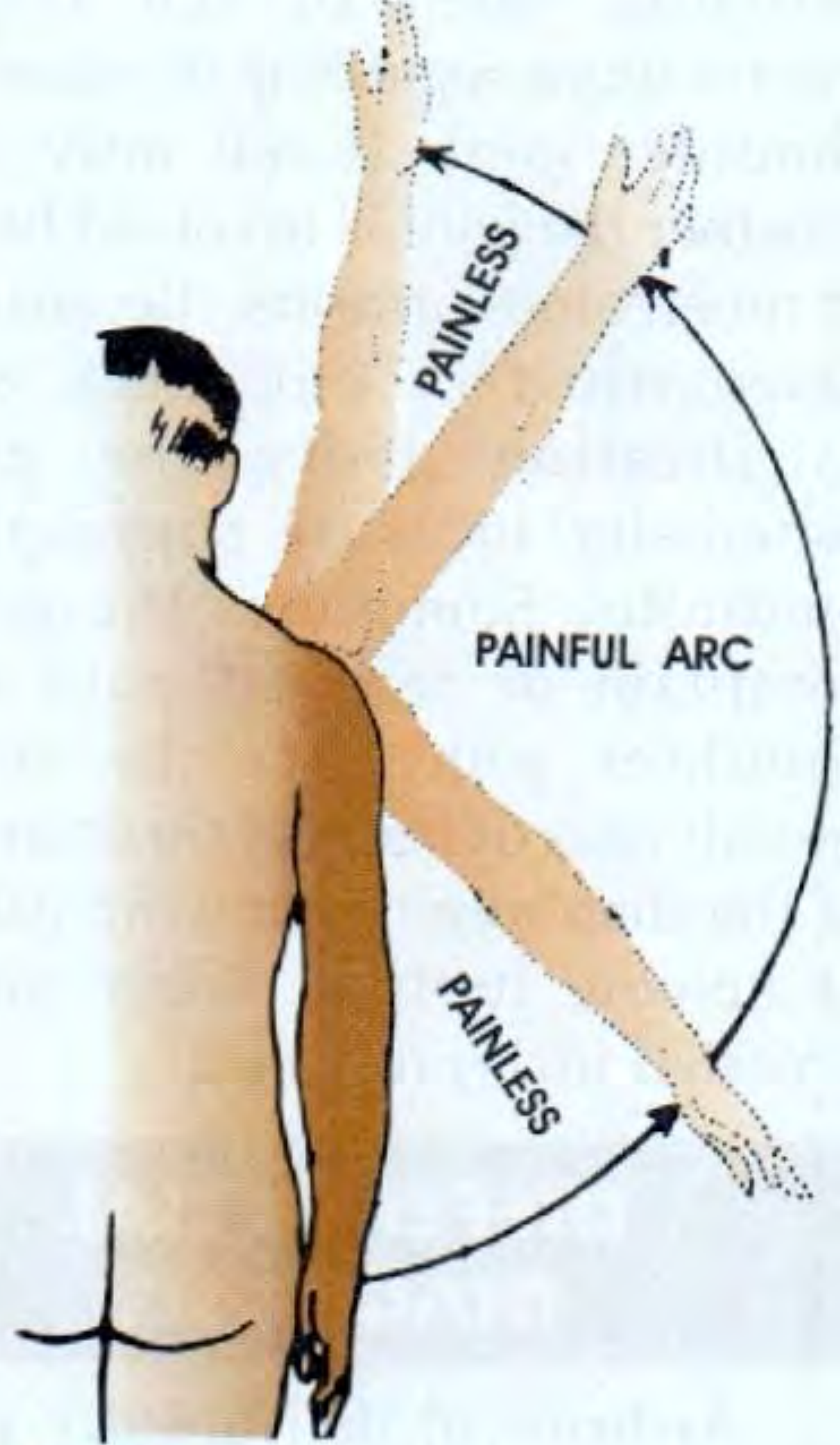
Classic painful arc: midrange abduction (60°–120°) is painful; both extremes are painless. (S Das, Manual on Clinical Surgery, Fig. 15.3)
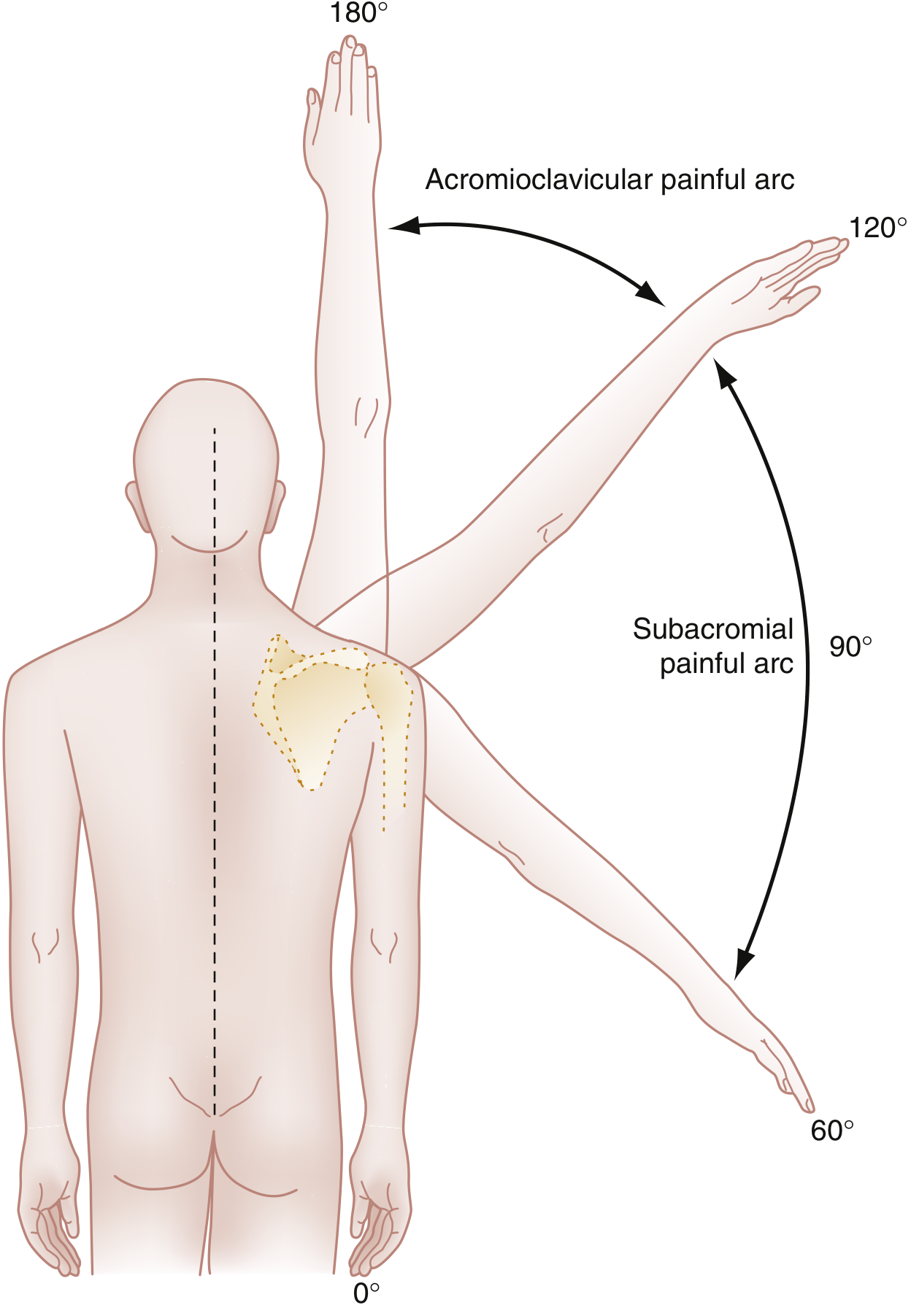
Arcs of painful abduction: Subacromial arc (60°–120°) vs Acromioclavicular arc (120°–180°). (Rosen's Emergency Medicine, Fig. 45.31)

Types of Painful Arc
The location of the painful arc tells you the structure involved - this is of great diagnostic value:
| Arc | Degrees | Structure Involved | Cause |
|---|---|---|---|
| Subacromial arc | 60°–120° | Supraspinatus tendon / subacromial bursa | Supraspinatus tendinitis, bursitis |
| Acromioclavicular arc | 120°–180° | AC joint | AC joint arthritis / osteophytes |
| Full arc (0°–180°) | All ranges painful | Glenohumeral joint | Arthritis, frozen shoulder (acute phase) |
| End-range only | >150° | AC joint / coracoclavicular ligament | AC joint pathology |
Causes / Conditions Producing Painful Arc
The painful arc is pathognomonic of chronic supraspinatus tendinitis but is also seen in:
- Chronic supraspinatus tendinitis - most common cause; degeneration of the tendon from deficient blood supply ± degenerative calcification
- Subdeltoid (subacromial) bursitis - inflamed bursa is compressed during mid-abduction
- Incomplete (partial thickness) rupture of the supraspinatus tendon
- Crack (stress) fracture of the greater tuberosity of the humerus
- AC joint arthritis - produces the higher-range painful arc (120°–180°)
- Calcific tendinitis - calcium hydroxyapatite deposits within the supraspinatus
Key distinction from other shoulder conditions:
- Acute supraspinatus tendinitis → whole range of abduction is painful
- Chronic supraspinatus tendinitis (painful arc syndrome) → only mid-range (60°–120°) is painful
- Frozen shoulder (acute) → all movements restricted and painful; NOT specific to abduction arc
- Complete supraspinatus rupture → patient shrugs the shoulder at the start of abduction (cannot initiate abduction without deltoid trick movement)
Pathology
The underlying process is degeneration of the supraspinatus tendon, typically triggered by:
- Overuse or repetitive overhead activity
- Deficient blood supply to the "critical zone" of the supraspinatus (1 cm from its insertion - hypovascular zone)
- Age-related changes (most common in males aged 45–60 years)
The swollen/thickened tendon is so situated that during mid-abduction (60°–120°), when the space between the greater tuberosity and the acromion is at its narrowest, the tendon is nipped between the two bones, producing pain. At the extremes of abduction, the geometry of the joint opens the space again, relieving the compression.
Clinical Features
History:
- Middle-aged to older adults (typically 45–60 years), males > females
- Pain localized to the anterolateral shoulder, radiating to the deltoid insertion
- Pain specifically during mid-range overhead activity (e.g., reaching to a shelf)
- Night pain - difficulty sleeping on the affected shoulder
- No significant stiffness at rest (distinguishes from frozen shoulder)
Examination:
- Painful arc of abduction: 60°–120° - the hallmark finding
- Localized tenderness over the supraspinatus insertion at the greater tuberosity and anterior acromion
- Full passive range of motion (no restriction - unlike frozen shoulder)
- Positive impingement tests (Neer, Hawkins-Kennedy)
- Radiograph: may show calcification in the supraspinatus tendon; hooked acromion
Differential Diagnosis Summary
| Condition | Arc/Range | Key Distinguishing Feature |
|---|---|---|
| Chronic supraspinatus tendinitis | 60°–120° painful | Pathognomonic painful arc; localized tendon tenderness |
| AC joint arthritis | 120°–180° painful | Tenderness over AC joint; scarf test positive |
| Subdeltoid bursitis | 60°–120° painful | Tenderness more diffuse; ultrasound shows bursal thickening |
| Frozen shoulder | All movements restricted | Global restriction, especially external rotation; all movements painful |
| Complete rotator cuff rupture | Painful + weak | Cannot initiate abduction; shrugs shoulder; drop arm sign |
| Glenohumeral OA | Full arc painful | Crepitus, X-ray changes, restricted ROM |
Investigations
- X-ray (AP + outlet view): May be normal early; hooked acromion; calcification in supraspinatus tendon; late - narrowed acromiohumeral interval
- Ultrasound: Supraspinatus tendon thickening, calcification, bursal fluid - dynamic assessment possible
- MRI: Gold standard for soft tissue; differentiates tendinitis from partial/full thickness tears
- Neer's injection test: 10 mL lidocaine into subacromial space - pain abolition confirms subacromial source
Management
Conservative (First Line)
- Rest and activity modification - avoid painful overhead activities
- NSAIDs - ibuprofen/diclofenac for analgesia and anti-inflammation
- Physiotherapy - rotator cuff strengthening, scapular stabilisation, posture; proven equivalent to surgery at 1, 2, and 5 years
- Subacromial corticosteroid injection - methylprednisolone + local anaesthetic; short-to-medium term relief; also diagnostic
- Extracorporeal shock wave therapy (ESWT) - for calcific tendinitis
Surgical (if conservative fails after 3–6 months)
- Arthroscopic subacromial decompression (acromioplasty) - removal of anteroinferior acromion, bursectomy, resection of coracoacromial ligament
- AC joint excision - if AC joint osteophytes are the primary source
- Calcific deposit needling/lavage - for refractory calcific tendinitis
Key Points to Remember
- Painful arc = 60°–120° - pathognomonic of chronic supraspinatus tendinitis
- Mechanism: supraspinatus tendon nipped between greater tuberosity and acromion at mid-abduction
- Extremes of abduction are painless - this distinguishes it from arthritis/frozen shoulder
- AC joint arc = 120°–180° - higher range, over shoulder level
- Other causes: subdeltoid bursitis, incomplete rupture, crack fracture of greater tuberosity
- Age group: 45–60 years, males more common
- Confirm with Neer's injection test (subacromial lidocaine)
- MRI is gold standard; X-ray may show calcification or hooked acromion
- First-line = physiotherapy + NSAIDs ± subacromial injection
- Surgery: arthroscopic acromioplasty if conservative treatment fails
Sources: S Das - A Manual on Clinical Surgery 13th Ed, pp. 148, 272–273; Rosen's Emergency Medicine (Concepts and Clinical Practice), p. 659; Campbell's Operative Orthopaedics 15th Ed 2026; Firestein & Kelley's Textbook of Rheumatology
This is a shared conversation. Sign in to Orris to start your own chat.