

These are present all over the body like hands, legs , stomach
molluscum contagiosum papule umbilicated skin lesion
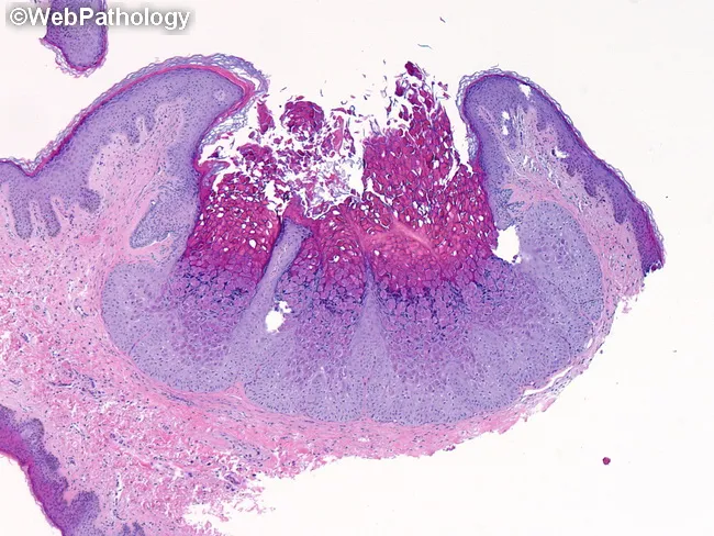
This image depicts a classic Molluscum contagiosum histopathology specimen from a cutaneous lesion examined under brightfield microscopy. Modality used: Light microscopy with Hematoxylin and Eosin staining, enabling visualization of epidermal architecture and cytoplasmic inclusions. Anatomical context: human skin, with an endophytic, cup-shaped, lobulated epidermal proliferation projecting into the dermis and producing a central depression. Morphology: well-circumscribed pseudotumor of thickened squamous epithelium displaying pronounced acanthosis and a central crater, at the edge of hyperplastic epithelium transitioning toward the lesion center. Cytopathic changes: numerous large intracytoplasmic molluscum bodies within keratinocytes of the basal to suprabasal layers; inclusions progress from eosinophilic early to basophilic with maturation and exert compressive effect on the nucleus. The viral inclusions impart a pale halo around nuclei and induce lobular rearrangement of the epithelium. Pathology significance: the presence of molluscum bodies and cup-shaped architecture is highly characteristic and often diagnostic of molluscum contagiosum; aids in distinguishing from verruca vulgaris and other epidermal proliferations. Clinically relevant contexts: pediatric skin lesions; immunosuppressed patients; sexually transmitted infections in adults; offers diagnostic confirmation and guides management (curettage, cryotherapy). Potential diagnostic use: histopathologic confirmation when clinical suspicion is uncertain; correlates with surface lesion appearance (umbilicated papule).
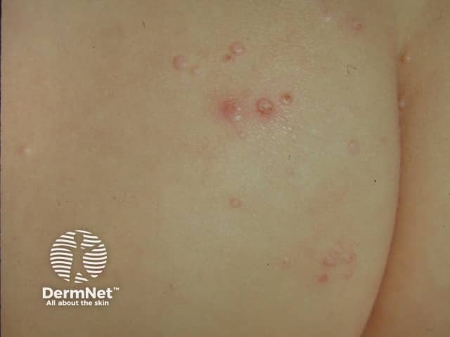
Dermatologic clinical photography capturing multiple molluscum contagiosum lesions on the gluteal skin. Modality: high‑resolution standardized digital photograph in a close‑up frontal view. Subject: human cutaneous tissue; epidermal and superficial dermal involvement. Imaging perspective: surface view of a non‑palpable, dome‑shaped papule cluster with central umbilication. Visual features include flesh‑colored to erythematous, umbilicated papules ranging approximately 2–5 mm in diameter, with smooth, shiny surfaces and a soft, pearly tactile quality. Distribution is localized to a single body region with minimal surrounding erythema; lesions are several in number, arranged in a scattered, clustered pattern typical of pediatric molluscum contagiosum. No surrounding edema or necrosis observed. Histology not available; diagnosis is clinical, supported by characteristic central indentations and fingertip tenderness when manipulated. The image provides rapid educational identification of molluscum contagiosum vs. differential diagnoses such as common warts (HPV), folliculitis, or keratinocyte nevi. Clinical significance includes contagiousness via direct contact and potential spontaneous resolution over months; treatment may be considered for cosmetically conspicuous or symptomatic lesions. Use cases include dermatology teaching, primary care triage, and teledermatology evaluation, with emphasis on recognizing pediatric cutaneous viral infections and communicating uncertainty to patients and caregivers. Document lesion count, distribution, and evolution over time to guide management decisions.
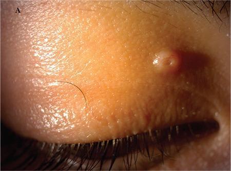
This clinical photograph shows a close-up of the right upper eyelid, depicting a classic presentation of Molluscum Contagiosum. The primary finding is a single, discrete, flesh-colored papule approximately 2x2 mm in size, located on the medial aspect of the eyelid near the lash line. The lesion exhibits a characteristic dome-shaped morphology with a distinct central pit or umbilication. The surface of the papule appears smooth and slightly waxy or shiny. The surrounding eyelid skin shows normal texture with subtle fine wrinkling and no significant surrounding erythema or edema in this view. This visual is an educational example used in ophthalmology and dermatology to illustrate a common viral skin infection that can cause secondary chronic follicular conjunctivitis if the lesion sheds viral particles into the tear film. The clinical significance lies in the recognition of the umbilicated center as a pathognomonic feature of the Poxvirus infection.
molluscum contagiosum treatment children
Molluscum Contagiosum
What is it?
Who gets it?
- Young children (peak ages 1-4 years) - spread through skin contact, shared towels, water play
- Sexually active young adults (ages 20-29) - sexually transmitted
- Immunosuppressed individuals (especially HIV) - can get severe, widespread disease
How do the lesions look?
- Small, firm, dome-shaped, pearly or flesh-colored papule, typically 3-5 mm
- A central umbilication (tiny pit or dimple at the top) is the hallmark feature - sometimes only visible under side-lighting
- Can have a white core if squeezed
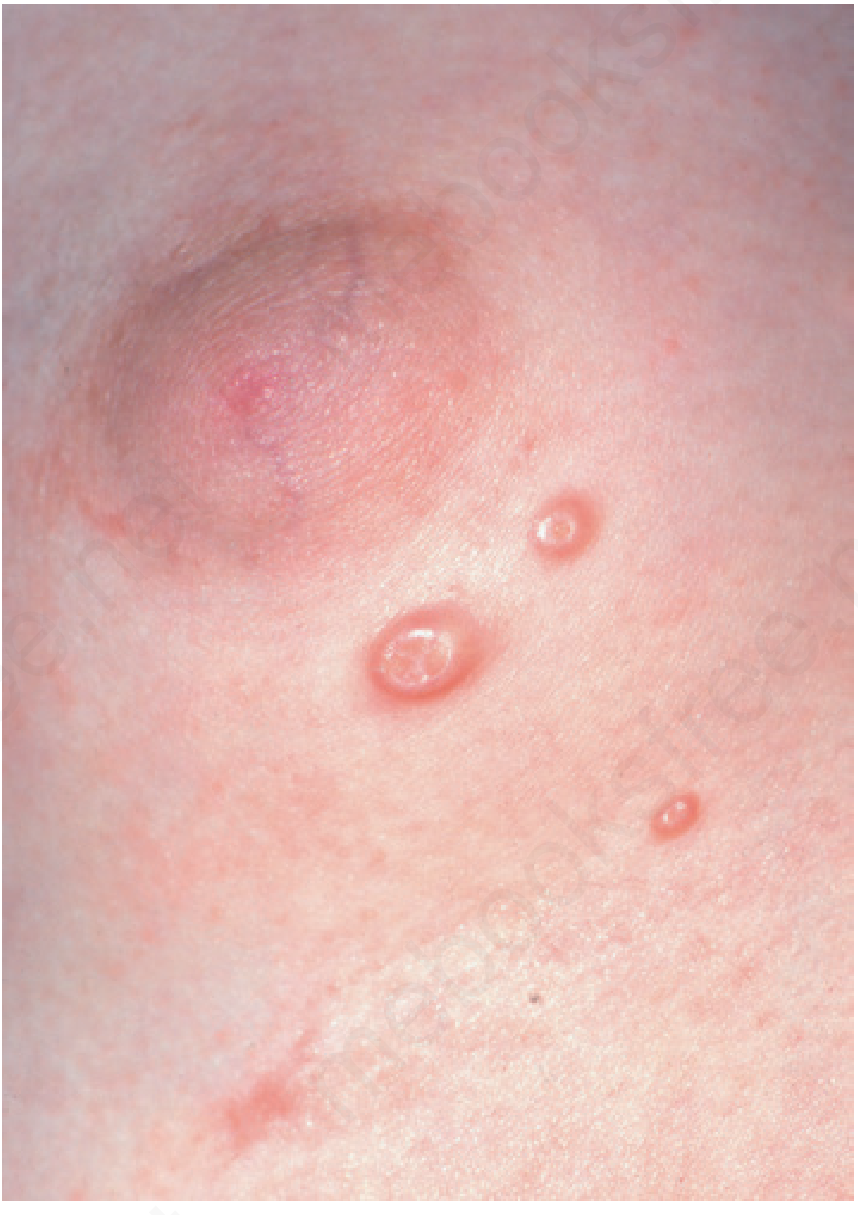
- Irritated lesions (like the one in your photo) become red, crusted, or even pustular, resembling a minor infection - this is actually a sign the immune system is reacting to the virus
- Surrounding redness and warmth can look like cellulitis
How does it spread?
- Direct skin-to-skin contact (most common)
- Shared wet towels, swimming pools, bath sponges
- Autoinoculation - touching one lesion then scratching elsewhere - explains why they spread all over the body
Does it go away on its own?
Treatment options (if desired)
| Treatment | How it works | Notes |
|---|---|---|
| Cantharidin (blister beetle extract) | Applied by doctor; causes blister that clears lesion | Fast, effective, no at-home risk |
| Curettage | Physical scraping of lesion | Works well but painful |
| Cryotherapy | Liquid nitrogen freeze | Effective, mild scarring risk |
| Imiquimod 5% cream | Immune stimulator, applied at home | Slower, good for widespread lesions |
| Berdazimer gel (nitric oxide-releasing) | Newer topical; systematic review PMID 37721050 shows good efficacy and safety | Not universally available yet |
| Observation only | Wait for spontaneous resolution | Valid first-line approach in children |
What to do now
Please see a dermatologist or your family doctor. Since the lesions are widespread across multiple body parts, a clinical examination is needed to:
- Confirm the diagnosis
- Rule out secondary bacterial infection in any crusted/red lesions
- Discuss treatment if the lesions are spreading rapidly, causing discomfort, or are cosmetically concerning
- Check if there is any underlying immune issue (relevant if the person is an adult with severe widespread disease)