
Exytensor carpi tendinopathy
"extensor carpi" AND tendinopathy
extensor carpi tendinopathy wrist lateral epicondylitis tennis elbow
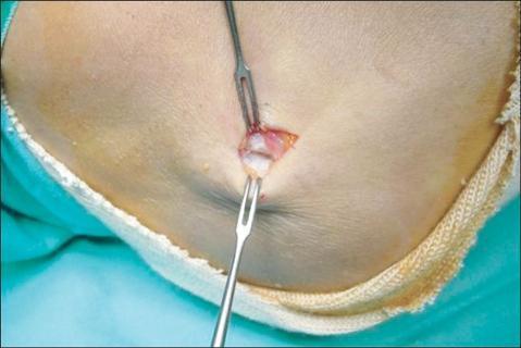
This clinical photograph captures an intraoperative view of a surgical procedure for lateral epicondylitis (tennis elbow) through a minimal skin incision. Two small toothed retractors are utilized to hold the wound edges open, exposing the origin of the extensor carpi radialis brevis (ECRB) muscle near the lateral epicondyle of the humerus. The exposed tendon tissue exhibits clear signs of tendinosis, characterized by a loss of normal glistening white appearance, irregular texture, and areas of brownish-gray discoloration. Visible pathology includes focal degeneration, scarring, and evidence of friable granulation tissue, indicating chronic repetitive microtrauma and a failed healing response. The image demonstrates the surgical approach for debriding degenerative tendon tissue and serves as an educational reference for orthopedic surgeons and residents studying the macroscopic presentation of chronic tendinopathy and the Nirschl surgical technique for lateral epicondylitis.
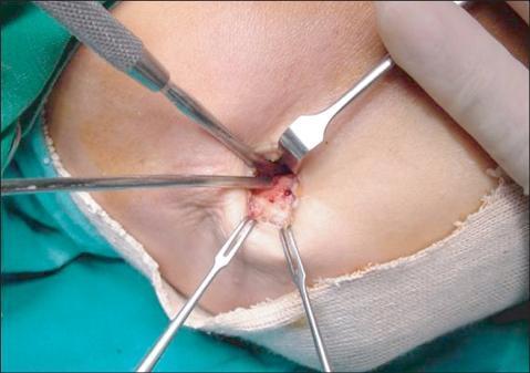
This clinical photograph captures an intraoperative view of a surgical procedure for lateral epicondylitis (tennis elbow). The image shows the lateral aspect of the elbow under local anesthesia with a 1.5 cm skin incision. Following the detachment and excision of the degenerative origin of the extensor carpi radialis brevis (ECRB) tendon, the cortical bone of the lateral epicondyle is exposed. Visible within the center of the surgical field are multiple small drill holes made using a microfracture technique. These holes demonstrate active punctate bleeding, an intentional surgical step designed to facilitate blood flow and promote the recruitment of mesenchymal stem cells to enhance fibrous tissue regeneration at the bony bed. Four metallic surgical instruments, including small retractors and a textured-handle probe, are used to provide visualization of the site. This image serves as a clinical demonstration of the Nirschl-type surgical approach for chronic tendinopathy, highlighting the transition from debridement to marrow stimulation.
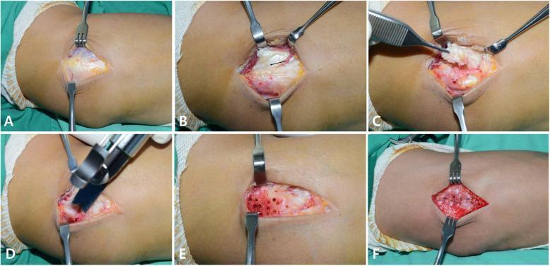
A series of six clinical photographs (A–F) demonstrating the surgical progression for lateral epicondylitis (tennis elbow). Panel A shows the initial exposure of the extensor muscle mass fascia and fibers of the extensor carpi radialis longus (ECRL) and brevis (ECRB). In Panel B, the ECRL is retracted to reveal the underlying pathologic ECRB tendinopathy, characterized by a dull, grayish, and friable appearance (indicated by a black arrow). Panel C illustrates the elliptical excision of this abnormal tissue, including the ECRB and extensor digitorum communis (EDC) tendon origins. Panel D shows decortication of the lateral humeral condyle using an oscillating saw. Panel E displays the decorticated bone with multiple small holes drilled via K-wire to create a vascular bed (marrow venting). Finally, Panel F demonstrates the anatomic repair of the interval between the ECRL and the common extensor aponeurosis with sutures, closing the site without reattaching the ECRB. The series provides an educational overview of orthopedic debridement and bony preparation for chronic tendinopathy.
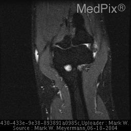
**Imaging Modality:** Magnetic Resonance Imaging (MRI), Coronal view. **Anatomical Region:** Elbow joint, specifically focusing on the humeroradial and humeroulnar articulations and surrounding soft tissues. **Observed Pathology:** The image demonstrates features consistent with **lateral epicondylitis** (tennis elbow). There is an increased signal intensity and thickening at the origin of the **common extensor tendon** near the lateral epicondyle of the humerus. **Characteristic Visual Features:** - **Signal Abnormality:** Hyperintense signal on T2-weighted/STIR-weighted imaging at the lateral aspect of the elbow, indicating edema or microtearing within the common extensor origin (primarily the extensor carpi radialis brevis). - **Landmarks:** Distinct visualization of the lateral and medial epicondyles, the radial head, and the capitellum. - **Secondary Findings:** Mild joint effusion may be present. The medial collateral ligament and medial epicondyle appear relatively unremarkable in this slice. **Key Diagnostic Features:** Focal signal alteration at the lateral epicondyle is the primary radiographic cue for tendinosis or tendinopathy of the wrist extensors, differentiating it from medial epicondylitis or intra-articular loose bodies.
Extensor Carpi Tendinopathy
- Lateral elbow tendinopathy (lateral epicondylitis / "tennis elbow") - primarily involving the extensor carpi radialis brevis (ECRB)
- Extensor carpi ulnaris (ECU) tendinopathy - at the ulnar wrist
Pathophysiology
- Disorganized collagen fibers
- Increased mucoid ground substance
- Prominent capillary proliferation (neovascularization)
- Increased fibroblasts and myofibroblasts
- Absence of inflammatory cells on histopathologic examination
- Textbook of Family Medicine, 9e, p. 798-819
1. Lateral Elbow Tendinopathy (Tennis Elbow / Lateral Epicondylitis)
Cause
Clinical Features
- Pain at the lateral aspect of the elbow
- Worsened by gripping, turning handles, lifting with palm-down (pronated) position
- Tenderness over the lateral epicondyle and proximal wrist extensors just distal to it
- Pain with resisted wrist extension and forearm supination
- Normal radial head, ligamentous stability, and neurovascular exam
- Plain X-rays not required for diagnosis unless trauma, motion loss, locking, or prolonged pain
Management
| Step | Treatment |
|---|---|
| Analgesia | Cryotherapy, ice massage, NSAIDs or acetaminophen |
| Bracing | Counterforce strap just distal to maximal tenderness - reduces tension on affected tendons |
| Exercise | Key stretch: elbow extended, forearm fully pronated, wrist flexed with traction on middle/ring fingers. Light-weight strengthening for wrist extension and supination |
| Injections | Corticosteroid: reduces peritendinous pain short-term but does not alter long-term outcome; may cause short-lived pain flare |
| Advanced | Prolotherapy, ESWT (extracorporeal shock wave therapy) - mixed short-term evidence; PRP not yet proven |
| Surgery | Reserved for recalcitrant cases - debridement of ECRB origin with marrow stimulation (Nirschl technique) |
- Textbook of Family Medicine, 9e, p. 819
2. Extensor Carpi Ulnaris (ECU) Tendinopathy
Clinical Features
- Pain, erythema, and tenderness on the ulnar border of the wrist
- Pain/crepitus with wrist range of motion
- May be associated with inflammatory arthropathy causing erosion of the ulnar styloid
- Must be distinguished from TFCC injury, DRUJ instability, hamate/triquetrum fracture
Management
-
Rest, splinting, NSAIDs
-
Corticosteroid injection into the ECU sheath
-
Surgery (rare) for refractory cases
-
Rosen's Emergency Medicine, p. 600; Grainger & Allison's Diagnostic Radiology, p. 986
3. Intersection Syndrome
-
Also called "crossover" or "oarsman's syndrome"
-
Mechanism: rowing, weightlifting, repetitive resistance pulling
-
Findings: pain, swelling, crepitus on radial dorsum of wrist
-
Treatment: wrist splint + anti-inflammatory medications; typically self-limited
-
Rosen's Emergency Medicine, p. 616
Imaging
- Increased signal intensity and thickening at the common extensor origin (ECRB) on T2/STIR
- Hyperintense signal at lateral epicondyle indicates edema or microtearing