
Ankle approaches give extensively
Surgical Approaches to the Ankle
Overview of Available Approaches
| Approach | Position | Primary Use | Structures at Risk |
|---|---|---|---|
| Anterolateral (Universal) | Supine | Pilon Fx, arthrodesis, talectomy | Superficial peroneal nerve, intermediate dorsal cutaneous branches |
| Extensile Anteromedial (Assal) | Supine | High-energy pilon fractures | Saphenous vein/nerve, tibialis anterior tendon |
| Anterior | Supine | Ankle arthrodesis, OCD access | Dorsalis pedis artery, deep peroneal nerve |
| Lateral to Distal Fibula | Supine | Lateral malleolus ORIF (most common) | Superficial peroneal nerve (~10 cm proximal to fibular tip) |
| Posterolateral | Prone/Lateral | Posterior malleolus + fibula fixation | Sural nerve, FHL, peroneal tendons |
| J-type Posterolateral (Choi) | Prone/Lateral | Posterior + lateral malleolus combined | Sural nerve |
| Transfibular Posterolateral (Gatellier & Chastang) | Prone | Large posterior malleolus Fx, OCD lateral talar dome, TAR | Distal fibular physis (children), PITFL |
| Anterolateral to Lateral Talar Dome (Tochigi) | Supine | Centrolateral OCD of talus | Lateral branch of superficial peroneal nerve |
| Posterior | Prone | Posterior ankle arthrodesis, blade plate | Posterior tibial vessels, tibial nerve |
| Medial Approach to Medial Malleolus | Supine | Medial malleolus fracture ORIF | Saphenous nerve and vein |
| Medial Approach to Tarsus (Knupp) | Supine | Hindfoot arthrodesis, PTT dysfunction | Anterior deltoid fibers |
| Transverse Medial (Koenig & Schaefer) | Supine | Fracture-dislocation of talus, OCD | Tibial vessels, tibial nerve |
| Medial Approach to Posterior Tibia (Broomhead/Colonna) | Supine | Medial posterior malleolus Fx | Neurovascular bundle |
| Kocher Lateral to Tarsus & Ankle | Supine | Triple arthrodesis, talectomy | Peroneal tendons, sural nerve, skin viability |
| Posteromedial via PTT Sheath (Bassett) | Supine/Prone | OCD posterior talar dome, posterior capsule | Deep deltoid, posteromedial NV structures |
1. Anterolateral Approach to the Ankle and Chopart Joint (Universal Incision)
- Incise the fascia and superior/inferior extensor retinacula down to periosteum and ankle capsule (usually divides anterolateral malleolar and lateral tarsal arteries)
- Find and protect intermediate dorsal cutaneous branches of the superficial peroneal nerve
- Divide extensor digitorum brevis in line with fibers, or detach from origin and reflect distally
- Retract extensor tendons, dorsalis pedis artery, and deep peroneal nerve medially and incise the capsule
- Expose talonavicular joint by transverse capsulotomy
- Continue dissection laterally through the calcaneocuboid joint capsule
- Incise fat lateral and inferior to talar neck to expose the subtalar joint
- Extend distally for access to cuboid-4th/5th metatarsal and navicular-3rd cuneiform joints
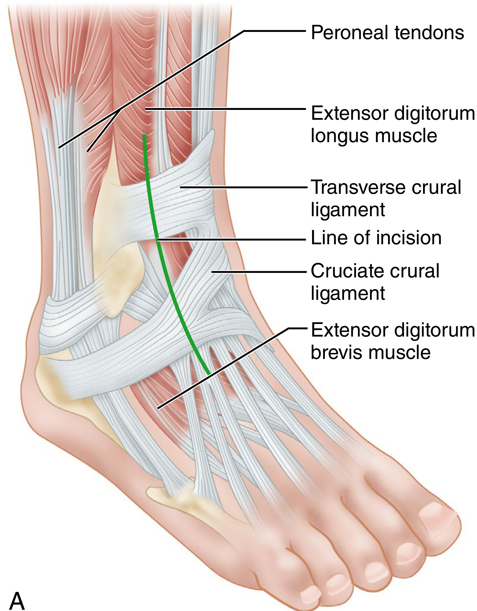
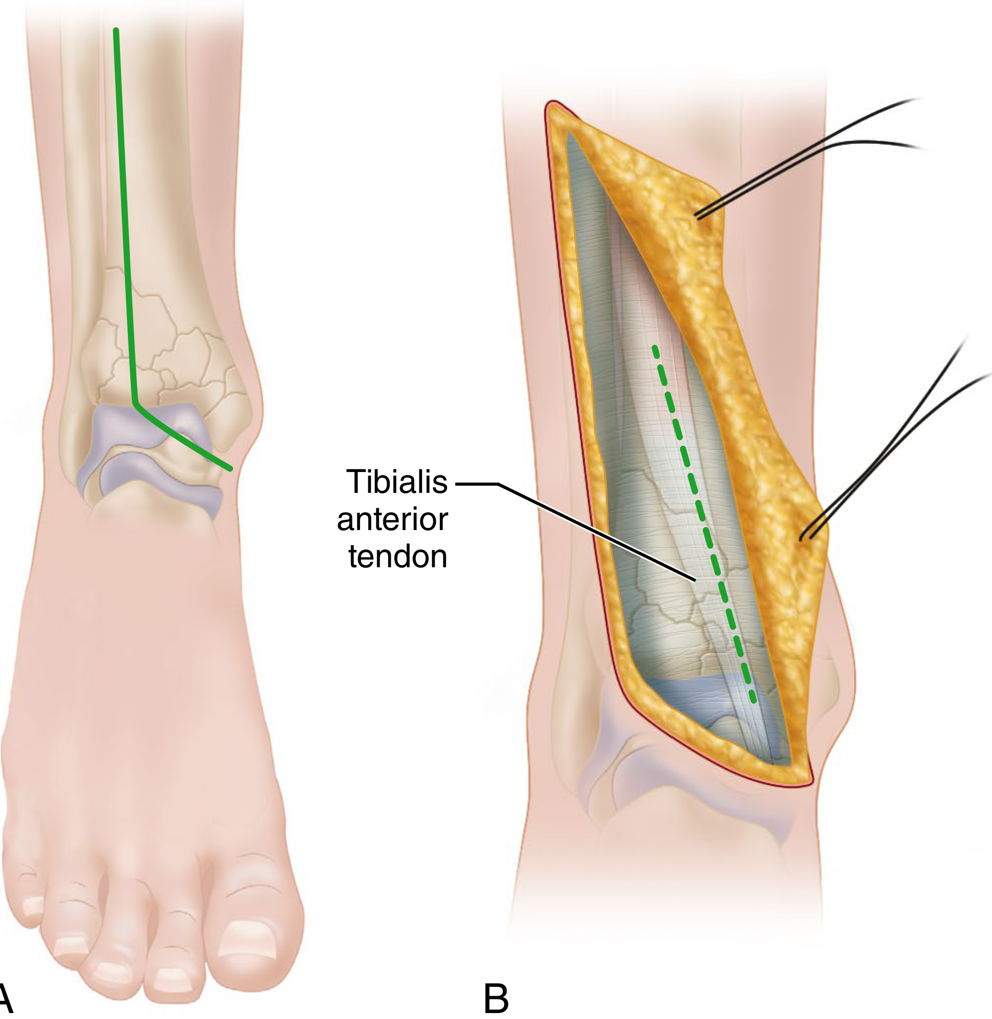
2. Extensile (Anteromedial) Approach to the Ankle - Assal, Ray & Stern
- Mark incision 0.5 cm lateral to tibialis anterior tendon, curve ending at medial malleolus tip (apex >120°)
- Incise skin, develop full-thickness flap to extensor retinaculum - expect crossing veins from saphenous vein near distal incision
- Incise extensor retinaculum medial to the tibialis anterior tendon
- Retract tibialis anterior tendon medially; identify and protect superficial peroneal nerve and dorsal veins
- Incise periosteum and capsule to access the anterior distal tibia
- Repair extensor retinaculum before skin closure
3. Anterior Approach to the Ankle Joint
- Begin 7.5-10 cm proximal to ankle on anterior aspect of leg, extend ~5 cm distal to the joint
- Divide deep fascia in line with skin incision
- Isolate, ligate, and divide anterolateral malleolar and lateral tarsal arteries; retract the neurovascular bundle laterally
- Incise periosteum, capsule, and synovium in line with incision - expose entire width of ankle anteriorly by subcapsular/subperiosteal dissection
4. Lateral Approach to the Distal Fibula (Most Commonly Used Ankle Approach)
- Mark skin incision based on fracture pattern and surgeon preference
- Elevate full-thickness flaps - protect superficial peroneal nerve (~10 cm proximal to distal fibula tip; range 3-18 cm)
- Incise fascia, elevate peroneal musculature posteriorly in supraperiosteal fashion
- For posterior/posterolateral plating - incise superior peroneal retinaculum partially
- For syndesmosis access - dissect anteriorly over fibula, partially incise extensor retinaculum, protecting AITFL fibers
- Layered closure; avoid suturing superficial peroneal nerve in anterior wound
Key Precautions: Superficial peroneal nerve location is highly variable (3-18 cm proximal to fibular tip). Avoid complete disruption of superior peroneal retinaculum when inserting plates. Protect AITFL fibers during extensor retinaculum incision for syndesmosis access.
5. Posterolateral Approach to the Posterior Malleolus and Fibula
- Mark posterior fibula and lateral Achilles tendon - incision at the midpoint between the two structures, just lateral to the midpoint
- Incise fascia just medial to the peroneal tendons
- Develop the interval between peroneal tendons and FHL
- Incise fascia overlying FHL along its lateral border
- Elevate FHL off posterior tibia, exposing the posterior malleolus
- CRITICAL: Avoid dissection into the interval between posterior malleolus and fibula to protect the PITFL
- For posterior fibular fixation - retract peroneal musculature laterally
- For lateral/posterolateral fibular fixation - create secondary deep window lateral to the peroneal tendons (may shift skin incision slightly lateral)
6. J-Type Single-Incision Posterolateral Approach (Choi et al.)
- 10-cm incision following the posterior edge of the fibula, curving posteriorly at the syndesmosis to end at the Achilles tendon insertion on the calcaneus. Dissect the sural nerve carefully.
- Take down peroneal tendons from the posterior lateral malleolus to expose the lateral malleolar fracture
- Develop interval between peroneal tendons and FHL
- Retract FHL and Achilles tendon medially, exposing the posterior malleolus
Warning: The sural nerve passes tangential to the skin incision - protect it carefully.
7. Transfibular Posterolateral Approach (Gatellier and Chastang, 1924)
- Incision: Begin 12 cm proximal to lateral malleolus tip along posterior fibular margin, extend to the malleolus tip, then curve anteriorly 2.5-4 cm along the peroneal tendons
- Expose fibula superiosteally; incise peroneal retinaculum sheaths; displace peroneal tendons anteriorly
- If fibula intact - divide it 10 cm proximal to the tip; divide interosseous membrane, anterior and posterior tibiofibular ligaments
- Preserve calcaneofibular and talofibular ligaments as the hinge
- Turn fibula laterally - expose lateral and posterior aspects of distal tibia and lateral ankle joint
- Closure: Replace fibula, fix with transverse screw (overdrill the fibula hole for compression across the syndesmosis). Dorsiflex the ankle while tightening the screw. Add plate if desired.
- Replace tendons, repair retinaculum sheaths.
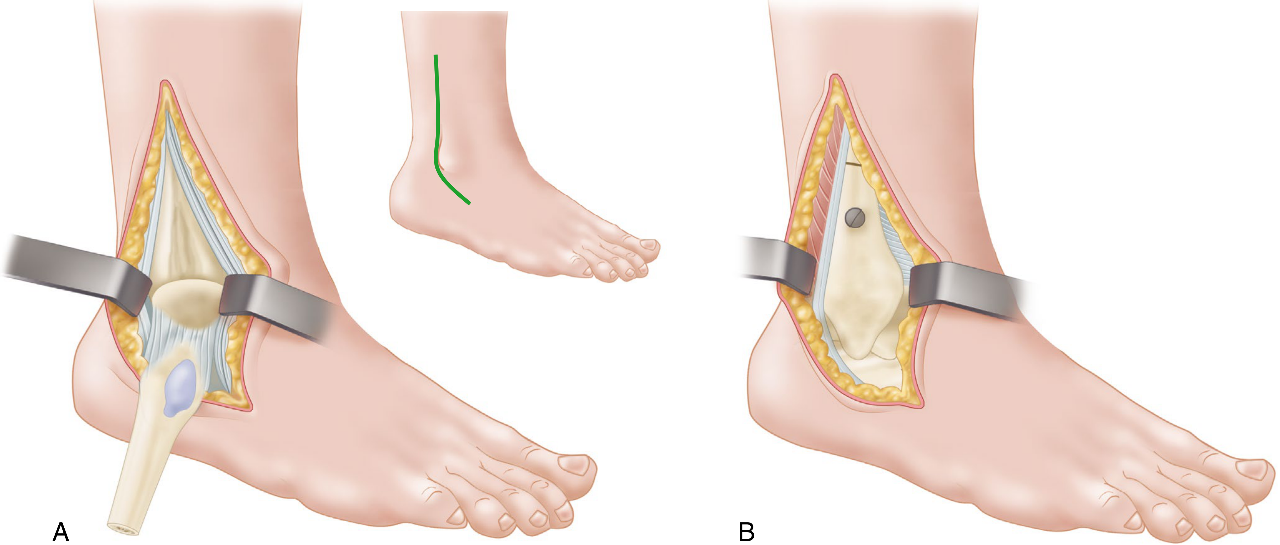
Key Precautions: In children with open physes, distal fibular physis is at risk during reflection. Failure to overdrill the fibula = no compression = syndesmosis widening = tibiotalar arthritis. Not commonly used now due to extensive soft-tissue disruption.
8. Anterolateral Approach to the Lateral Dome of the Talus (Tochigi et al.)
- 10-cm vertical incision along anterolateral corner of ankle (avoid lateral branch of superficial peroneal nerve)
- Outline anterolateral tibial osteotomy to include the anterior tibiofibular ligament - cortical fragment must be ≥1 cm²
- Predrill fixation screw holes before osteotomy
- Rotate osteotomy fragment on its planned pivot (articular cartilage "cracked" as it rotates)
- Closure: Rotate fragment back, fix with a 4-mm cancellous screw and washer
9. Posterior Approach to the Ankle
- 12-cm incision along the posterolateral border of the Achilles tendon, down to calcaneal insertion
- Divide superficial and deep fasciae; divide Achilles tendon by Z-plasty (or split centrally - Hammit modification)
- Develop fat and areolar tissue to posterior tibia surface in the space between FHL and peroneal tendons
- Retract FHL tendon medially to expose:
- Distal 2.5 cm of tibia
- Posterior ankle joint
- Posterior end of talus
- Subtalar joint
- Posterior superior calcaneus
Safety Note: Keeping dissection lateral to FHL protects the posterior tibial vessels and tibial nerve, which lie medial to FHL.
10. Medial Approach to the Medial Malleolus
- Curved incision starting along the anterior aspect of the medial malleolus, ending at the tip of the anterior colliculus. For associated talar fracture: apex-posterior incision extending into anteromedial talar approach.
- Identify and protect the saphenous nerve and vein
- Incise through anteromedial joint capsule along the anterior border of the medial malleolus
- Excise injured periosteum from fracture site
- Gently retract medial malleolus, inspect and irrigate talar dome
- Reduce and fix with plate or screws
Author's Preference (Campbell's): Apex-anterior curved incision to anterior colliculus tip. For talar fracture, use apex-posterior incision instead.
11. Medial Approach to the Tarsus (Knupp et al.)
- 4-cm incision from center of medial malleolus toward the navicular, 5 mm above and parallel to the posterior tibial tendon. Extend as needed to reach the cuneiform.
- Open subtalar joint capsule carefully - avoid damaging anterior fibers of the deltoid ligament
12. Transverse Incision Medial Approach with Medial Malleolar Osteotomy (Koenig and Schaefer)
- Curve incision just proximal to the medial malleolus; divide malleolus with osteotome or power saw; preserve deltoid ligament attachment
- Subluxate talus and malleolus laterally to reach joint surfaces
- Pre-drill screw holes before osteotomy; insert then remove screws; at end of operation - reinsert screws
- Use two screws to prevent rotation of the osteotomized malleolus; use interfragmentary technique for compression
13. Medial Approach to the Posterior Tibia (Broomhead; Modified by Colonna and Ralston)
- Begin ~10 cm proximal and 2.5 cm posterior to the medial malleolus
- Curve anteriorly and inferiorly across the center of the medial malleolus
- Continue inferiorly and posteriorly 4 cm toward the heel
- Expose medial malleolus by reflecting periosteum; preserve deltoid ligament
- Divide flexor retinaculum; retract FHL tendon and neurovascular bundle posteriorly and laterally
- Retract tibialis posterior and FDL tendons medially and anteriorly to expose the posterior tibial fracture
14. Kocher Lateral Approach to the Tarsus and Ankle
- Incision from just lateral and distal to the talar head, curving 2.5 cm inferior to the lateral malleolus tip, then posteriorly and proximally, ending 2.5 cm posterior to fibula and 5 cm proximal to lateral malleolus tip (can extend 5-7 cm further proximally)
- Incise fascia to peroneal tendons; retract them posteriorly (protect lesser saphenous vein and sural nerve posterior to the incision)
- For larger field - divide tendons by Z-plasty
- Deepen dissection distally, divide calcaneofibular ligament, expose subtalar joint; calcaneocuboid and talonavicular joints can be reached distally
- Divide talofibular ligaments and dislocate ankle by medial traction for access to entire articular surface
15. Posteromedial Approach Through Posterior Tibial Tendon Sheath (Bassett et al.)
- Palpate medial malleolus and Achilles tendon
- Make a 5-8 cm incision overlying the posterior tibial tendon behind the medial malleolus, curving distally and slightly anteriorly following the contour of the medial malleolus
- Enter the posterior tibial tendon sheath to access the posteromedial ankle compartment
Summary: Approach Selection by Clinical Scenario
| Clinical Scenario | Recommended Approach |
|---|---|
| Ankle fracture - lateral malleolus | Lateral to distal fibula |
| Ankle fracture - posterior malleolus | Posterolateral |
| Ankle fracture - medial malleolus | Medial approach to medial malleolus |
| High-energy pilon fracture | Extensile anteromedial (± lateral fibula approach) |
| Ankle arthrodesis | Anterolateral or Anterior |
| Posterior ankle arthrodesis | Posterior |
| Triple or pantalar arthrodesis | Anterolateral (universal), Kocher, or medial (Knupp) |
| Large posterior malleolus + lateral OCD talar dome | Transfibular posterolateral (Gatellier & Chastang) |
| Centrolateral OCD of talus (extensive) | Anterolateral talar dome (Tochigi) |
| Medial talar dome OCD / talar fracture-dislocation | Transverse medial (Koenig & Schaefer) or posteromedial (Bassett) |
| PTT dysfunction / hindfoot arthrodesis | Medial to tarsus (Knupp) |
| Posterior tibial fracture (posterior malleolus, medial part) | Medial to posterior tibia (Colonna & Ralston) |
What are the sources unused?
Sources Used in the Previous Answer
| Book | Speciality | Status |
|---|---|---|
| Campbell's Operative Orthopaedics, 15th Ed 2026 | Orthopedics | ✅ Used |
Sources NOT Used (by Speciality)
Anatomy
- Color Atlas of Ultrasound Anatomy, 3e
- Color Atlas of Human Anatomy Vol 1, 2, 3
- General Anatomy and Musculoskeletal System (THIEME Atlas)
- Gray's Anatomy for Students
- Imaging Anatomy Text and Atlas Volume 3 (Bones, Joints, Vessels and Nerves) - most relevant to ankle anatomy
- Netter's Atlas of Human Anatomy
Anesthesiology
- Barash, Cullen & Stoelting's Clinical Anesthesia, 9e
- Miller's Anesthesia, 10e
- Morgan & Mikhail's Clinical Anesthesiology, 7e
Biochemistry
- Basic Medical Biochemistry - A Clinical Approach, 6e
- Biochemistry, 8th Ed - Lippincott Illustrated Reviews
- Harper's Illustrated Biochemistry, 32nd Ed
Cardiology
- Braunwald's Heart Disease, 2 Vol Set
- Fuster and Hurst's The Heart, 15th Ed
- Textbook of Clinical Echocardiography
Community Medicine
- Park's Textbook of Preventive and Social Medicine
Dermatology
- Andrews' Diseases of the Skin Clinical Atlas, 2e
- Andrews' Diseases of the Skin - Clinical Dermatology
- Dermatology 2-Volume Set, 5e
- Fitzpatrick's Dermatology, Vol 1 & 2
Embryology
- Langman's Medical Embryology
- The Developing Human - Clinically Oriented Embryology
Emergency Medicine
- Rosen's Emergency Medicine, 9e
- Roberts and Hedges' Clinical Procedures in Emergency Medicine
- Tintinalli's Emergency Medicine - A Comprehensive Study Guide
ENT
- Cummings Otolaryngology Head and Neck Surgery
- K J Lee's Essential Otolaryngology
- Scott-Brown's Otorhinolaryngology Head & Neck Surgery (Vol 1 & 2)
- Shambaugh Surgery of the Ear
Family Medicine
- Pfenninger and Fowler's Procedures for Primary Care, 3e
- Swanson's Family Medicine Review
- Textbook of Family Medicine, 9e
Forensic Medicine
- Brogdon's Forensic Radiology
- DiMaio's Forensic Pathology, 3rd Ed
- Forensic Anthropology - A Comprehensive Introduction, 2e
- P C Dikshit Textbook of Forensic Medicine and Toxicology
- Parikh's Textbook of Medical Jurisprudence
- The Essentials of Forensic Medicine and Toxicology, 36th Ed
Gastroenterology / GI Surgery
- Clinical Gastrointestinal Endoscopy, 3e
- Sleisenger and Fordtran's GI and Liver Disease
- Yamada's Textbook of Gastroenterology, 7e
- Maingot's Abdominal Operations
General Surgery
- Pye's Surgical Handicraft, 22nd Ed
- Bailey and Love's Short Practice of Surgery, 28th Ed
- Current Surgical Therapy, 14e
- Fischer's Mastery of Surgery, 8e
- Mulholland and Greenfield's Surgery, 7e
- S Das A Manual on Clinical Surgery, 13th Ed
- Schwartz's Principles of Surgery, 11th Ed
- Sabiston Textbook of Surgery
Genetics
- Emery's Elements of Medical Genetics and Genomics
- Thompson & Thompson Genetics and Genomics in Medicine, 9e
Histology
- Histology: A Text and Atlas - Correlated Cell and Molecular Biology
- Junqueira's Basic Histology, 17e
Immunology
- Cellular and Molecular Immunology
- Janeway's Immunobiology, 10e
- Roitt's Essential Immunology
Internal Medicine / Rheumatology
- Firestein & Kelley's Textbook of Rheumatology, 2-Vol Set
- Frameworks for Internal Medicine
- Goldman-Cecil Medicine, 2 Vol Set
- Harrison's Principles of Internal Medicine, 22e (2025)
- Rheumatology, 2-Volume Set (2022, Elsevier)
- Symptom to Diagnosis - An Evidence Based Guide, 4e
- The Washington Manual of Medical Therapeutics
Laboratory Medicine
- Henry's Clinical Diagnosis and Management by Laboratory Methods
- Quick Compendium of Clinical Pathology, 5e
- Tietz Textbook of Laboratory Medicine, 7e
Microbiology
- Jawetz, Melnick & Adelberg's Medical Microbiology, 28e
- Medical Microbiology, 9e
- Sherris & Ryan's Medical Microbiology, 8e
Nephrology
- Brenner and Rector's The Kidney, 2-Vol Set
- Comprehensive Clinical Nephrology, 7e
- National Kidney Foundation Primer on Kidney Diseases, 8e
Neurology / Neuroscience
- Adams and Victor's Principles of Neurology, 12e
- Bradley and Daroff's Neurology in Clinical Practice
- Localization in Clinical Neurology, 8e
- Plum and Posner's Diagnosis and Treatment of Stupor and Coma
- Eric Kandel's Principles of Neural Science, 6e
- Neuroanatomy through Clinical Cases, 3e
- Neuroscience: Exploring the Brain, 5e
Obstetrics & Gynecology
- Berek & Novak's Gynecology
- Creasy & Resnik's Maternal-Fetal Medicine
Ophthalmology
- Kanski's Clinical Ophthalmology, 10e
- The Wills Eye Manual
Orthopedics (Unused)
- Miller's Review of Orthopaedics, 9th Ed ← most relevant, not used
- Rockwood and Green's Fractures in Adults, 10th Ed 2025 ← directly relevant to ankle fracture approaches, not used
Pathology
- Robbins & Kumar Basic Pathology
- Robbins, Cotran & Kumar Pathologic Basis of Disease
Pediatrics
- Red Book 2021
- The Harriet Lane Handbook, 23e
Pharmacology
- Goodman & Gilman's The Pharmacological Basis of Therapeutics
- Katzung's Basic and Clinical Pharmacology, 16e
- Lippincott Illustrated Reviews: Pharmacology
Physiology
- Costanzo Physiology, 7e
- Ganong's Review of Medical Physiology, 26e
- Guyton and Hall Textbook of Medical Physiology
- Medical Physiology
Psychiatry
- Kaplan & Sadock's Comprehensive Textbook of Psychiatry
- Kaplan and Sadock's Synopsis of Psychiatry
- Stahl's Essential Psychopharmacology
- The Clozapine Handbook
- The Maudsley Deprescribing Guidelines
- The Maudsley Prescribing Guidelines in Psychiatry, 15e
Pulmonology
- Fishman's Pulmonary Diseases and Disorders, 2-Vol Set
- Murray & Nadel's Textbook of Respiratory Medicine, 2-Vol Set
Radiology
- Grainger & Allison's Diagnostic Radiology
Urology
- Campbell Walsh Wein Urology, 3-Vol Set
- Hinman's Atlas of Urologic Surgery
- Smith and Tanagho's General Urology, 19e
Taxation / CA Exam (Navkar & BB Virtual Skills)
- CA Final GST (Navkar)
- Hitesh Sir ACCA (Navkar)
- CA/CMA Final DT - Bhanwar Borana
- DT-Final
Search in those books too and search in stanley hoppenfield also
Hoppenfield Status
From Rockwood and Green's Fractures in Adults, 10th Ed 2025
Applied Anatomy Note (Anterior Ankle)
1. Medial Approach (Rockwood & Green)
- Straight longitudinal incision directly over the malleolus - simplest, easy access to fracture and screw start-point at malleolar tip
- Curvilinear incision further anteriorly over the front of the medial malleolus, curving posteriorly distal to it - allows visualization of the medial corner of the tibial plafond, and accommodates screw or plate placement
2. Posteromedial Approach (Rockwood & Green)
- Window between FHL and flexor digitorum longus
- Mobilize tibialis posterior and carefully retract neurovascular bundle laterally
3. Lateral Approach (Rockwood & Green)
- Superficial peroneal nerve is the main risk - increasingly vulnerable proximally; a substantial branch lies within 5 cm of the malleolar tip in 20% of patients
- Use blunt dissection through fat; elevate periosteum from fracture margins only enough for anatomical reduction
- Strategic perforations in anterior fascia allow reduction clamp placement without excessive dissection
- Can curve incision anteriorly distally for arthrotomy, ankle joint inspection, or access to the syndesmosis and tubercle of Chaput
- For posterior plating of fibula: Align incision with the posterior border - peroneal tendons retracted away from posterior fibula. More powerful mechanically, but prevents access to the AITFL, ankle joint, and Chaput tubercle fracture.
4. Posterolateral Approach (Rockwood & Green)
From Miller's Review of Orthopaedics, 9th Ed
Key Mnemonic
"Tom, Dick, and Very Angry Nervous Harry" = Tibialis posterior, flexor Digitorum longus, posterior tibial Vein, posterior tibial Artery, tibial Nerve, flexor Hallucis longus
1. Anterior Approach to the Ankle (Miller's)
- Interval: Between EHL and EDL, or tibialis anterior (both deep peroneal nerve)
- Dissection: Before incising extensor retinaculum, carefully protect the superficial peroneal nerve (courses superficially)
- Risks: Deep peroneal nerve and anterior tibial artery lie directly in this interval - must be retracted medially along with EHL
2. Posteromedial Approach to the Ankle and Foot (Miller's)
- Used for: Release of clubfoot in children
- Dissection: Begin medial to the Achilles tendon, follow the curve distally along the medial border of the foot; use the posterior tibialis tendon as a landmark for the subluxated navicular
- Risks: Posterior tibial nerve and artery and their branches
- Anterior approach landmark: Saphenous nerve and the long saphenous vein
3. Lateral Approach to the Ankle (Miller's)
- Dissection: Subcutaneous approach for ORIF of distal fibula fractures
- Risks: Sural nerve (posterolateral) and the superficial peroneal nerve (anterior)
4. Posterolateral Approach to the Ankle (Miller's)
- Interval: Between peroneus brevis and FHL
- Dissection:
- Identify peroneal tendons (brevis is more muscular and lies anterior to longus directly behind the fibula)
- Incise peroneal retinaculum to mobilize tendons laterally, exposing FHL
- Retract FHL medially to expose tibia and ankle joint
- Risks: Lesser saphenous vein and sural nerve posterior to the lateral malleolus
5. Lateral Approach to the Hindfoot (Miller's)
- Used for: Triple arthrodesis
- Interval: Between peroneus tertius (deep peroneal nerve) and peroneal tendons (superficial peroneal nerve)
- Dissection: Remove fat pad covering the sinus tarsi; reflect extensor digitorum brevis (EDB) from its origin to expose the joints
- Risks:
- Lateral branch of deep peroneal nerve (supplies EDB) must be protected
- Deep penetration can injure the FHL
6. Anterolateral Approach to the Midfoot (Miller's)
- Used for: Excision of calcaneonavicular bar; release EDB
- Risk: Calcaneonavicular (spring) ligament may be injured
Integrated Summary Table (All 3 Sources)
| Approach | Campbell's | Rockwood & Green | Miller's | Key Addition |
|---|---|---|---|---|
| Anterior | ✅ (Technique 1.16) | ✅ (anatomy plane) | ✅ (interval EHL/EDL) | Safe plane = TA/EHL interval; retract NV bundle medially |
| Anterolateral (Universal) | ✅ (Technique 1.15) | - | - | "Universal incision"; exposes almost all tarsal joints |
| Extensile Anteromedial (Assal) | ✅ (Technique 1.14) | - | - | Pilon fractures; apex >120° |
| Lateral to Distal Fibula | ✅ (Technique 1.17) | ✅ (fracture detail) | ✅ | SPN in 20% within 5 cm of tip; posterior plating sacrifices AITFL/Chaput access |
| Medial to Medial Malleolus | ✅ (Technique 1.25) | ✅ (2 variants) | - | Straight or curvilinear; saphenous vein/nerve at risk |
| Posteromedial | - | ✅ (gaining popularity) | ✅ (clubfoot) | FHL-peroneal interval; midway between MM and Achilles |
| Posterolateral | ✅ (Technique 1.18) | ✅ | ✅ (peroneus brevis/FHL) | Prone; FHL elevated medially; sural nerve risk |
| J-type Posterolateral (Choi) | ✅ (Technique 1.19) | - | - | Single incision for posterior + lateral malleolus |
| Transfibular Posterolateral (Gatellier) | ✅ (Technique 1.22) | - | - | Historical; fibula osteotomized/swung lateral |
| Posterolateral to Hindfoot | - | - | ✅ | Triple arthrodesis; EDB reflected off sinus tarsi |
| Posterior | ✅ (Technique 1.24) | - | - | Z-plasty Achilles; FHL medially |
| Kocher Lateral | ✅ (Technique 1.20) | - | - | Triple arthrodesis; skin slough risk |
| Transverse Medial (Koenig) | ✅ (Technique 1.27) | - | - | Medial malleolar osteotomy; OCD |
| Posteromedial via PTT Sheath (Bassett) | ✅ | - | - | Posterior talar dome; through PTT sheath |
Can u give this in slide manner for ppt
ls -la /tmp/workspace/ 2>/dev/null || mkdir -p /tmp/workspace && echo "created"
mkdir -p /tmp/workspace/ankle-approaches-ppt
~/ankle-approaches-ppt/build_ppt.py
from pptx import Presentation
from pptx.util import Inches, Pt, Emu
from pptx.dml.color import RGBColor
from pptx.enum.text import PP_ALIGN, MSO_ANCHOR
from pptx.oxml.ns import qn
from pptx.util import Inches, Pt
import copy
from lxml import etree
# ── Colour palette (medical / academic theme) ──────────────────────────────
C_DARK_BLUE = RGBColor(0x1A, 0x3A, 0x5C) # header background
C_ACCENT = RGBColor(0x2E, 0x86, 0xAB) # accent / bullet icons
C_LIGHT_BG = RGBColor(0xF0, 0xF6, 0xFC) # slide background
C_WHITE = RGBColor(0xFF, 0xFF, 0xFF)
C_TEXT = RGBColor(0x1A, 0x1A, 0x2E)
C_SUBHEAD = RGBColor(0x2E, 0x86, 0xAB)
C_TABLE_HDR = RGBColor(0x1A, 0x3A, 0x5C)
C_TABLE_ALT = RGBColor(0xE8, 0xF4, 0xFD)
C_TABLE_ROW = RGBColor(0xFF, 0xFF, 0xFF)
C_HIGHLIGHT = RGBColor(0xFF, 0xC1, 0x07) # warning / key point yellow
prs = Presentation()
prs.slide_width = Inches(13.333)
prs.slide_height = Inches(7.5)
blank = prs.slide_layouts[6]
# ── Helper functions ────────────────────────────────────────────────────────
def fill_shape(shape, color):
sp = shape._element
spPr = sp.find(qn('p:spPr'))
solidFill = etree.SubElement(spPr, qn('a:solidFill'))
srgb = etree.SubElement(solidFill, qn('a:srgbClr'))
srgb.set('val', f'{color.rgb:06X}')
def add_rect(slide, x, y, w, h, color, alpha=None):
shape = slide.shapes.add_shape(
1, # MSO_SHAPE_TYPE.RECTANGLE
Inches(x), Inches(y), Inches(w), Inches(h)
)
shape.line.fill.background()
fill = shape.fill
fill.solid()
fill.fore_color.rgb = color
return shape
def add_text(slide, text, x, y, w, h, size=18, bold=False, color=None,
align=PP_ALIGN.LEFT, italic=False, wrap=True, font="Calibri"):
tb = slide.shapes.add_textbox(Inches(x), Inches(y), Inches(w), Inches(h))
tf = tb.text_frame
tf.word_wrap = wrap
tf.margin_left = Inches(0.05)
tf.margin_right = Inches(0.05)
tf.margin_top = Inches(0.02)
tf.margin_bottom= Inches(0.02)
p = tf.paragraphs[0]
p.alignment = align
run = p.add_run()
run.text = text
run.font.name = font
run.font.size = Pt(size)
run.font.bold = bold
run.font.italic = italic
if color:
run.font.color.rgb = color
return tf
def add_para(tf, text, size=14, bold=False, color=None,
align=PP_ALIGN.LEFT, italic=False, space_before=0, indent=0,
font="Calibri"):
p = tf.add_paragraph()
p.alignment = align
if space_before:
p.space_before = Pt(space_before)
if indent:
p.level = indent
run = p.add_run()
run.text = text
run.font.name = font
run.font.size = Pt(size)
run.font.bold = bold
run.font.italic = italic
if color:
run.font.color.rgb = color
return p
def slide_bg(slide, color=C_LIGHT_BG):
bg = slide.background
fill = bg.fill
fill.solid()
fill.fore_color.rgb = color
def header_bar(slide, title, subtitle=None):
"""Top dark-blue header bar with title."""
add_rect(slide, 0, 0, 13.333, 1.1, C_DARK_BLUE)
add_text(slide, title, 0.3, 0.1, 10.5, 0.7,
size=28, bold=True, color=C_WHITE, align=PP_ALIGN.LEFT, font="Calibri")
if subtitle:
add_text(slide, subtitle, 0.3, 0.75, 12, 0.35,
size=13, bold=False, color=RGBColor(0xAD, 0xD8, 0xE6),
align=PP_ALIGN.LEFT, font="Calibri")
# Accent line
add_rect(slide, 0, 1.08, 13.333, 0.05, C_ACCENT)
def footer_bar(slide, text="Surgical Approaches to the Ankle | Orthopaedics"):
add_rect(slide, 0, 7.2, 13.333, 0.3, C_DARK_BLUE)
add_text(slide, text, 0.2, 7.22, 13, 0.26,
size=9, color=RGBColor(0xAD, 0xD8, 0xE6), align=PP_ALIGN.LEFT)
def bullet_box(slide, bullets, x, y, w, h, title=None,
bg_color=C_WHITE, title_color=C_DARK_BLUE, bullet_size=13.5,
title_size=15):
"""Card with optional title and bullet points."""
add_rect(slide, x, y, w, h, bg_color)
# thin left accent bar
add_rect(slide, x, y, 0.06, h, C_ACCENT)
start_y = y + 0.08
if title:
add_text(slide, title, x+0.12, start_y, w-0.2, 0.32,
size=title_size, bold=True, color=title_color, font="Calibri")
start_y += 0.32
tb = slide.shapes.add_textbox(
Inches(x+0.15), Inches(start_y),
Inches(w-0.25), Inches(h - (start_y - y) - 0.05)
)
tf = tb.text_frame
tf.word_wrap = True
tf.margin_left = Inches(0.0)
tf.margin_top = Inches(0.0)
first = True
for b in bullets:
if first:
p = tf.paragraphs[0]
first = False
else:
p = tf.add_paragraph()
p.space_before = Pt(3)
run = p.add_run()
run.text = b
run.font.name = "Calibri"
run.font.size = Pt(bullet_size)
run.font.color.rgb = C_TEXT
return tf
def make_table(slide, headers, rows, x, y, w, h, col_widths=None):
"""Simple table via python-pptx add_table."""
n_cols = len(headers)
n_rows = len(rows) + 1
tbl = slide.shapes.add_table(
n_rows, n_cols,
Inches(x), Inches(y), Inches(w), Inches(h)
).table
if col_widths:
for i, cw in enumerate(col_widths):
tbl.columns[i].width = Inches(cw)
def fmt_cell(cell, text, bg, fg, bold=False, sz=11, align=PP_ALIGN.LEFT):
cell.fill.solid()
cell.fill.fore_color.rgb = bg
tf = cell.text_frame
tf.word_wrap = True
p = tf.paragraphs[0]
p.alignment = align
run = p.add_run()
run.text = text
run.font.name = "Calibri"
run.font.size = Pt(sz)
run.font.bold = bold
run.font.color.rgb = fg
for ci, h_txt in enumerate(headers):
fmt_cell(tbl.cell(0, ci), h_txt, C_TABLE_HDR, C_WHITE, bold=True,
sz=12, align=PP_ALIGN.CENTER)
for ri, row in enumerate(rows):
bg = C_TABLE_ALT if ri % 2 == 0 else C_TABLE_ROW
for ci, val in enumerate(row):
fmt_cell(tbl.cell(ri+1, ci), val, bg, C_TEXT, sz=11)
return tbl
# ════════════════════════════════════════════════════════════════════════════
# SLIDE 1 — TITLE SLIDE
# ════════════════════════════════════════════════════════════════════════════
slide = prs.slides.add_slide(blank)
slide_bg(slide, C_DARK_BLUE)
# Big background accent rectangle
add_rect(slide, 0, 2.6, 13.333, 2.8, C_ACCENT)
add_text(slide, "SURGICAL APPROACHES", 0.6, 1.2, 12.2, 1.1,
size=44, bold=True, color=C_WHITE, align=PP_ALIGN.CENTER, font="Calibri")
add_text(slide, "TO THE ANKLE", 0.6, 2.15, 12.2, 0.9,
size=38, bold=True, color=C_HIGHLIGHT, align=PP_ALIGN.CENTER, font="Calibri")
add_text(slide, "Anatomy · Indications · Technique · Hazards", 0.6, 2.75, 12.2, 0.55,
size=18, bold=False, color=C_WHITE, align=PP_ALIGN.CENTER, font="Calibri")
add_text(slide, "Based on: Campbell's Operative Orthopaedics 15e (2026) | Rockwood & Green's Fractures in Adults 10e (2025) | Miller's Review of Orthopaedics 9e",
0.6, 3.4, 12.2, 0.5,
size=11, color=RGBColor(0xE0,0xF0,0xFF), align=PP_ALIGN.CENTER)
add_text(slide, "Orthopaedic Surgery · Foot & Ankle", 0.6, 6.8, 12.2, 0.5,
size=13, color=RGBColor(0xAD,0xD8,0xE6), align=PP_ALIGN.CENTER)
# ════════════════════════════════════════════════════════════════════════════
# SLIDE 2 — OVERVIEW / TABLE OF CONTENTS
# ════════════════════════════════════════════════════════════════════════════
slide = prs.slides.add_slide(blank)
slide_bg(slide)
header_bar(slide, "Overview of Ankle Approaches", "Classification by anatomical direction")
footer_bar(slide)
approaches = [
("ANTERIOR", ["1. Anterior Approach", "2. Extensile Anteromedial (Assal)", "3. Anterolateral (Universal Incision)", "4. Anterolateral to Lateral Talar Dome (Tochigi)"]),
("LATERAL", ["5. Lateral Approach to Distal Fibula", "6. Kocher Lateral Approach"]),
("POSTERIOR", ["7. Posterolateral Approach", "8. J-type Posterolateral (Choi)", "9. Transfibular Posterolateral (Gatellier)", "10. Posterior Approach"]),
("MEDIAL", ["11. Medial Approach to Medial Malleolus", "12. Medial Approach to Tarsus (Knupp)", "13. Transverse Medial / Osteotomy (Koenig)", "14. Medial to Posterior Tibia (Colonna)", "15. Posteromedial via PTT Sheath (Bassett)"]),
]
colors = [RGBColor(0xE8,0xF4,0xFD), RGBColor(0xFFF3E0), RGBColor(0xE8,0xF5,0xE9), RGBColor(0xFCE4EC)]
acc = [C_ACCENT, RGBColor(0xFF,0x98,0x00), RGBColor(0x43,0xA0,0x47), RGBColor(0xE5,0x39,0x35)]
positions = [(0.3,1.3,3.0,5.6),(3.55,1.3,3.0,5.6),(6.8,1.3,3.0,5.6),(10.05,1.3,3.0,5.6)]
for i,(direction, items) in enumerate(approaches):
x,y,w,h = positions[i]
add_rect(slide, x, y, w, h, colors[i])
add_rect(slide, x, y, w, 0.45, acc[i])
add_text(slide, direction, x+0.1, y+0.05, w-0.15, 0.38,
size=15, bold=True, color=C_WHITE, font="Calibri")
tb = slide.shapes.add_textbox(Inches(x+0.12), Inches(y+0.55),
Inches(w-0.2), Inches(h-0.65))
tf = tb.text_frame
tf.word_wrap = True
first = True
for it in items:
p = tf.paragraphs[0] if first else tf.add_paragraph()
first = False
p.space_before = Pt(5)
run = p.add_run()
run.text = "• " + it
run.font.name = "Calibri"
run.font.size = Pt(12)
run.font.color.rgb = C_TEXT
# ════════════════════════════════════════════════════════════════════════════
# SLIDE 3 — ANTERIOR APPROACH
# ════════════════════════════════════════════════════════════════════════════
slide = prs.slides.add_slide(blank)
slide_bg(slide)
header_bar(slide, "1. Anterior Approach to the Ankle Joint", "Technique 1.16 | Campbell's Operative Orthopaedics")
footer_bar(slide)
# Left column
bullet_box(slide,
["Interval: between EHL & EDL (or tibialis anterior & EHL)",
"Incision: 7.5–10 cm proximal to ankle, extend 5 cm distal",
"Divide deep fascia in line with incision",
"Ligate anterolateral malleolar & lateral tarsal arteries",
"Retract neurovascular bundle LATERALLY",
"Incise periosteum, capsule & synovium",
"Expose entire anterior ankle width by subperiosteal dissection"],
0.3, 1.25, 6.0, 5.6,
title="⚙ TECHNIQUE", bg_color=RGBColor(0xE8,0xF4,0xFD), bullet_size=13)
# Right column
bullet_box(slide,
["Deep peroneal nerve & anterior tibial artery — lie directly in interval → must be retracted MEDIALLY with EHL",
"Superficial peroneal nerve — superficial, protect before incising retinaculum",
"Dorsalis pedis artery — lies lateral to EHL"],
6.6, 1.25, 6.4, 2.6,
title="⚠ STRUCTURES AT RISK", bg_color=RGBColor(0xFF,0xF3,0xE0), bullet_size=13)
bullet_box(slide,
["Ankle arthrodesis (medial compartment access)",
"OCD — medial corner of tibial plafond",
"Anterior ankle debridement",
"Difficult to access medial facet via anterolateral approach"],
6.6, 4.1, 6.4, 2.75,
title="✔ INDICATIONS", bg_color=RGBColor(0xE8,0xF5,0xE9), bullet_size=13)
# ════════════════════════════════════════════════════════════════════════════
# SLIDE 4 — EXTENSILE ANTEROMEDIAL APPROACH (ASSAL)
# ════════════════════════════════════════════════════════════════════════════
slide = prs.slides.add_slide(blank)
slide_bg(slide)
header_bar(slide, "2. Extensile Anteromedial Approach (Assal, Ray & Stern)", "Technique 1.14 | Campbell's Operative Orthopaedics")
footer_bar(slide)
bullet_box(slide,
["Incision: 0.5 cm LATERAL to tibialis anterior tendon",
"Curve at ankle level → tip of medial malleolus",
"⚠ Apex must be > 120° to avoid apex necrosis",
"Develop full-thickness flap to extensor retinaculum",
"Expect crossing veins from saphenous vein near distal incision",
"Incise extensor retinaculum MEDIAL to tibialis anterior",
"Retract tibialis anterior medially",
"Protect superficial peroneal nerve & dorsal veins",
"Incise periosteum & capsule to expose anterior distal tibia",
"Repair extensor retinaculum before skin closure"],
0.3, 1.25, 6.0, 5.6,
title="⚙ TECHNIQUE", bg_color=RGBColor(0xE8,0xF4,0xFD), bullet_size=12.5)
bullet_box(slide,
["High-energy pilon fractures",
"Allows both anterolateral AND medial plate application",
"Best for: distal tibial metaphyseal fractures"],
6.6, 1.25, 6.4, 2.0,
title="✔ INDICATIONS", bg_color=RGBColor(0xE8,0xF5,0xE9), bullet_size=13)
bullet_box(slide,
["Saphenous vein & saphenous nerve (near distal incision)",
"Superficial peroneal nerve",
"Tibialis anterior tendon integrity"],
6.6, 3.5, 6.4, 1.7,
title="⚠ STRUCTURES AT RISK", bg_color=RGBColor(0xFF,0xF3,0xE0), bullet_size=13)
bullet_box(slide,
["Less ideal for ankle fusion (tibialis anterior blocks anterior talar access)",
"Can be combined with lateral fibula approach — plan skin bridges carefully"],
6.6, 5.45, 6.4, 1.4,
title="📌 NOTES", bg_color=RGBColor(0xFCE4EC), bullet_size=12)
# ════════════════════════════════════════════════════════════════════════════
# SLIDE 5 — ANTEROLATERAL (UNIVERSAL) APPROACH
# ════════════════════════════════════════════════════════════════════════════
slide = prs.slides.add_slide(blank)
slide_bg(slide)
header_bar(slide, "3. Anterolateral Approach — The 'Universal Incision'", "Technique 1.15 | Campbell's Operative Orthopaedics")
footer_bar(slide)
bullet_box(slide,
["Begin: anterolateral leg, medial to fibula, 5 cm proximal to ankle",
"Extend over anterolateral talus → calcaneocuboid joint → base of 4th metatarsal",
"Incise fascia & superior/inferior extensor retinacula down to periosteum",
"Divide anterolateral malleolar & lateral tarsal arteries",
"Protect intermediate dorsal cutaneous branches of superficial peroneal nerve",
"Divide extensor digitorum brevis in line with fibers (or reflect distally)",
"Retract ext. tendons + dorsalis pedis + deep peroneal nerve MEDIALLY",
"Incise ankle capsule → talonavicular joint (transverse capsulotomy)",
"Continue to calcaneocuboid joint (same plane)",
"Incise fat lateral to talar neck → expose subtalar joint",
"Extend distally: cuboid-4th/5th MT and navicular-3rd cuneiform"],
0.3, 1.25, 6.2, 5.6,
title="⚙ TECHNIQUE", bg_color=RGBColor(0xE8,0xF4,0xFD), bullet_size=12)
bullet_box(slide,
["Ankle joint (tibiotalar)",
"Talonavicular joint",
"Subtalar joint",
"Calcaneocuboid joint",
"Cuboid-4th & 5th MT joints",
"Navicular-3rd cuneiform joint",
"❌ Cannot reach: navicular-1st & 2nd cuneiform joints"],
6.75, 1.25, 6.2, 3.2,
title="🦴 STRUCTURES EXPOSED", bg_color=RGBColor(0xE8,0xF5,0xE9), bullet_size=12.5)
bullet_box(slide,
["Pilon fractures", "Excision of entire talus", "Triple arthrodesis (single incision)",
"Pantalar arthrodesis", "Osteochondral lesions of talus"],
6.75, 4.65, 6.2, 2.2,
title="✔ INDICATIONS", bg_color=RGBColor(0xFF,0xF3,0xE0), bullet_size=12.5)
# ════════════════════════════════════════════════════════════════════════════
# SLIDE 6 — LATERAL APPROACH TO DISTAL FIBULA
# ════════════════════════════════════════════════════════════════════════════
slide = prs.slides.add_slide(blank)
slide_bg(slide)
header_bar(slide, "4. Lateral Approach to the Distal Fibula (Most Common)", "Technique 1.17 | Campbell's · Rockwood & Green")
footer_bar(slide)
bullet_box(slide,
["Mark incision along anterior or posterior border of distal fibula (or central)",
"Elevate FULL-THICKNESS flaps",
"Incise fascia; elevate peroneal musculature posteriorly (supraperiosteal)",
"For posterior plating: incise superior peroneal retinaculum partially",
"For syndesmosis access: dissect anteriorly, partially incise extensor retinaculum",
" → Protect AITFL fibers if intact",
"Layered closure; avoid suturing superficial peroneal nerve in anterior wound"],
0.3, 1.25, 6.0, 5.6,
title="⚙ TECHNIQUE", bg_color=RGBColor(0xE8,0xF4,0xFD), bullet_size=12.5)
bullet_box(slide,
["Superficial peroneal nerve (SPN):",
" • Highly variable location: 3–18 cm proximal to fibular tip",
" • Substantial branch within 5 cm of tip in 20% of patients (R&G)",
" • Risk increases as you go PROXIMAL",
"AITFL — do not disrupt if intact when accessing syndesmosis",
"Superior peroneal retinaculum — avoid complete disruption when inserting plate"],
6.6, 1.25, 6.4, 3.2,
title="⚠ STRUCTURES AT RISK", bg_color=RGBColor(0xFF,0xF3,0xE0), bullet_size=12.5)
bullet_box(slide,
["ORIF of lateral malleolus (most commonly used ankle approach)",
"Fibula plating — anterolateral or posterior",
"Syndesmosis repair / screw fixation",
"Ankle joint inspection (arthrotomy distally)",
"Access to tubercle of Chaput (anterior extension)"],
6.6, 4.65, 6.4, 2.2,
title="✔ INDICATIONS", bg_color=RGBColor(0xE8,0xF5,0xE9), bullet_size=12.5)
# ════════════════════════════════════════════════════════════════════════════
# SLIDE 7 — POSTEROLATERAL APPROACH
# ════════════════════════════════════════════════════════════════════════════
slide = prs.slides.add_slide(blank)
slide_bg(slide)
header_bar(slide, "5. Posterolateral Approach to Posterior Malleolus & Fibula", "Technique 1.18 | Campbell's · Rockwood & Green · Miller's")
footer_bar(slide)
bullet_box(slide,
["Position: PRONE or Lateral decubitus",
"Incision: midway between posterior fibular border & lateral Achilles tendon",
"Blunt dissection → avoid sural nerve",
"Incise deep fascia JUST MEDIAL to peroneal tendons",
"Internervous plane: Peroneal tendons (SPN) ↔ FHL (tibial nerve)",
"Incise fascia overlying FHL along its LATERAL border",
"Elevate FHL off posterior tibia → expose posterior malleolus",
"⚠ DO NOT dissect into interval between posterior malleolus & fibula (protects PITFL)",
"For posterior fibular fixation: retract peroneal musculature LATERALLY",
"For lateral fibular fixation: create secondary window LATERAL to peroneal tendons"],
0.3, 1.25, 6.1, 5.6,
title="⚙ TECHNIQUE", bg_color=RGBColor(0xE8,0xF4,0xFD), bullet_size=12)
bullet_box(slide,
["Sural nerve & lesser saphenous vein (posterior to lateral malleolus)",
"PITFL — posterior inferior tibiofibular ligament (avoid deep dissection between PM and fibula)",
"FHL tendon itself"],
6.65, 1.25, 6.3, 2.4,
title="⚠ STRUCTURES AT RISK", bg_color=RGBColor(0xFF,0xF3,0xE0), bullet_size=13)
bullet_box(slide,
["Posterior malleolus fracture fixation (ORIF)",
"Combined posterior malleolus + fibula fixation",
"Posterior tibial plafond exposure"],
6.65, 3.85, 6.3, 1.7,
title="✔ INDICATIONS", bg_color=RGBColor(0xE8,0xF5,0xE9), bullet_size=13)
bullet_box(slide,
["Peroneus brevis: more muscular, lies ANTERIOR to longus directly behind fibula (Miller's mnemonic)"],
6.65, 5.75, 6.3, 1.1,
title="📌 MILLER'S KEY POINT", bg_color=RGBColor(0xFCE4EC), bullet_size=12.5)
# ════════════════════════════════════════════════════════════════════════════
# SLIDE 8 — J-TYPE POSTEROLATERAL (CHOI) + TRANSFIBULAR (GATELLIER)
# ════════════════════════════════════════════════════════════════════════════
slide = prs.slides.add_slide(blank)
slide_bg(slide)
header_bar(slide, "6–7. J-type Posterolateral (Choi) & Transfibular Posterolateral (Gatellier)", "Techniques 1.19, 1.22 | Campbell's Operative Orthopaedics")
footer_bar(slide)
# Left half — Choi
add_rect(slide, 0.2, 1.25, 6.3, 5.6, RGBColor(0xE8,0xF4,0xFD))
add_rect(slide, 0.2, 1.25, 6.3, 0.4, C_ACCENT)
add_text(slide, "J-type Single-Incision Posterolateral (Choi et al.)", 0.35, 1.28, 6.1, 0.35,
size=13, bold=True, color=C_WHITE)
tb = slide.shapes.add_textbox(Inches(0.4), Inches(1.75), Inches(6.0), Inches(4.9))
tf = tb.text_frame; tf.word_wrap = True
for b in [
"Position: Prone or lateral",
"Incision: 10 cm along posterior fibular edge → curves posteriorly at syndesmosis → Achilles tendon insertion on calcaneus",
"Dissect sural nerve carefully (passes tangential to incision)",
"Take down peroneal tendons off posterior lateral malleolus",
"Develop interval: peroneal tendons ↔ FHL",
"Retract FHL + Achilles tendon MEDIALLY → expose posterior malleolus",
"─────────────────────────────",
"INDICATIONS: Posterior + lateral malleolus fracture (combined, single incision); improved access to lateral fibula & anterior syndesmosis",
"⚠ RISK: Sural nerve — tangential to incision",
]:
p = tf.paragraphs[0] if b == "Position: Prone or lateral" else tf.add_paragraph()
p.space_before = Pt(3)
run = p.add_run(); run.text = b
run.font.name = "Calibri"; run.font.size = Pt(12); run.font.color.rgb = C_TEXT
# Right half — Gatellier
add_rect(slide, 6.8, 1.25, 6.3, 5.6, RGBColor(0xFCE4EC))
add_rect(slide, 6.8, 1.25, 6.3, 0.4, RGBColor(0xE5,0x39,0x35))
add_text(slide, "Transfibular Posterolateral (Gatellier & Chastang, 1924)", 6.95, 1.28, 6.1, 0.35,
size=13, bold=True, color=C_WHITE)
tb2 = slide.shapes.add_textbox(Inches(7.0), Inches(1.75), Inches(5.9), Inches(4.9))
tf2 = tb2.text_frame; tf2.word_wrap = True
for b in [
"Incision: 12 cm proximal to lateral malleolus tip → along posterior fibular margin → tip of malleolus → curves anteriorly 2.5–4 cm along peroneal tendons",
"Expose fibula superiosteally; displace peroneal tendons ANTERIORLY",
"If fibula intact: osteotomize 10 cm proximal to tip; divide interosseous membrane + AITFL + PITFL",
"Preserve calcaneofibular & talofibular ligaments as HINGE",
"Turn fibula LATERALLY → expose lateral & posterior distal tibia + lateral ankle",
"Closure: Replace fibula; fix with transverse screw",
" → OVERDRILL fibula hole for compression across syndesmosis",
" → DORSIFLEX ankle while tightening (talar dome wider anteriorly)",
"─────────────────────────────",
"INDICATIONS: Large laterally-situated posterior malleolus Fx, lateral talar dome OCD, ankle osteochondromatosis, TAR (laterally-based)",
"⚠ RISKS: Distal fibular physis (children), syndesmosis widening if not overdrilled → tibiotalar arthritis",
]:
p = tf2.paragraphs[0] if b.startswith("Incision") else tf2.add_paragraph()
p.space_before = Pt(2)
run = p.add_run(); run.text = b
run.font.name = "Calibri"; run.font.size = Pt(11.5); run.font.color.rgb = C_TEXT
# ════════════════════════════════════════════════════════════════════════════
# SLIDE 9 — POSTERIOR APPROACH
# ════════════════════════════════════════════════════════════════════════════
slide = prs.slides.add_slide(blank)
slide_bg(slide)
header_bar(slide, "8. Posterior Approach to the Ankle", "Technique 1.24 | Campbell's Operative Orthopaedics")
footer_bar(slide)
bullet_box(slide,
["Position: PRONE",
"Incision: 12 cm along posterolateral border of Achilles tendon → calcaneal insertion",
"Divide superficial & deep fasciae",
"Divide Achilles tendon by Z-PLASTY (or central split — Hammit modification, lower complication rate)",
"Develop fat & areolar tissue to posterior tibia: space between FHL and peroneal tendons",
"Retract FHL tendon MEDIALLY → exposes:",
" • Distal 2.5 cm of tibia",
" • Posterior ankle joint",
" • Posterior talus",
" • Subtalar joint",
" • Posterior superior calcaneus",
"Keeping dissection LATERAL to FHL protects posterior tibial vessels & tibial nerve"],
0.3, 1.25, 6.0, 5.6,
title="⚙ TECHNIQUE", bg_color=RGBColor(0xE8,0xF4,0xFD), bullet_size=12.5)
bullet_box(slide,
["Posterior tibial vessels & tibial nerve (medial to FHL — protected if dissection stays lateral to FHL)",
"Achilles tendon integrity (Z-plasty required)"],
6.6, 1.25, 6.4, 2.2,
title="⚠ STRUCTURES AT RISK", bg_color=RGBColor(0xFF,0xF3,0xE0), bullet_size=13)
bullet_box(slide,
["Posterior ankle arthrodesis (blade plate)",
"Posterior fracture fixation (less commonly)",
"Access to posterior subtalar joint & calcaneus"],
6.6, 3.65, 6.4, 1.9,
title="✔ INDICATIONS", bg_color=RGBColor(0xE8,0xF5,0xE9), bullet_size=13)
bullet_box(slide,
["Hammit modification (central Achilles split): lower wound complication rate than standard approach without sacrificing exposure"],
6.6, 5.75, 6.4, 1.1,
title="📌 MODIFICATION", bg_color=RGBColor(0xFCE4EC), bullet_size=12)
# ════════════════════════════════════════════════════════════════════════════
# SLIDE 10 — MEDIAL APPROACHES (3 on one slide)
# ════════════════════════════════════════════════════════════════════════════
slide = prs.slides.add_slide(blank)
slide_bg(slide)
header_bar(slide, "9–11. Medial Approaches to the Ankle", "Techniques 1.25–1.28 | Campbell's · Rockwood & Green")
footer_bar(slide)
# Medial Malleolus
add_rect(slide, 0.2, 1.25, 4.1, 5.6, RGBColor(0xE8,0xF4,0xFD))
add_rect(slide, 0.2, 1.25, 4.1, 0.4, C_ACCENT)
add_text(slide, "Medial Approach to Medial Malleolus", 0.35, 1.28, 3.9, 0.35,
size=12, bold=True, color=C_WHITE)
tb = slide.shapes.add_textbox(Inches(0.38), Inches(1.75), Inches(3.85), Inches(4.85))
tf = tb.text_frame; tf.word_wrap = True
for b in [
"Internervous plane: Dorsiflexors (DPN) ↔ Invertors/plantarflexors (PTN)",
"Variant 1 (R&G): Straight longitudinal directly over malleolus",
"Variant 2 (R&G): Curvilinear anteriorly over front of malleolus → curves posteriorly distally (for plafond visualization)",
"Campbell's preferred: Apex-ANTERIOR curved incision to tip of anterior colliculus",
"For talar fracture: Apex-POSTERIOR incision → anteromedial talar approach",
"Protect saphenous nerve & vein (anterior to malleolus in subcut. fat)",
"Incise through anteromedial joint capsule",
"Excise injured periosteum; irrigate ankle joint",
"Fix with plate or screws through same incision",
"─",
"INDICATIONS: Medial malleolus ORIF",
"⚠ RISK: Great saphenous vein & saphenous nerve",
]:
p = tf.paragraphs[0] if b.startswith("Internervous") else tf.add_paragraph()
p.space_before = Pt(2)
run = p.add_run(); run.text = b
run.font.name = "Calibri"; run.font.size = Pt(11); run.font.color.rgb = C_TEXT
# Medial to Posterior Tibia (Broomhead / Colonna)
add_rect(slide, 4.55, 1.25, 4.1, 5.6, RGBColor(0xFFF3E0))
add_rect(slide, 4.55, 1.25, 4.1, 0.4, RGBColor(0xFF,0x98,0x00))
add_text(slide, "Medial Approach to Posterior Tibia (Colonna & Ralston)", 4.7, 1.28, 3.9, 0.35,
size=12, bold=True, color=C_WHITE)
tb2 = slide.shapes.add_textbox(Inches(4.73), Inches(1.75), Inches(3.85), Inches(4.85))
tf2 = tb2.text_frame; tf2.word_wrap = True
for b in [
"Incision: begin 10 cm proximal & 2.5 cm posterior to medial malleolus",
"Curve anteriorly & inferiorly across center of medial malleolus",
"Continue inferiorly & posteriorly 4 cm toward heel",
"Expose medial malleolus by reflecting periosteum (PRESERVE deltoid ligament)",
"Divide flexor retinaculum",
"Retract FHL + neurovascular bundle POSTERIORLY & LATERALLY",
"Retract tibialis posterior + FDL MEDIALLY & ANTERIORLY",
"→ Exposes posterior tibial fracture",
"─",
"INDICATIONS: Medial posterior malleolus Fx (Broomhead); ORIF of posteromedial tibial fragment",
"⚠ RISK: Posterior tibial NV bundle",
]:
p = tf2.paragraphs[0] if b.startswith("Incision") else tf2.add_paragraph()
p.space_before = Pt(2)
run = p.add_run(); run.text = b
run.font.name = "Calibri"; run.font.size = Pt(11); run.font.color.rgb = C_TEXT
# Transverse Medial (Koenig)
add_rect(slide, 8.9, 1.25, 4.2, 5.6, RGBColor(0xE8,0xF5,0xE9))
add_rect(slide, 8.9, 1.25, 4.2, 0.4, RGBColor(0x43,0xA0,0x47))
add_text(slide, "Transverse Medial Approach + Osteotomy (Koenig & Schaefer)", 9.05, 1.28, 4.0, 0.35,
size=12, bold=True, color=C_WHITE)
tb3 = slide.shapes.add_textbox(Inches(9.08), Inches(1.75), Inches(3.95), Inches(4.85))
tf3 = tb3.text_frame; tf3.word_wrap = True
for b in [
"Transverse incision just proximal to medial malleolus",
"Divide malleolus with osteotome / power saw",
"PRESERVE deltoid ligament attachment",
"Pre-drill screw holes BEFORE osteotomy; insert then remove",
"Subluxate talus & malleolus LATERALLY to reach joint surfaces",
"At closure: reinsert screws → use interfragmentary compression",
"Use TWO screws to prevent rotation of osteotomized malleolus",
"─",
"INDICATIONS: Fracture-dislocations of talus, OCD of medial talar dome, traumatic ankle lesions",
"⚠ RISK: Proximity of tibial vessels & nerve; transverse incision not popular",
]:
p = tf3.paragraphs[0] if b.startswith("Transverse") else tf3.add_paragraph()
p.space_before = Pt(2)
run = p.add_run(); run.text = b
run.font.name = "Calibri"; run.font.size = Pt(11); run.font.color.rgb = C_TEXT
# ════════════════════════════════════════════════════════════════════════════
# SLIDE 11 — POSTEROMEDIAL + KOCHER + TOCHIGI + KNUPP
# ════════════════════════════════════════════════════════════════════════════
slide = prs.slides.add_slide(blank)
slide_bg(slide)
header_bar(slide, "12–15. Remaining Approaches", "Bassett · Knupp · Kocher · Tochigi | Campbell's Operative Orthopaedics")
footer_bar(slide)
pairs = [
("Posteromedial via PTT Sheath (Bassett et al.)",
["Patient: Supine (hip/knee flexed, externally rotated) or prone",
"5–8 cm incision overlying posterior tibial tendon behind medial malleolus",
"Curve distally & slightly anteriorly, following contour of medial malleolus",
"Enter posterior tibial tendon sheath",
"Exposes: talar dome, tibial articular surface of posterior joint, posterior capsule",
"Protects: posteromedial tendons, NV structures, deep posterior deltoid fibers",
"INDICATIONS: OCD posterior talar dome, posterior capsule pathology"],
RGBColor(0xE8,0xF4,0xFD), C_ACCENT),
("Medial Approach to Tarsus (Knupp et al.)",
["Patient: Supine, involved foot externally rotated",
"4 cm incision from center of medial malleolus toward navicular",
"5 mm above & parallel to posterior tibial tendon",
"Extend as needed to reach cuneiform",
"Open subtalar joint capsule",
"⚠ Do NOT damage anterior fibers of deltoid ligament",
"INDICATIONS: Hindfoot arthrodesis in PTT dysfunction"],
RGBColor(0xFFF3E0), RGBColor(0xFF,0x98,0x00)),
("Kocher Lateral Approach to Tarsus & Ankle",
["Incision: from lateral/distal to talar head → 2.5 cm inferior to lateral malleolus tip → posteriorly & proximally",
"Ends 2.5 cm posterior to fibula, 5 cm proximal to malleolus tip",
"Retract peroneal tendons posteriorly; protect sural nerve & lesser saphenous vein",
"Divide calcaneofibular lig. → expose subtalar joint",
"Divide talofibular ligs. → dislocate ankle medially for full articular access",
"DISADVANTAGE: Skin slough risk; peroneal tendons must be divided",
"INDICATIONS: Triple arthrodesis, talectomy (anterolateral approach preferred)"],
RGBColor(0xE8,0xF5,0xE9), RGBColor(0x43,0xA0,0x47)),
("Anterolateral to Lateral Talar Dome (Tochigi et al.)",
["10 cm vertical incision along anterolateral corner of ankle",
"Avoid lateral branch of superficial peroneal nerve",
"Outline anterolateral tibial osteotomy to include AITFL",
"Cortical fragment ≥ 1 cm²",
"Pre-drill fixation screw holes before osteotomy",
"Rotate fragment; articular cartilage 'cracked' as fragment rotates",
"Closure: rotate back, fix with 4-mm cancellous screw + washer",
"Exposes: all but posterior 1/4 of lateral talar dome",
"INDICATIONS: Extensive centrolateral OCD of talus (alternative to lateral malleolar osteotomy)"],
RGBColor(0xFCE4EC), RGBColor(0xE5,0x39,0x35)),
]
positions2 = [(0.2,1.25,6.3,5.6),(6.8,1.25,6.3,5.6),(0.2,1.25,6.3,5.6),(6.8,1.25,6.3,5.6)]
# Use 2x2 grid
positions2 = [
(0.2, 1.25, 6.3, 2.65),
(6.8, 1.25, 6.3, 2.65),
(0.2, 4.1, 6.3, 2.75),
(6.8, 4.1, 6.3, 2.75),
]
for i,(title_t, bullets, bg, ac) in enumerate(pairs):
x,y,w,h = positions2[i]
add_rect(slide, x, y, w, h, bg)
add_rect(slide, x, y, w, 0.38, ac)
add_text(slide, title_t, x+0.12, y+0.05, w-0.2, 0.32, size=11.5, bold=True, color=C_WHITE)
tb = slide.shapes.add_textbox(Inches(x+0.15), Inches(y+0.46), Inches(w-0.25), Inches(h-0.55))
tf = tb.text_frame; tf.word_wrap = True
first=True
for b in bullets:
p = tf.paragraphs[0] if first else tf.add_paragraph()
first = False
p.space_before = Pt(1)
run = p.add_run(); run.text = "• " + b
run.font.name = "Calibri"; run.font.size = Pt(10.5); run.font.color.rgb = C_TEXT
# ════════════════════════════════════════════════════════════════════════════
# SLIDE 12 — MILLER'S KEY POINTS & MNEMONICS
# ════════════════════════════════════════════════════════════════════════════
slide = prs.slides.add_slide(blank)
slide_bg(slide)
header_bar(slide, "Key Points & Mnemonics (Miller's Review of Orthopaedics)", "High-yield summary for exams")
footer_bar(slide)
add_rect(slide, 0.3, 1.25, 12.7, 1.2, C_DARK_BLUE)
add_text(slide, '🧠 Mnemonic — Structures behind medial malleolus (Tom, Dick, and Very Angry Nervous Harry)',
0.5, 1.3, 12.4, 0.45, size=15, bold=True, color=C_HIGHLIGHT)
add_text(slide, '"Tibialis posterior | flexor Digitorum longus | posterior tibial Vein | posterior tibial Artery | tibial Nerve | flexor Hallucis longus"',
0.5, 1.72, 12.4, 0.55, size=13.5, bold=False, color=C_WHITE)
rows2 = [
("Anterior","EHL ↔ EDL or TA","DPN & ant. tibial artery → retract MEDIALLY; protect SPN before retinaculum incision"),
("Lateral","Subcutaneous over fibula","SPN (variable; 20% have branch within 5 cm of tip); sural nerve posterolateral"),
("Posterolateral","Peroneus brevis ↔ FHL","Lesser saphenous vein; sural nerve; peroneus brevis = more muscular, lies anterior to longus"),
("Posteromedial","FHL ↔ FDL (or retract PTN)","Posterior tibial nerve & artery and branches"),
("Lateral Hindfoot","Peroneus tertius ↔ Peroneal tendons","Lateral branch of DPN (to EDB); deep penetration → FHL injury"),
]
make_table(slide,
["Approach","Internervous Plane","Key Hazard / Note"],
rows2,
x=0.3, y=2.65, w=12.7, h=4.3,
col_widths=[2.5, 4.0, 6.2])
# ════════════════════════════════════════════════════════════════════════════
# SLIDE 13 — QUICK REFERENCE: APPROACH SELECTION
# ════════════════════════════════════════════════════════════════════════════
slide = prs.slides.add_slide(blank)
slide_bg(slide)
header_bar(slide, "Quick Reference: Approach Selection by Clinical Scenario", "Integrated from all three sources")
footer_bar(slide)
sel_rows = [
("Lateral malleolus fracture ORIF", "Lateral to distal fibula (most common approach)"),
("Posterior malleolus fracture", "Posterolateral approach"),
("Combined posterior + lateral malleolus", "J-type posterolateral (Choi) — single incision"),
("Large laterally-placed posterior malleolus / lateral talar OCD", "Transfibular posterolateral (Gatellier & Chastang)"),
("Medial malleolus fracture", "Medial approach to medial malleolus"),
("High-energy pilon fracture", "Extensile anteromedial (Assal) ± lateral fibula approach"),
("Ankle arthrodesis — anterior", "Anterior approach or Anterolateral (Universal)"),
("Posterior ankle arthrodesis", "Posterior approach (blade plate)"),
("Triple or pantalar arthrodesis", "Anterolateral (universal), Kocher, or Medial (Knupp)"),
("Centrolateral OCD of talus (extensive)", "Anterolateral lateral talar dome (Tochigi)"),
("Medial OCD talar dome / talar fracture-dislocation", "Transverse medial + osteotomy (Koenig & Schaefer)"),
("Hindfoot arthrodesis in PTT dysfunction", "Medial to tarsus (Knupp et al.)"),
("Posteromedial tibial fragment", "Medial to posterior tibia (Broomhead / Colonna & Ralston)"),
("Posterior talar dome OCD / posterior capsule", "Posteromedial via PTT sheath (Bassett et al.)"),
("Clubfoot release (children)", "Posteromedial approach (Cincinnati incision)"),
]
make_table(slide,
["Clinical Scenario", "Recommended Approach"],
sel_rows,
x=0.3, y=1.25, w=12.7, h=5.7,
col_widths=[6.0, 6.7])
# ════════════════════════════════════════════════════════════════════════════
# SLIDE 14 — COMPLICATIONS & KEY HAZARDS SUMMARY
# ════════════════════════════════════════════════════════════════════════════
slide = prs.slides.add_slide(blank)
slide_bg(slide)
header_bar(slide, "Complications & Key Hazards Summary", "Across all ankle surgical approaches")
footer_bar(slide)
hazard_rows = [
("Superficial peroneal nerve (SPN)", "Lateral / Extensile anteromedial / Anterolateral", "Variable course; 3–18 cm from fibular tip; 20% have branch within 5 cm (R&G)"),
("Deep peroneal nerve + ant. tibial artery", "Anterior approach", "Lie directly in EHL/EDL interval — must be retracted MEDIALLY"),
("Sural nerve", "Posterolateral / Transfibular / Kocher / J-type", "Posterior to lateral malleolus; tangential to Choi J-incision"),
("Saphenous vein + nerve", "Medial / Extensile anteromedial", "Anterior to medial malleolus in subcutaneous fat"),
("Posterior tibial NV bundle", "Posteromedial / Posterior / Colonna", "Medial to FHL — protected if dissection stays lateral to FHL"),
("AITFL", "Lateral fibula / Gatellier", "Protect if intact during syndesmosis access; divided in transfibular approach"),
("PITFL", "Posterolateral", "Avoid dissection between posterior malleolus and fibula"),
("Apex skin necrosis", "Extensile anteromedial (Assal)", "Ensure apex of curved incision > 120° at all times"),
("Skin slough", "Kocher lateral", "Especially if ankle dislocation performed"),
("Syndesmosis widening (Gatellier)", "Transfibular posterolateral", "Overdrill fibula hole; dorsiflex ankle while tightening syndesmosis screw"),
("Fibular physis fracture", "Transfibular posterolateral (children)", "Extra care during fibular reflection over the physis"),
("Spring ligament injury", "Anterolateral midfoot", "Can be injured during calcaneonavicular bar excision"),
]
make_table(slide,
["Structure at Risk", "Approach(es) Involved", "Key Note / Prevention"],
hazard_rows,
x=0.3, y=1.2, w=12.7, h=5.8,
col_widths=[3.2, 4.0, 5.5])
# ════════════════════════════════════════════════════════════════════════════
# SLIDE 15 — SOURCES
# ════════════════════════════════════════════════════════════════════════════
slide = prs.slides.add_slide(blank)
slide_bg(slide, C_DARK_BLUE)
header_bar(slide, "References & Sources", "")
add_rect(slide, 0, 1.08, 13.333, 0.05, C_HIGHLIGHT)
footer_bar(slide)
srcs = [
("1.", "Campbell's Operative Orthopaedics, 15th Edition (2026)", "Chapters 1 & 94 — Approaches to the ankle. Techniques 1.13–1.28, 94.11"),
("2.", "Rockwood and Green's Fractures in Adults, 10th Edition (2025)", "Chapter 65 — Ankle Fractures: Surgical Approaches for Ankle Fractures section (pp. 3289–3291)"),
("3.", "Miller's Review of Orthopaedics, 9th Edition", "Chapter 2 — Surgical Approaches: Foot and Ankle subsection (pp. 224–227)"),
("4.", "Hoppenfield S, deBoer P. Surgical Exposures in Orthopaedics: The Anatomic Approach", "NOT AVAILABLE IN LIBRARY — classic reference for layered dissection of ankle approaches"),
]
y = 1.5
for num, title_s, detail in srcs:
add_text(slide, num, 0.4, y, 0.4, 0.35, size=16, bold=True, color=C_HIGHLIGHT)
add_text(slide, title_s, 0.85, y, 11.5, 0.38, size=16, bold=True, color=C_WHITE)
add_text(slide, detail, 0.85, y+0.37, 11.5, 0.38, size=12, color=RGBColor(0xAD,0xD8,0xE6))
y += 1.05
out = "/tmp/workspace/ankle-approaches-ppt/Ankle_Surgical_Approaches.pptx"
prs.save(out)
print(f"Saved: {out}")
print(f"Total slides: {len(prs.slides)}")
cd /tmp/workspace/ankle-approaches-ppt && python build_ppt.py
~/ankle-approaches-ppt/build_ppt.py
Successfully replaced 1 instance(s) of the string in '/tmp/workspace/ankle-approaches-ppt/build_ppt.py'
cd /tmp/workspace/ankle-approaches-ppt && python build_ppt.py 2>&1
grep -n "RGBColor(0x[A-F0-9][A-F0-9][A-F0-9][A-F0-9][A-F0-9][A-F0-9])" /tmp/workspace/ankle-approaches-ppt/build_ppt.py
sed -i 's/RGBColor(0xFCE4EC)/RGBColor(0xFC,0xE4,0xEC)/g; s/RGBColor(0xFFF3E0)/RGBColor(0xFF,0xF3,0xE0)/g' /tmp/workspace/ankle-approaches-ppt/build_ppt.py && python /tmp/workspace/ankle-approaches-ppt/build_ppt.py 2>&1
Ankle Surgical Approaches
Presentation · PPTX
| Slide | Content |
|---|---|
| 1 | Title slide |
| 2 | Overview / classification by direction (4-column card layout) |
| 3 | Anterior Approach - technique, risks, indications |
| 4 | Extensile Anteromedial (Assal) |
| 5 | Anterolateral Universal Incision |
| 6 | Lateral Approach to Distal Fibula (most common) |
| 7 | Posterolateral Approach |
| 8 | J-type Posterolateral (Choi) + Transfibular (Gatellier) side-by-side |
| 9 | Posterior Approach |
| 10 | Medial Approaches - three on one slide (medial malleolus, posterior tibia, Koenig osteotomy) |
| 11 | Remaining 4 approaches - Bassett, Knupp, Kocher, Tochigi in 2x2 grid |
| 12 | Miller's mnemonics + internervous plane table (Tom Dick & Very Angry Nervous Harry) |
| 13 | Quick reference - approach selection by clinical scenario (table) |
| 14 | Complications & hazards summary table |
| 15 | References (Campbell's 15e, Rockwood & Green 10e, Miller's 9e, Hoppenfield note) |