
Epidermoid cyst
Reading File
An epidermoid cyst is a benign, slow-growing cyst lined by stratified squamous epithelium and filled with keratin debris. It is commonly found in the skin/subcutaneous tissue but can also occur intracranially and in other locations.
Searching sources...
Finding Sources
Searching PubMed
"epidermoid cyst" pathology treatment
Reading File
Reading File
Reading File
Reading File
Loading Image
Loading Image
Reading File
Here is a comprehensive overview of epidermoid cysts, drawn from Fitzpatrick's Dermatology and Dermatology 2-Volume Set (5e):
Epidermoid Cyst
Synonyms: Epidermal inclusion cyst, infundibular cyst, epidermal cyst, keratin cyst
(Note: "Sebaceous cyst" is a misnomer - these cysts do not involve sebaceous glands or contain sebum.)
Etiology and Pathogenesis
Epidermoid cysts most commonly result from plugged pilosebaceous units. They are lined by epithelium resembling the infundibulum of the hair follicle (hence the synonym "infundibular cyst"), expressing the same cytokeratin profile.
On non-hair-bearing sites (palms, soles), they are thought to result from:
- Traumatic implantation of epidermal cells into deeper tissues
- Cyst formation in an eccrine duct
- HPV types 57 and 60 have been detected in palmoplantar epidermoid cysts
Multiple cysts may arise in individuals with a history of significant acne vulgaris (due to follicular disruption) or in the context of specific syndromes (see Epidemiology below). - Fitzpatrick's Dermatology, p. 1843
Clinical Features
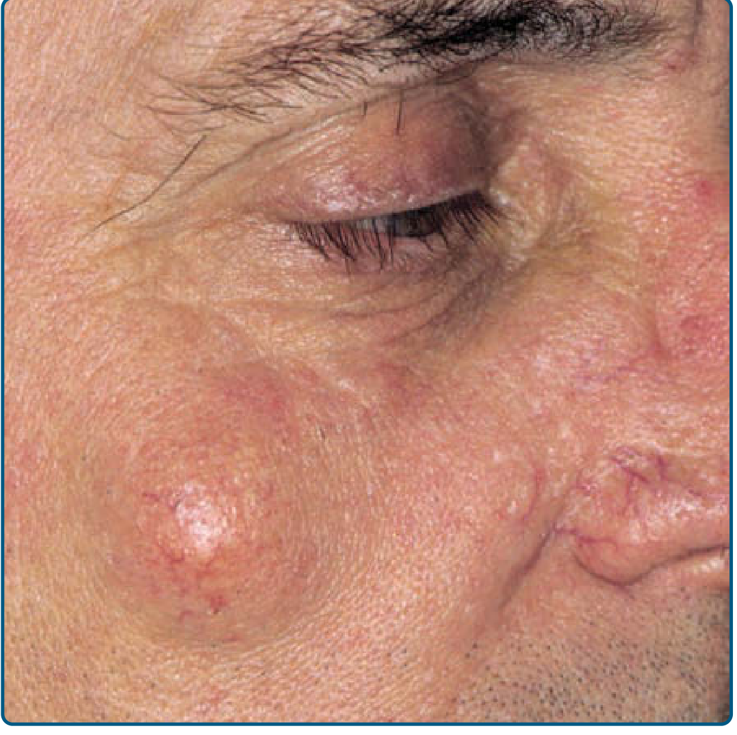
- Dermal or subcutaneous, mobile, dome-shaped nodules
- Central punctum (when present) = plugged pilosebaceous unit from which foul-smelling "cheesy" keratin debris can be expressed
- Most common sites: face, upper chest, upper back, neck, scalp
- Traumatic/implantation cysts: palms, soles, buttocks
- Typically slow-growing and asymptomatic
- Range from a few mm to several cm
Complications:
- Cyst rupture - very common; releases keratin into dermis triggering an intensely painful foreign body granulomatous inflammatory reaction
- Infection - common organisms include S. aureus, group A streptococcus, E. coli, Peptostreptococcus spp., Bacteroides spp. (polymicrobial most common)
- Malignant transformation (rare) - BCC, SCC, and epithelioid carcinoma have been reported arising within epidermoid cysts
Epidemiology
- Most common type of cutaneous cyst
- Found in adults of both sexes
- Associated genetic syndromes predisposing to multiple cysts:
- Gardner syndrome (familial adenomatous polyposis) - cysts may contain shadow cells (pilomatricoma-like) on histology
- Gorlin syndrome (nevoid BCC syndrome)
- Pachyonychia congenita type 2 (Jackson-Lawler type)
- Basal cell nevus syndrome
- Drug-induced multiple cysts/milia: cyclosporine, BRAF inhibitors (sorafenib, vemurafenib)
- Multiple scrotal epidermoid cysts can undergo dystrophic calcification causing scrotal calcinosis
Histopathology
The hallmark features are:
| Feature | Description |
|---|---|
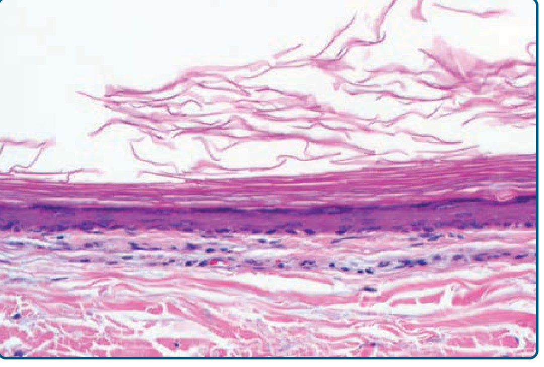
| Wall lining | Stratified squamous epithelium with a granular layer |
| Contents | Orthokeratotic (laminated) keratin debris |
| Rupture response | Foreign body reaction with multinucleated giant cells and histiocytes; suppurative + chronic granulomatous inflammation |
| Gardner syndrome variant | Columns of pilomatricoma-like shadow cells projecting into the cyst cavity |
Differential Diagnosis
| Lesion | Distinguishing features |
|---|---|
| Trichilemmal (pilar) cyst | Predominantly on scalp; wall lacks a granular layer; contents more compact; firmer |
| Steatocystoma | Expresses liquid oily material (not cheese-like); sebaceous gland lobule in wall |
| Lipoma | Softer, deeper; no punctum |
| Dermoid cyst | Along embryonic fusion planes (e.g., lateral eyebrow); contains adnexal structures |
| Milia | Tiny (1-2 mm) superficial epidermoid cysts |
Treatment
Definitive: Complete surgical excision - removes the entire cyst wall to prevent recurrence. Can be performed by:
- Simple excision (elliptical excision including overlying skin and punctum)
- Minimal incision technique (punch biopsy incision, express contents, extract wall)
Key principles:
- Excision is best performed when the cyst is "cold" (non-inflamed)
- If inflamed/ruptured: defer excision until inflammation subsides (reduces wound dehiscence risk)
- For acutely inflamed/purulent cysts: incision and drainage first; wound cultures if atypical presentation
- For small inflamed cysts: intralesional triamcinolone may speed resolution
- Incomplete removal of the cyst wall = recurrence
Proliferating Epidermoid Cyst
A rare variant in which the cyst lining undergoes proliferative change. Favors the pelvic/anogenital region. Carries a small risk of malignant transformation to SCC. Distinguished histologically by epithelial proliferation and atypia within the cyst wall. - Dermatology 2-Volume Set 5e
Sources: Fitzpatrick's Dermatology (Editors Vol. 1 & 2), p. 1843 | Dermatology 2-Volume Set 5e, Chapter 110
This is a shared conversation. Sign in to Orris to start your own chat.