
Microscopic and macro features of adult cortical bone
I'll read the medical library skill first, then search for cortical bone content in parallel.
Reading File
Now I'll run parallel searches across the library and images simultaneously.
Finding Sources
Finding Sources
Searching Images
cortical bone microscopic histology osteon Haversian system
Good results. Now I'll read the most relevant sections in parallel - the Imaging Anatomy book on cortical/trabecular bone, the Medical Physiology section, the Rheumatology microscopic level section, and Miller's orthopaedics bone section.
Reading File
Reading File
Reading File
Reading File
Excellent content found. Let me now read more of the Rheumatology microscopic section and Miller's cortical bone remodeling section, plus fetch the key histology images.
Reading File
Reading File
Loading Image
Loading Image
Excellent - I have rich content and two excellent diagrams. Let me also fetch the cortical bone remodeling image from Miller's and the macro anatomy diagram from Rheumatology, plus read the Rheumatology composition section.
Loading Image
Loading Image
Reading File
I now have comprehensive, well-sourced content with excellent diagrams. I have enough to write a thorough, well-illustrated answer.
Adult Cortical Bone: Macroscopic and Microscopic Features
Macroscopic Features
General anatomy and distribution
Cortical bone (also called compact or lamellar bone) forms the outer shell of all bones and constitutes the bulk of long bones such as the femur and tibia. It accounts for approximately 80% of total bone mass in the adult skeleton. The inner surface of cortical bone (endocortical surface) borders the marrow cavity, while the outer surface is covered by the periosteum.
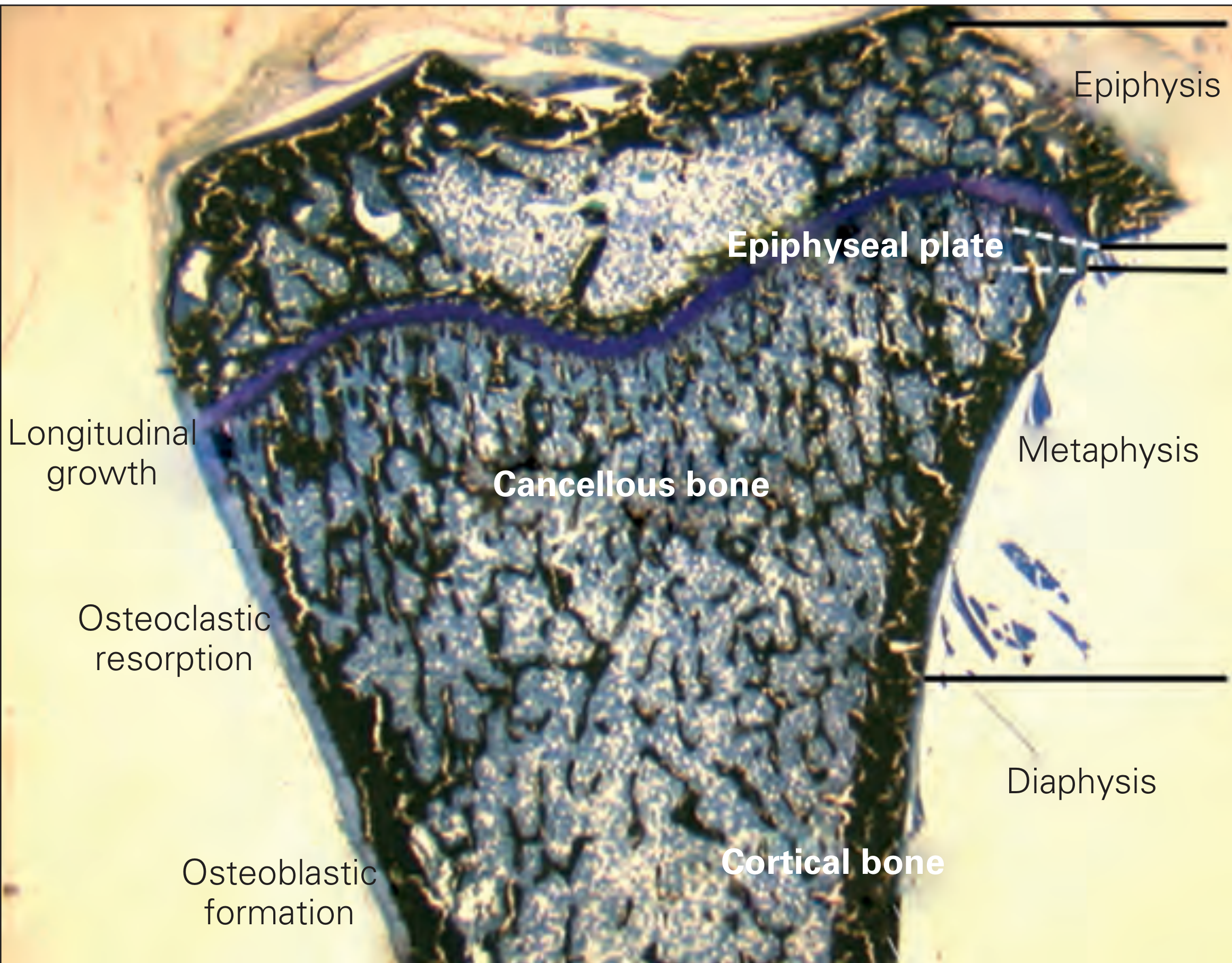
The long bone is divided into three regions where cortical bone plays different structural roles:
- Diaphysis (shaft): Almost entirely cortical bone, providing maximum mechanical strength for weight bearing
- Metaphysis: Transition zone where cortical walls thin and trabecular bone becomes more prominent
- Epiphysis: Thin cortical shell surrounding predominantly cancellous bone
Rheumatology (Firestein et al., 2022 Elsevier) - Fig. 3.1:
Gross appearance
Cortical bone is ivory-white, very dense, and hard - cut surfaces appear smooth without visible pores to the naked eye. It is surrounded on its outer aspect by the periosteum (two layers: an outer fibrous layer and an inner cambial/osteogenic layer). The inner surface lines the medullary canal (endosteum).
Mechanical properties
Cortical bone is anisotropic - it is stiffer parallel to its osteon orientation (Young's modulus E ≈ 17 GPa longitudinally) than in the transverse direction (E ≈ 12 GPa). This transverse anisotropy reflects the longitudinal alignment of osteons along the long axis of bone. It provides the primary structural support for weight bearing in long bones.
Microscopic Features
The Fundamental Unit: The Osteon (Haversian System)
The fundamental structural unit of cortical bone is the secondary osteon (Haversian system) - a tube-like structure oriented parallel to the long axis of the bone.
Diagrams of cortical bone microscopic structure:
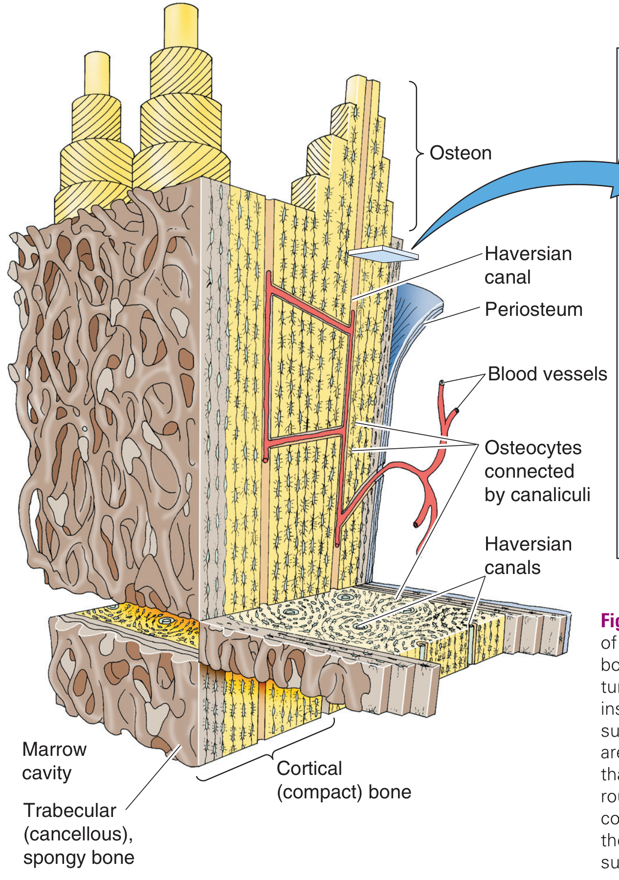
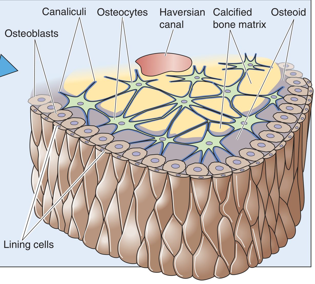
Structure of a secondary osteon:
| Component | Details |
|---|---|
| Haversian canal | Central vascular canal, 50-90 µm in diameter |
| Concentric lamellae | Rings of mineralized collagen surrounding the canal (typically 5-20 rings per osteon) |
| Lacunae | Lens-shaped spaces within lamellae housing osteocytes |
| Canaliculi | Microscopic channels radiating from lacunae, connecting osteocytes to each other and to the canal surface |
| Cement line | Outer boundary of the osteon, composed largely of sulfated glycosaminoglycans; effectively arrests microcracks |
Canals and vasculature:
- Haversian canals run longitudinally along the bone's long axis
- Volkmann's canals (perforating canals) run obliquely/transversely, connecting Haversian canals to each other and to the periosteal and endosteal surfaces
- The blood vessels in Haversian canals are fenestrated capillaries lined by an incomplete endothelial layer - they lack smooth muscle. About 60% of canals also contain 2-7 unmyelinated or poorly myelinated nerve fibers (sensory and sympathetic), which express neuropeptides involved in bone remodeling
Lamellar Organization
Each osteon is built from concentric lamellae - sheets of highly organized, mineralized collagen. Within each lamella, collagen fibrils run parallel to one another but at a different angle from adjacent lamellae (the "twisted plywood" or orthogonal arrangement). This alternating fiber orientation gives cortical bone its resistance to multidirectional stresses and is the structural basis of its birefringence under polarized light microscopy.
Types of lamellae in cortical bone:
- Haversian lamellae - the concentric rings around the central canal of each osteon
- Circumferential lamellae - outer (subperiosteal) and inner (endocortical) sheets running around the entire bone shaft; formed by primary lamellar apposition
- Interstitial lamellae - remnants of older, partially resorbed osteons filling the gaps between newer osteons; they appear as irregular, incomplete arcs
Bone Cells
Three cell types are embedded within or on the surfaces of cortical bone:
- Osteoblasts: Cuboidal cells on bone surfaces; secrete osteoid (unmineralized organic matrix), which mineralizes to become new bone. Principal target of PTH for anabolic bone stimulation.
- Osteocytes: Former osteoblasts entombed within lacunae in the mineralized matrix. They extend processes through canaliculi, forming a communication network. They act as mechanosensors and regulate osteoblast/osteoclast activity via Wnt signaling (stimulatory) and sclerostin/Dkk1 (inhibitory). They also facilitate calcium transfer from bone interior to surface (osteocytic osteolysis).
- Osteoclasts: Large, multinucleated cells on bone surfaces responsible for resorption. Activated by RANK ligand and cytokines.
- Lining cells: Quiescent, flattened osteoblasts covering Haversian canal walls when remodeling is not active.
Chemical Composition
| Component | Proportion by weight |
|---|---|
| Mineral (carbonated hydroxyapatite) | ~65-67% |
| Organic matrix | ~22-25% |
| Water | ~10% |
The organic matrix is ~90% type I collagen (triple helix of 2α1 + 1α2 chains). The mineral - nanocrystalline carbonated apatite [Ca₁₀(PO₄)₆(OH)₂] - nucleates within the hole zones between collagen molecules and also forms extrafibrillar plates (~75% of total mineral). In cortical bone, the long axis of mineral crystals orients parallel to the long axis of the osteon. Secondary mineralization (crystal growth and maturation) continues over approximately one year after initial bone deposition.
Cortical Bone Remodeling (The Cutting Cone)
Cortical bone is continuously renewed through the basic multicellular unit (BMU), forming a "cutting cone" (also called a bone remodeling unit):
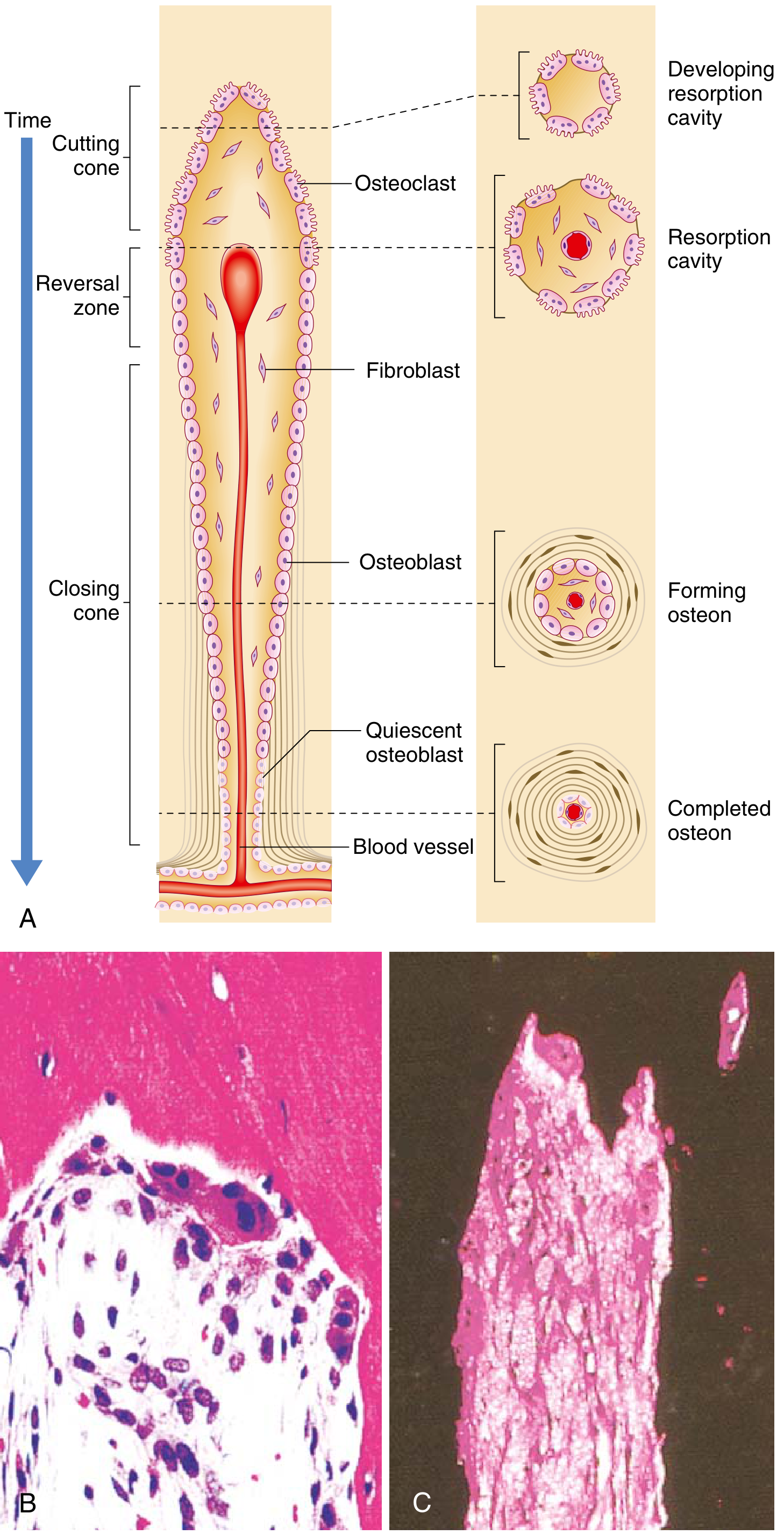
The sequence:
- Cutting cone - A team of osteoclasts tunnels longitudinally through existing cortical bone, creating a cylindrical resorption cavity (~200 µm diameter)
- Reversal zone - Osteoclasts are replaced by fibroblasts and then osteoblasts
- Closing cone - Osteoblasts line the cavity wall and progressively lay down concentric lamellae inward, eventually leaving only the narrow central Haversian canal (~50-90 µm)
- Cement line forms the outer boundary of the completed secondary osteon
The ratio of osteon diameter to canal diameter reflects the balance between resorption and formation. A secondary osteon can be distinguished from a primary osteon by the presence of a distinct cement line (primary osteons lack this).
Summary Table
| Feature | Cortical Bone |
|---|---|
| Proportion of total bone mass | ~80% |
| Main location | Diaphysis of long bones, outer shell of all bones |
| Fundamental unit | Secondary osteon (Haversian system) |
| Central canal diameter | 50-90 µm |
| Lamellar types | Haversian, circumferential, interstitial |
| Key structural protein | Type I collagen |
| Mineral | Carbonated hydroxyapatite |
| Mineral content | ~65-67% by weight |
| Vascularity | Haversian + Volkmann's canals |
| Mechanical behavior | Anisotropic; stiffer longitudinally (~17 GPa) than transversely (~12 GPa) |
| MRI signal | Low on all sequences (dense, slow-moving protons) |
Sources: Medical Physiology (Boron & Boulpaep), Rheumatology 2022 (Firestein et al.), Miller's Review of Orthopaedics 9th ed., Rockwood & Green's Fractures in Adults 10th ed. 2025.
.
Is there something you'd like to ask or follow up on?
Continue
Let me know what you'd like to continue with. A few options based on our previous topic:
- Trabecular (cancellous) bone - macro and microscopic features for comparison
- Bone composition in depth - collagen fibril structure, mineralization, noncollagenous proteins
- Bone cells in detail - osteoblast/osteoclast/osteocyte biology and signaling
- Bone remodeling - BMU cycle, coupling factors, Wolff's law
- Age-related changes in cortical bone (osteoporosis, cortical thinning, osteon accumulation)
- Something else entirely
Which direction would you like to go?
This is a shared conversation. Sign in to Orris to start your own chat.