Endometroid carcinoma

Reading File
Finding Sources
Finding Sources
Searching PubMed

"endometrioid carcinoma" AND endometrium

Reading File
Reading File
Searching Images

endometrioid carcinoma histology uterus

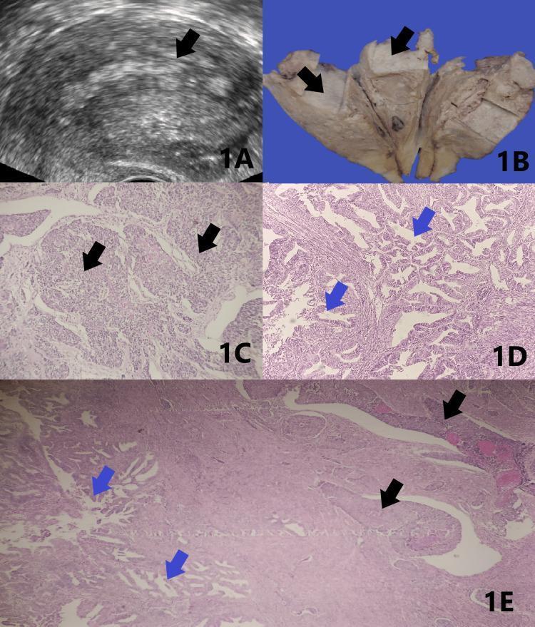
This composite figure illustrates the multi-modal diagnostic findings of dedifferentiated endometrioid carcinoma of the uterus. Panel 1A presents a pelvic ultrasonogram demonstrating a thickened, homogenous myometrium with a poorly defined endo-myometrial junction (indicated by a black arrow). Panel 1B shows a gross pathological specimen of a bivalved uterus; the cut surface reveals an irregular, grey-white endometrial tumor mass (black arrows) with deep myometrial invasion. Panels 1C, 1D, and 1E are H&E-stained photomicrographs detailing the tumor's biphasic histology. Panel 1C (black arrows) shows the undifferentiated component, characterized by solid sheets of monomorphic tumor cells lacking architectural differentiation. Panel 1D (blue arrows) depicts the well-differentiated component, featuring organized glandular structures characteristic of low-grade endometrioid adenocarcinoma. Panel 1E captures the hallmark features of this entity: an abrupt transition and juxtaposition between the well-differentiated glandular areas (blue arrows) and the solid, undifferentiated sheets (black arrows). This combination of imaging and histopathology is critical for differentiating this aggressive carcinoma from standard FIGO Grade 2 or 3 endometrial cancers.

This composite figure illustrates the multi-modal diagnostic findings of dedifferentiated endometrioid carcinoma of the uterus. Panel 1A presents a pelvic ultrasonogram demonstrating a thickened, homogenous myometrium with a poorly defined endo-myometrial junction (indicated by a black arrow). Panel 1B shows a gross pathological specimen of a bivalved uterus; the cut surface reveals an irregular, grey-white endometrial tumor mass (black arrows) with deep myometrial invasion. Panels 1C, 1D, and 1E are H&E-stained photomicrographs detailing the tumor's biphasic histology. Panel 1C (black arrows) shows the undifferentiated component, characterized by solid sheets of monomorphic tumor cells lacking architectural differentiation. Panel 1D (blue arrows) depicts the well-differentiated component, featuring organized glandular structures characteristic of low-grade endometrioid adenocarcinoma. Panel 1E captures the hallmark features of this entity: an abrupt transition and juxtaposition between the well-differentiated glandular areas (blue arrows) and the solid, undifferentiated sheets (black arrows). This combination of imaging and histopathology is critical for differentiating this aggressive carcinoma from standard FIGO Grade 2 or 3 endometrial cancers.

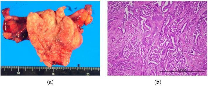
This dual-panel image displays the macroscopic and microscopic features of Grade 3 endometrioid adenocarcinoma of the uterus. Panel (a) is a clinical photograph of a gross surgical specimen (total abdominal hysterectomy) featuring a voluminous, exophytic tumor mass within the uterine corpus. The tumor exhibits a variegated light-tan to pale-red appearance with a nodular, lobulated surface and areas of apparent hemorrhage. A metric ruler indicates the primary mass is approximately 7 cm in maximum dimension. Panel (b) shows a light microscopy image (H&E stain) of the tumor at low magnification (1.0 mm scale bar). The histology demonstrates high-grade (G3) features, characterized by a complex, disordered growth pattern and back-to-back irregular glandular structures with significant loss of intervening stroma. The cellular architecture reveals significant atypia, solid growth components, and architectural complexity consistent with a high-grade endometrial malignancy. The images together illustrate the correlation between a large, invasive gross tumor and its poorly differentiated histological characteristics in the context of stage 3A endometrial carcinoma.

This dual-panel image displays the macroscopic and microscopic features of Grade 3 endometrioid adenocarcinoma of the uterus. Panel (a) is a clinical photograph of a gross surgical specimen (total abdominal hysterectomy) featuring a voluminous, exophytic tumor mass within the uterine corpus. The tumor exhibits a variegated light-tan to pale-red appearance with a nodular, lobulated surface and areas of apparent hemorrhage. A metric ruler indicates the primary mass is approximately 7 cm in maximum dimension. Panel (b) shows a light microscopy image (H&E stain) of the tumor at low magnification (1.0 mm scale bar). The histology demonstrates high-grade (G3) features, characterized by a complex, disordered growth pattern and back-to-back irregular glandular structures with significant loss of intervening stroma. The cellular architecture reveals significant atypia, solid growth components, and architectural complexity consistent with a high-grade endometrial malignancy. The images together illustrate the correlation between a large, invasive gross tumor and its poorly differentiated histological characteristics in the context of stage 3A endometrial carcinoma.

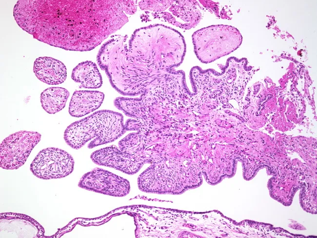
Imaging modality: Histology (Light microscopy) of uterine tissue section stained with Hematoxylin and Eosin. Specimen: uterine lesion in a paraffin-embedded tissue block; section thickness 4–5 μm; magnification range from 4x to 40x to evaluate architecture and cytology. Location: uterus, involving endometrium with adjacent fibromatous stroma; papillary fronds project into glandular spaces; stalks are sessile or papillary; epithelium is endometrioid, endocervical, or tubal type and shows no cytologic atypia. Tumor architecture is predominantly benign with fibromatous stroma supporting papillary fronds, consistent with adenofibroma. Rarely, ducts or glands may harbor foci of adenocarcinoma; presence of malignancy should be carefully assessed for invasion versus incidental focal carcinoma. Myometrial invasion is not a prominent feature in classic adenofibroma. Immunohistochemistry is not routinely required but can aid in differential diagnosis (estrogen/progesterone receptor positivity in glandular epithelium; stromal markers). Clinical relevance: benign neoplasm most often seen in postmenopausal women presenting with abnormal uterine bleeding; critical to distinguish from endometrial adenocarcinoma and atypical polypoid adenomyoma. Ethical considerations: ensure accurate reporting to prevent overtreatment. Use: educational reference for gynecologic pathology; supports differential diagnosis and surgical decision-making. This description emphasizes tissue architecture, epithelial lineage, and stromal context essential for accurate grading, prognosis, and patient management decisions overall.

Imaging modality: Histology (Light microscopy) of uterine tissue section stained with Hematoxylin and Eosin. Specimen: uterine lesion in a paraffin-embedded tissue block; section thickness 4–5 μm; magnification range from 4x to 40x to evaluate architecture and cytology. Location: uterus, involving endometrium with adjacent fibromatous stroma; papillary fronds project into glandular spaces; stalks are sessile or papillary; epithelium is endometrioid, endocervical, or tubal type and shows no cytologic atypia. Tumor architecture is predominantly benign with fibromatous stroma supporting papillary fronds, consistent with adenofibroma. Rarely, ducts or glands may harbor foci of adenocarcinoma; presence of malignancy should be carefully assessed for invasion versus incidental focal carcinoma. Myometrial invasion is not a prominent feature in classic adenofibroma. Immunohistochemistry is not routinely required but can aid in differential diagnosis (estrogen/progesterone receptor positivity in glandular epithelium; stromal markers). Clinical relevance: benign neoplasm most often seen in postmenopausal women presenting with abnormal uterine bleeding; critical to distinguish from endometrial adenocarcinoma and atypical polypoid adenomyoma. Ethical considerations: ensure accurate reporting to prevent overtreatment. Use: educational reference for gynecologic pathology; supports differential diagnosis and surgical decision-making. This description emphasizes tissue architecture, epithelial lineage, and stromal context essential for accurate grading, prognosis, and patient management decisions overall.

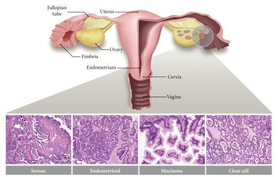
Educational medical illustration and histology panel depicting the major subtypes of epithelial ovarian carcinoma and their anatomical context. The upper section features a coronal cross-section of the female reproductive tract, including labels for the uterus, fallopian tube, fimbria, ovary, endometrium, cervix, and vagina. A shaded circle on the right ovary highlights the hypothesized site of origin for these carcinomas, showing maturing follicles and a developing tumor. The lower section consists of four Hematoxylin and Eosin (H&E) stained micrographs representing the primary histologic subtypes: 1. Serous: exhibiting complex papillary architecture and high-grade nuclear features reminiscent of fallopian tube epithelium. 2. Endometrioid: showing glandular structures resembling endometrial glands. 3. Mucinous: displaying organized glands lined by mucin-producing columnar cells similar to endocervical epithelium. 4. Clear cell: demonstrating characteristic cells with abundant clear cytoplasm. This composite image serves to correlate anatomical origin with the divergent histological manifestations of ovarian cancer.

Educational medical illustration and histology panel depicting the major subtypes of epithelial ovarian carcinoma and their anatomical context. The upper section features a coronal cross-section of the female reproductive tract, including labels for the uterus, fallopian tube, fimbria, ovary, endometrium, cervix, and vagina. A shaded circle on the right ovary highlights the hypothesized site of origin for these carcinomas, showing maturing follicles and a developing tumor. The lower section consists of four Hematoxylin and Eosin (H&E) stained micrographs representing the primary histologic subtypes: 1. Serous: exhibiting complex papillary architecture and high-grade nuclear features reminiscent of fallopian tube epithelium. 2. Endometrioid: showing glandular structures resembling endometrial glands. 3. Mucinous: displaying organized glands lined by mucin-producing columnar cells similar to endocervical epithelium. 4. Clear cell: demonstrating characteristic cells with abundant clear cytoplasm. This composite image serves to correlate anatomical origin with the divergent histological manifestations of ovarian cancer.

Reading File
Loading Image
Finding Sources
Reading File
Reading File
Here is a comprehensive overview of endometrioid carcinoma based on authoritative textbook sources:

Endometrioid Carcinoma

Overview

Endometrioid carcinoma is the most common type of endometrial carcinoma, accounting for approximately 80-85% of cases. Most are well-differentiated and microscopically mimic proliferative endometrial glands - the basis of the name. It typically arises in the setting of endometrial hyperplasia and is strongly associated with conditions of unopposed estrogenic stimulation of the endometrium. - Robbins, Cotran & Kumar Pathologic Basis of Disease, p. 933

Risk Factors / Associations

FactorNotes
ObesityMajor risk factor; particularly high in Western populations
HypertensionCommonly co-occurs
Type 2 DiabetesCo-occurs via obesity link
Unopposed estrogenExogenous or endogenous excess
Nulliparity
Anovulatory cycles
Lynch syndrome~3-5% of endometrial cancers; associated DNA mismatch repair defects
Peak incidence is in postmenopausal females aged 55-65; the incidence in younger females is increasing.

Pathogenesis & Molecular Alterations

Development follows stepwise acquisition of mutations in tumor suppressor genes and oncogenes. A hallmark is increased signaling through the PI3K/AKT pathway:
GeneFrequencyRole
PTEN (tumor suppressor)30-80%Loss of function; most common mutation
PIK3CA (oncogene)~40%Activating mutation; encodes catalytic subunit of PI3K
KRAS~25%Activating mutation; also stimulates PI3K/AKT
ARID1A~1/3Loss of function; chromatin regulator; also mutated in ovarian endometrioid/clear cell
DNA mismatch repair (MLH1)~20% sporadicEpigenetic silencing via promoter hypermethylation; prevalent in Lynch syndrome
DNA polymerase ε (POLE)<10%"Ultramutated" phenotype; highest somatic mutation burden of any cancer
PI3K/AKT signaling also augments estrogen receptor-dependent gene expression, linking molecular and hormonal pathogenesis. - Robbins p. 933

Morphology

Grossly, endometrioid carcinoma can present as a localized polypoid mass or diffusely involve the endometrial lining.
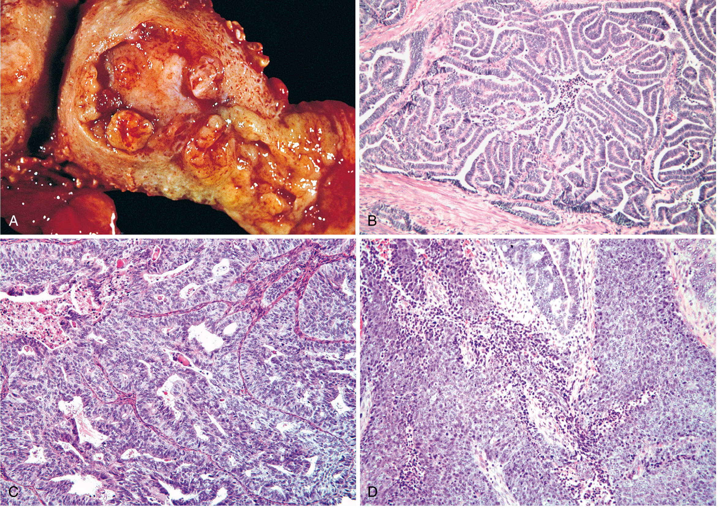
Endometrioid carcinoma - gross and histologic grades
Fig. 22.24: (A) Fungating mass in the fundus. (B) Grade 1 - well-differentiated glands, no intervening stroma. (C) Grade 2 - glands mixed with solid areas (≤50% solid). (D) Grade 3 - predominantly solid (>50% solid). - Robbins, Cotran & Kumar

Histologic Grading (FIGO)

GradeDescription
Grade 1 (Low-grade)Well-formed glands only; <5% solid non-squamous growth
Grade 2 (Low-grade)Well-formed glands + solid areas (6-50% solid)
Grade 3 (High-grade)>50% solid growth pattern
  • Grades 1 and 2 are often combined into a low-grade binary category; Grade 3 is high-grade.
  • Up to 20% contain foci of squamous differentiation (adenoacanthoma when benign-appearing squamous elements are present). Grading is based on the glandular component only.
  • Tumors with POLE mutations or DNA mismatch repair defects often show large numbers of infiltrating T cells (tumor-infiltrating lymphocytes).

Variants of Endometrioid Carcinoma

VariantFeatures
Villoglandular~2%; papillary arrangement along fibrovascular stalks; always well-differentiated; good prognosis
Secretory~1%; early postmenopausal; intracytoplasmic vacuoles like secretory endometrium; excellent prognosis; must be distinguished from clear cell carcinoma
With squamous differentiationUp to 20%; grading based on glandular component alone

Spread

  1. Myometrial invasion (depth is the key staging parameter)
  2. Direct extension to adjacent structures (broad ligaments, cervix, adnexa)
  3. Regional lymph node involvement
  4. Distant metastases (lungs, liver, bone) in late stages

Clinical Features

  • Postmenopausal vaginal bleeding - the hallmark symptom and often leads to early detection
  • May be asymptomatic early in course
  • Diagnosis by endometrial biopsy, pipelle sampling, or hysteroscopic biopsy + histologic examination
  • At diagnosis, Lynch syndrome testing (DNA mismatch repair) should be performed - 3-5% of cases have Lynch syndrome, conferring high risk for colorectal cancer

FIGO Staging (Surgical-Pathological)

StageDescriptionMRI Finding
IaConfined to corpus; <50% myometrial invasionSignal extends into <50% myometrium
IbConfined to corpus; ≥50% myometrial invasionSignal extends into ≥50% myometrium
IICervical stromal invasion (not beyond uterus)Disruption of low-signal cervical stroma
IIIaSerosa/adnexa involvedIrregular outer uterine contour
IIIbVaginal/parametrial involvementLoss of hypointense vaginal wall
IIIc1/IIIc2Pelvic/para-aortic lymph node metastasisNodes >1 cm or abnormal signal
IVaBladder/bowel mucosa invasionDisruption of bladder/rectal mucosa signal
IVbDistant metastasesTumor in distant sites
MRI is the key imaging modality for staging - it accurately assesses depth of myometrial invasion, cervical stromal invasion, and pelvic organ involvement. - Grainger & Allison's Diagnostic Radiology, p. 872

Prognosis

Stage/Grade5-Year Survival
Stage I (grade 1/2)~90%
Stage I (grade 3)~75%
Stage II-III≤50%
In the USA, ~80% of tumors are Stage I, well- or moderately differentiated at presentation - the reason for the generally favorable prognosis.

Treatment

ScenarioApproach
Localized endometrioidTotal abdominal hysterectomy + bilateral salpingo-oophorectomy (TAH + BSO) ± adjuvant radiotherapy
Adjuvant radiationReduces local recurrence in higher-risk early-stage disease
Spread beyond uterusChemotherapy added
Hypermutated tumors (MSI-H / POLE)Immune checkpoint inhibitors (e.g., pembrolizumab)
HER2-amplified (25-30% of cases)HER2-targeted therapy

Endometrioid Carcinoma vs. Serous Carcinoma (Key Comparison)

FeatureEndometrioidSerous
Frequency80-85%~15%
Hormone associationEstrogen-drivenEstrogen-independent
PrecursorEndometrial hyperplasiaEndometrial atrophy
Key mutationPTEN, PIK3CA, KRASTP53 (>90%)
GradeUsually low-gradeHigh-grade
PrognosisGenerally favorablePoor (5-year survival 18-27%)
Age at presentationYounger (55-65)Older (~10 yrs more)

Endometrioid Carcinoma of the Ovary (Bonus)

Endometrioid carcinoma also accounts for approximately 20% of all ovarian cancers. About 15-30% coexist with endometriosis, and ~15-20% coexist simultaneously with uterine endometrioid carcinoma. The peak incidence is a decade earlier than that of uterine endometrioid carcinomas associated with endometriosis. - Robbins p. 3296
This is a shared conversation. Sign in to Orris to start your own chat.