
Facial recess
Reading File
The facial recess is a key surgical landmark in the middle ear - a triangular space posterior to the tympanic membrane, bounded by the facial nerve, the chorda tympani, and the fossa incudis. It's the primary access route for posterior tympanotomy in cochlear implant and stapedectomy surgery.
Searching sources...
Finding Sources
Finding Sources
Reading File
Reading File
Reading File
Reading File
Reading File
Loading Image
Loading Image
Facial Recess
Definition and Anatomy
The facial recess (also called the posterior tympanotomy) is a triangular space located in the posterior wall of the tympanic cavity, accessible via the mastoid. Its three borders are:
| Border | Structure |
|---|---|
| Medial (posterior) | Facial nerve (vertical/mastoid segment) |
| Lateral (anterior) | Chorda tympani nerve |
| Superior | Fossa incudis (incus buttress) |
The size of the recess varies among individuals, depending on where the chorda tympani branches from the facial nerve and the degree of pneumatization of the region.
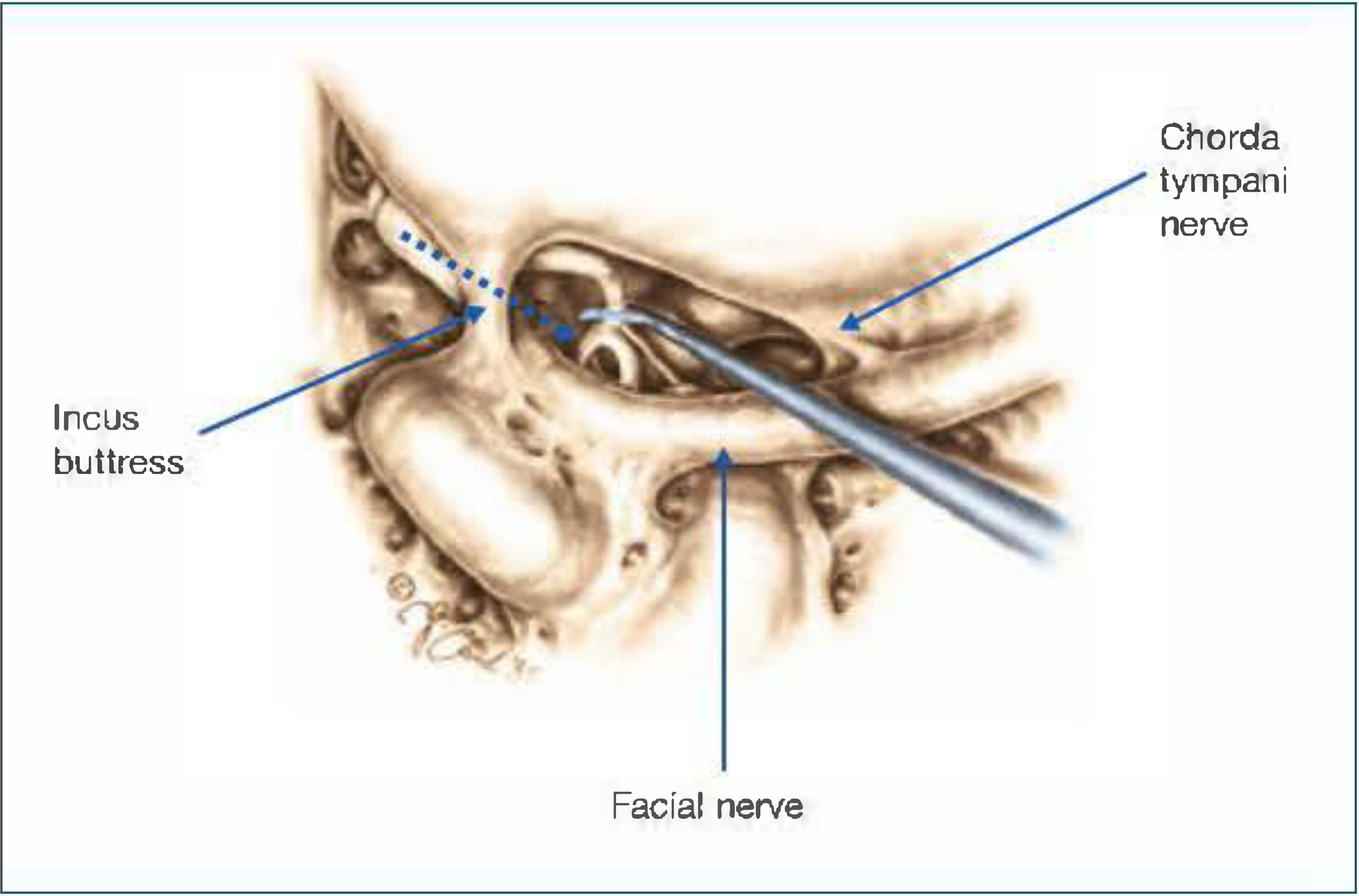
Illustration of the facial recess - triangular space bordered by the incus buttress superiorly, chorda tympani nerve anterolaterally, and facial nerve posteriorly.
Surgical Landmarks and Approach
Before opening the facial recess, several key landmarks must be identified:
- Lateral semicircular canal - lies just superior to the facial nerve at the second genu
- Short process of the incus - points directly toward the facial recess and serves as a reliable guide
- Digastric ridge - another landmark for the vertical segment of the facial nerve
- "Herald" air cell - a single air cell typically encountered just lateral to the second genu of the facial nerve, even in sclerotic bones
Technique:
- Thin the posterior canal wall first
- Expose the facial nerve using a large diamond burr with copious irrigation, always stroking parallel to the nerve - never perpendicular
- Identify the chorda tympani as it branches from the vertical facial nerve segment, and trace it superiorly toward the incus
- Open the recess with a 2-mm diamond burr, starting superiorly where the space is widest
- Drill in an anteromedial direction, preserving a small bone sliver just inferior to the incus to protect it from the burr
Caution: Even light contact of the drill with the incus (when the ossicular chain is intact) can cause significant sensorineural hearing loss. Using a small curette or pick for the final bone removal near the incus is advisable.
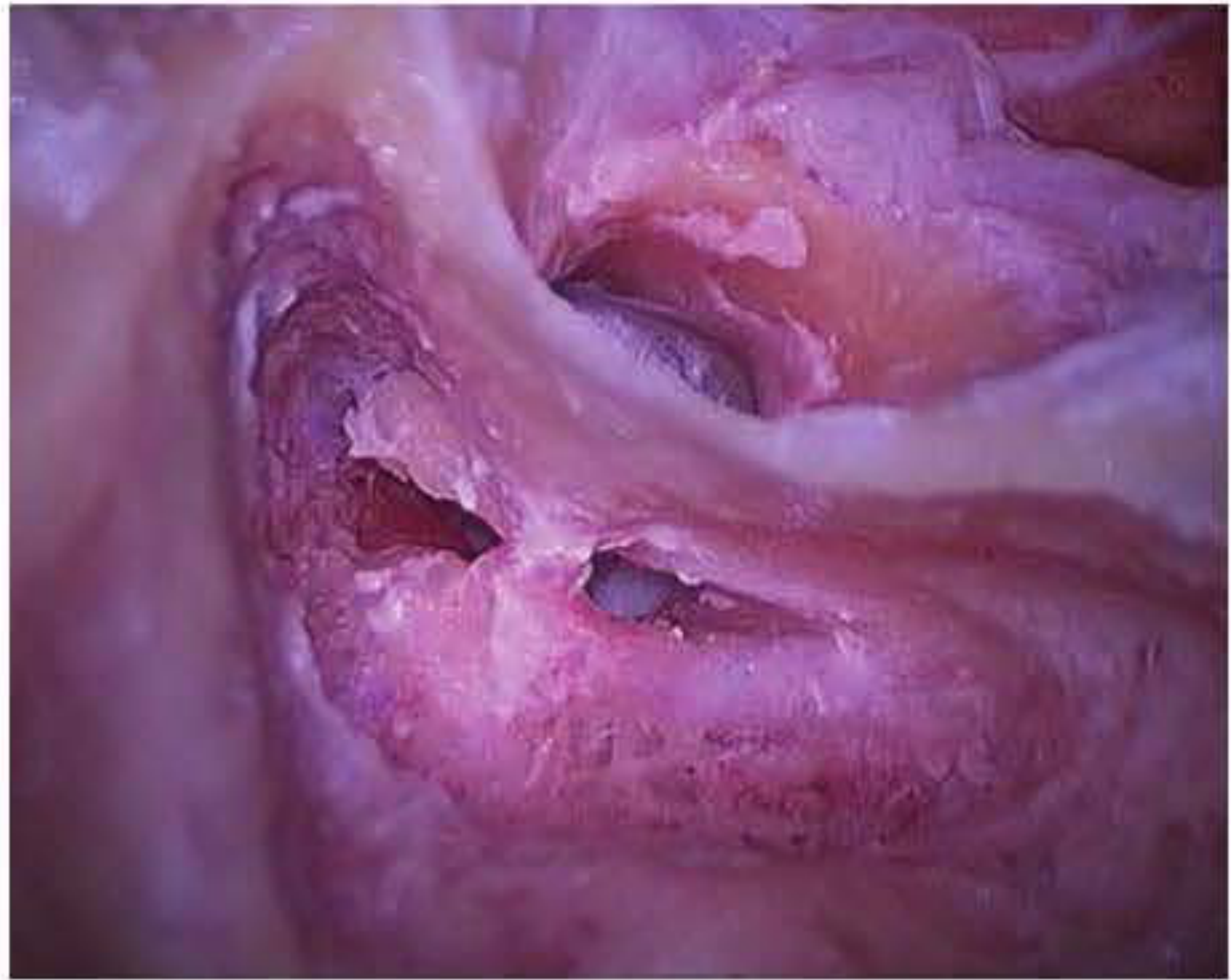
Intraoperative view: facial recess fully developed between the facial and chorda tympani nerves.
Structures Visible After Opening
Once the facial recess is opened, the following middle ear structures can be visualized:
- Pyramidal process and stapedial tendon
- Stapes superstructure
- Long process of the incus
- Oval and round windows
- Promontory
- Incudostapedial joint
- Cochleariform process
- Medial surface of the tympanic membrane
- Eustachian tube orifice
The sinus tympani remains hidden and cannot be seen through this approach.
Extended Facial Recess
For greater access to the hypotympanum, the facial recess can be extended by sacrificing the chorda tympani nerve:
- The chorda is transected at its origin from the facial nerve
- Drilling continues inferiorly along the fallopian canal with a small diamond burr and copious irrigation
- The lateral limit of dissection becomes the tympanic annulus
- The facial nerve courses laterally at the inferior extent, so superior-to-inferior drilling direction avoids injury
Indications for the extended facial recess include:
- Access to the hypotympanum
- Jugular bulb region (e.g., resection of glomus tympanicum tumors)
Clinical Uses
| Indication | Role of Facial Recess |
|---|---|
| Cochlear implantation | Standard transmastoid-facial recess approach to reach the round window for electrode insertion |
| Cholesteatoma (CWU mastoidectomy) | Used when disease extends into the mesotympanum via this route |
| Ossiculoplasty | Allows accurate disarticulation of the incudostapedial joint |
| Glomus tympanicum | Extended facial recess for hypotympanum access |
| Cochlear malformations | Majority can be implanted via the standard transmastoid-facial recess technique |
Sources: Shambaugh Surgery of the Ear, p. 534; p. 807 | Cummings Otolaryngology Head and Neck Surgery, Cochlear Implantation chapter
This is a shared conversation. Sign in to Orris to start your own chat.