
Describe and discuss function of protein and structure-function relationship of haemoglobin.
I'll start by reading the medical library skill, then search for comprehensive information on haemoglobin structure and function.
Reading File
Now I'll run parallel searches across the library for haemoglobin structure, function, and cooperative binding.
Finding Sources
Finding Sources
Searching Images
haemoglobin structure quaternary subunits heme oxygen binding
Excellent. I have four highly relevant textbook sections. Let me read them all in parallel for the most comprehensive coverage.
Reading File
Reading File
Reading File
Reading File
I have rich content. Let me read the remaining truncated sections and also fetch the key structural images.
Reading File
Reading File
Reading File
Loading Image
Excellent. I now have detailed content on structure and cooperativity. Let me get the remaining content on allosteric effectors (Bohr effect, 2,3-BPG) and fetch key images.
Reading File
Loading Image
Loading Image
I now have comprehensive content. Let me get the rest of the allosteric effectors section (Bohr effect, 2,3-BPG, CO2) and the final Harper's section.
Reading File
Reading File
I have comprehensive material. Let me get the 2,3-BPG section and fetch the iron-movement diagram.
Reading File
Loading Image
I now have all the information needed for a thorough, well-illustrated answer. Let me compile the comprehensive response.
Haemoglobin: Structure, Function, and Structure-Function Relationships
1. Overview and Biological Role
Haemoglobin (Hb) is the principal oxygen-carrying protein found exclusively in red blood cells (RBCs). Its key functions are:
- O2 transport: carries four molecules of O2 from the lungs to peripheral tissues
- CO2 transport: returns CO2 (as carbamino compounds and by facilitating bicarbonate formation) from tissues to the lungs
- H+ buffering: acts as a significant blood buffer via the Bohr effect
- Proton transport: carries H+ generated in metabolically active tissues back to the lungs
O2 is only slightly soluble in aqueous media. Without haemoglobin, diffusion alone would limit organismal size. The circulatory system overcomes this limit only when paired with a transport protein that can load O2 at the lung and unload it precisely where it is needed.
2. Primary and Secondary Structure
Each haemoglobin polypeptide chain (globin) contains stretches of alpha-helical secondary structure - classically 8 helical segments (labelled A-H) in each beta-chain, and 7 in each alpha-chain. These helices fold to create a hydrophobic pocket that cradles the heme prosthetic group. The primary structures of the beta, gamma, and delta chains are highly conserved across human haemoglobin variants, reflecting the functional constraints on the heme-binding pocket.
3. Tertiary Structure: The Globin Fold and the Heme Group
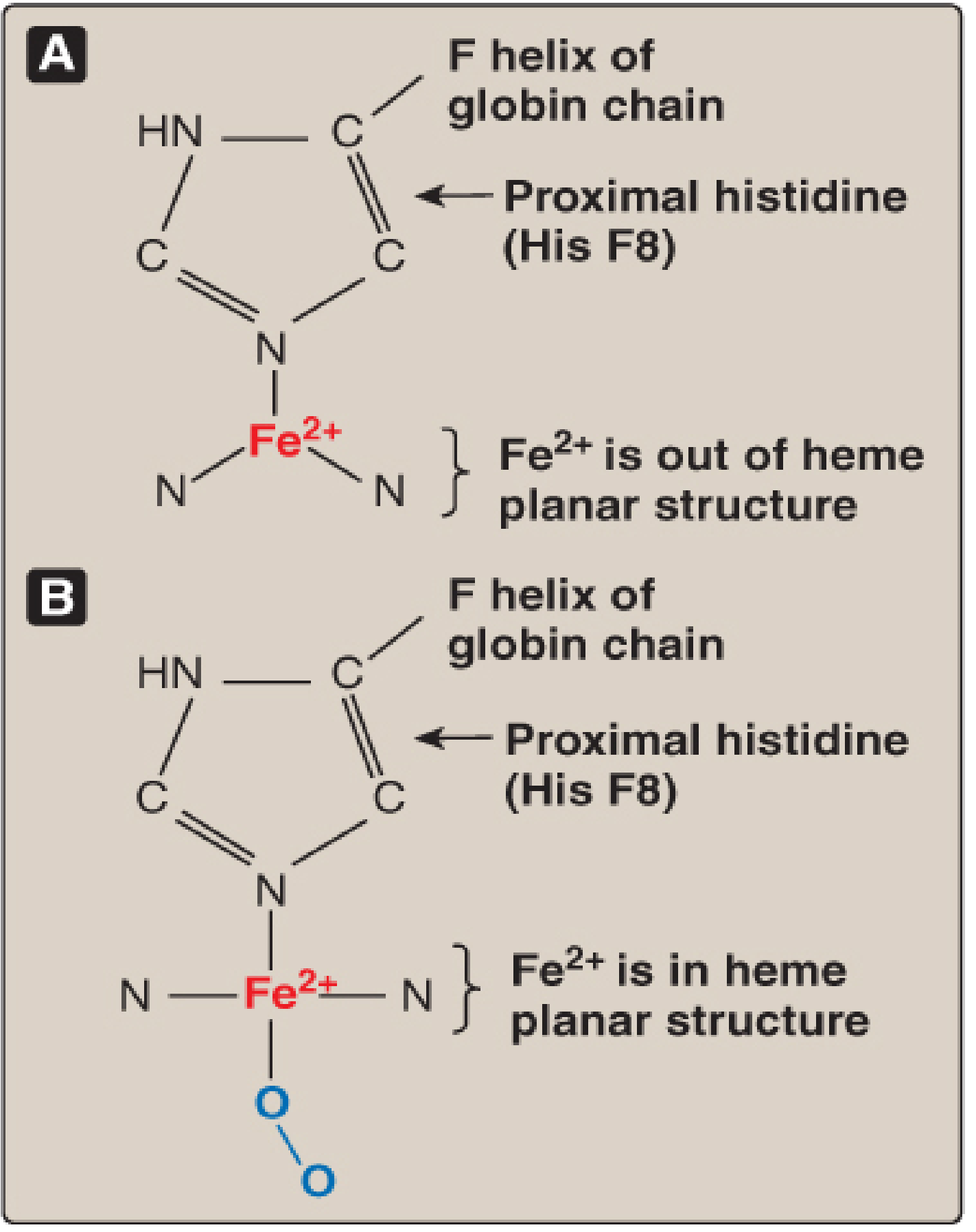
Each subunit folds into a compact, roughly globular tertiary structure. The heme group - a protoporphyrin ring containing a central ferrous iron atom (Fe²⁺) - is tucked into a hydrophobic cleft. The iron has six coordination bonds:
- Four bonds to the pyrrole nitrogens of the porphyrin ring
- One bond to the imidazole nitrogen of the proximal histidine (His F8) of the globin chain (position 87 in alpha-chains; 92 in beta-chains)
- One reversibly bonds to O2 (the sixth coordinate position, on the distal side)
The proximal histidine is the structural linchpin connecting the heme iron to the globin polypeptide. In the deoxygenated state, the Fe²⁺ is slightly domed out of the plane of the porphyrin ring. Upon O2 binding, the iron is pulled into the plane - a small movement (~0.04 nm) that is physically transmitted through the proximal histidine and the F-helix to the entire protein, triggering the conformational change that underlies cooperativity.
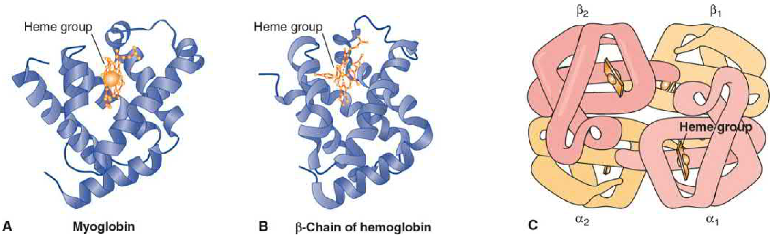
The tertiary structure of the beta-chain of haemoglobin is almost identical to that of myoglobin, despite differences in amino acid sequence - a striking example of how tertiary fold determines function.
4. Quaternary Structure: The Tetramer
Haemoglobin A (HbA), the predominant adult form, is a tetramer composed of two alpha (α) and two beta (β) polypeptide chains (α₂β₂), giving a molecular weight of ~64,500 Da. The tetramer is best understood as two identical αβ dimers held together with different bond types:
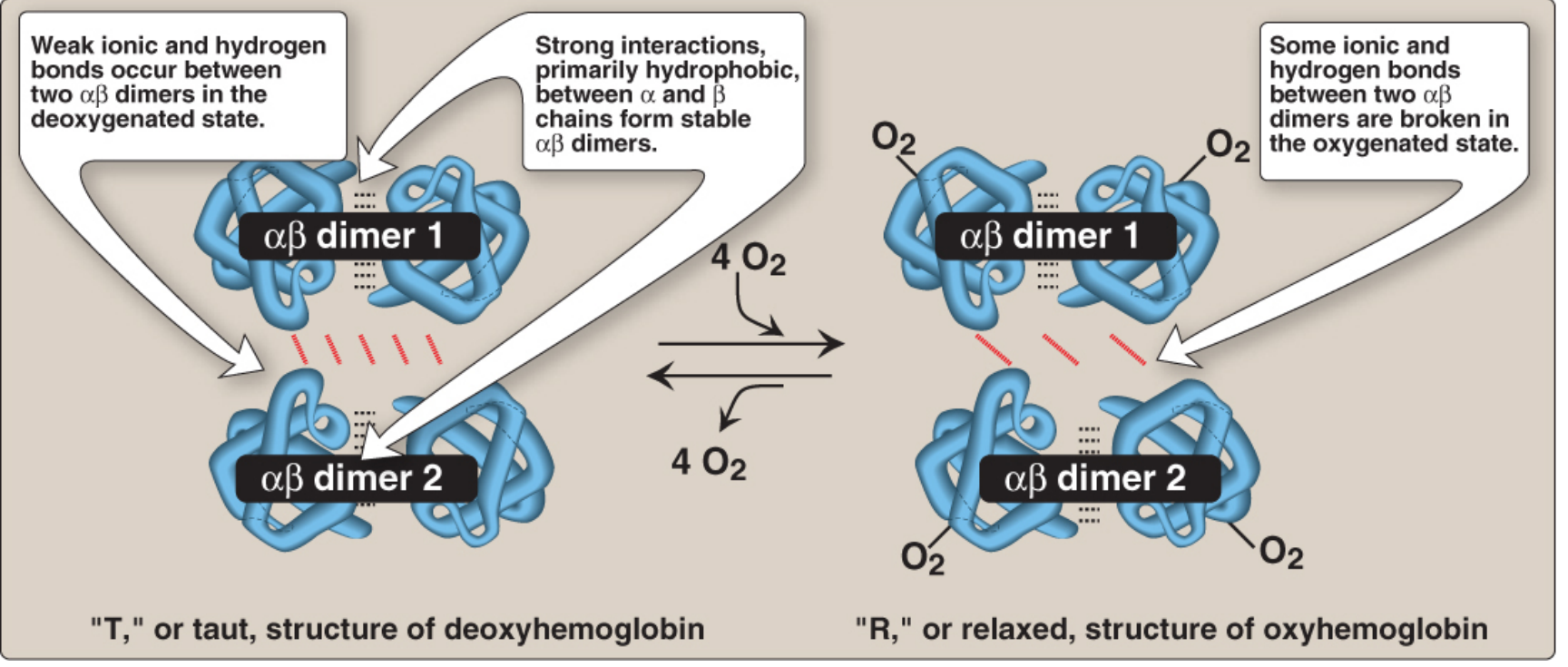
- Within each αβ dimer: primarily strong hydrophobic interactions between the α and β chains - these contacts are tight and stable
- Between the two αβ dimers: primarily polar bonds (ionic bonds and hydrogen bonds) - these are weaker and allow relative movement
This asymmetry is functionally critical: the weak inter-dimer interface permits the two dimers to rotate relative to each other during oxygenation, generating the allosteric conformational switch.
Major Haemoglobin Types
| Form | Composition | Context |
|---|---|---|
| HbA | α₂β₂ | Normal adult (major form) |
| HbA₂ | α₂δ₂ | Minor adult form (~2.5%) |
| HbF | α₂γ₂ | Fetal haemoglobin |
| HbS | α₂βS₂ | Sickle-cell haemoglobin |
The fetal form (HbF) has higher O2 affinity (P50 ~20 mmHg vs 26 mmHg for HbA) because its gamma chains have lower affinity for 2,3-BPG. This enables HbF to extract O2 from maternal HbA across the placenta.
5. The T and R States: Conformational Switch
The defining feature of haemoglobin's quaternary structure is that it exists in two interconvertible conformations:
| State | Name | Character | Oxygen Affinity |
|---|---|---|---|
| Deoxy | T (Taut/Tense) | Ionic bonds + H-bonds constrain both αβ dimers; Fe²⁺ out of heme plane | Low |
| Oxy | R (Relaxed) | Polar inter-dimer bonds rupture; Fe²⁺ moves into heme plane; dimers rotate 15° | High |
When the first O2 binds to heme Fe²⁺, the iron moves into the porphyrin plane, dragging the proximal histidine and F-helix with it. This mechanical signal is propagated through the subunit interface, rupturing some of the ionic salt bridges that locked the T state. The dimers rotate ~15° relative to each other, compacting the tetramer into the R state. Each subsequent O2 binding requires rupture of fewer remaining salt bridges, so affinity progressively increases.
6. Cooperative O2 Binding and the Sigmoidal Dissociation Curve
Myoglobin vs Haemoglobin
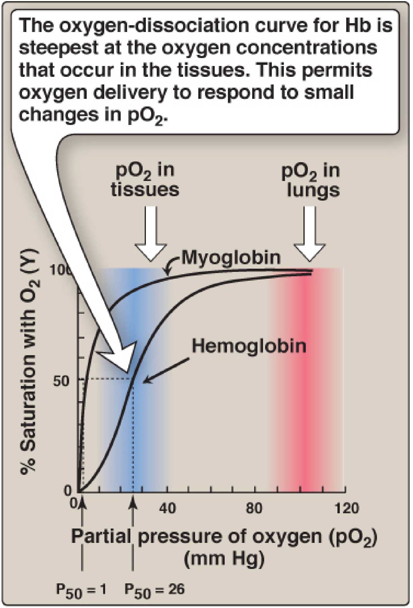
Myoglobin is a monomer with a hyperbolic O2-dissociation curve (P50 ~1 mmHg). It binds O2 with high affinity at all pO2 values and stores O2 in muscle. Haemoglobin's curve is sigmoidal (S-shaped), reflecting its cooperative, allosteric behaviour.
Why the Sigmoidal Curve Matters
The sigmoidal curve confers a dramatic physiological advantage:
- At pO2 ~100 mmHg (lungs): Hb is ~98% saturated - efficiently loads O2
- At pO2 ~40 mmHg (resting tissue): Hb drops to ~75% saturation - releases ~25% of its O2
- At pO2 ~20 mmHg (exercising muscle): saturation falls to ~30%, releasing much more O2
The steep slope of the curve precisely in the range of tissue pO2 values means that small decreases in pO2 (from increased metabolic activity) trigger large increases in O2 unloading. A hyperbolic curve (like myoglobin) would remain nearly saturated throughout this range and deliver very little O2 to the tissues.
The net effect of cooperativity is that haemoglobin's affinity for the last O2 molecule bound is approximately 300 times greater than its affinity for the first.
7. Allosteric Effectors
Allosteric effectors bind at sites distinct from the heme and alter O2 affinity by stabilising either the T or R conformation.
7a. Oxygen itself
O2 is its own allosteric effector. Each bound O2 molecule progressively shifts the remaining subunits toward the R state (higher affinity). This positive cooperativity underpins the sigmoidal curve.
7b. The Bohr Effect (H⁺ and CO2)
Lowering pH (raising H⁺) or raising pCO2 decreases O2 affinity, shifting the dissociation curve to the right (stabilises T state). This is the Bohr effect.
Mechanism: Deoxyhemoglobin has a greater affinity for H⁺ than oxyhemoglobin. Key ionisable groups (particularly specific histidine side chains) have a higher pKa in the T state. When H⁺ concentration rises (as in active tissues where CO2 is produced), these residues become protonated and form ionic salt bridges that lock the T conformation and reduce O2 affinity.
In metabolically active capillaries:
- CO2 produced by respiration enters RBCs
- Carbonic anhydrase converts it: CO2 + H2O → H2CO3 → HCO3⁻ + H⁺
- The H⁺ lowers intracellular pH, enhancing O2 release
- In the lungs, CO2 is exhaled, pH rises, and O2 affinity is restored
This differential pH gradient between lungs (pH ~7.4) and active tissues (lower pH) is a self-regulating mechanism: the more a tissue respires, the more O2 haemoglobin delivers to it.
Schematically: HbO2 + H⁺ ⇌ HbH⁺ + O2
CO2 is also carried directly on haemoglobin as carbamino compounds, binding to the N-terminal amino groups of the globin chains (not the heme iron) and further stabilising the T state.
7c. 2,3-Bisphosphoglycerate (2,3-BPG)
2,3-BPG is the most abundant organic phosphate in RBCs and is synthesised from an intermediate of glycolysis (1,3-BPG via bisphosphoglycerate mutase).
- 2,3-BPG binds in the central cavity of the deoxyhemoglobin tetramer, in a positively charged pocket formed by the two β-chains (Lys 82β, His 143β, and His 2β on each chain)
- It does not fit in the narrower cavity of oxyhemoglobin (R state), so it is released upon oxygenation
- Net effect: 2,3-BPG preferentially stabilises the T (deoxy) state, lowering O2 affinity and shifting the curve to the right
Schematically: HbO2 + 2,3-BPG ⇌ Hb-2,3-BPG + O2
Physiological significance of 2,3-BPG:
- Stagnant hypoxia (e.g. heart failure), anaemia, high altitude: 2,3-BPG levels rise, rightward shift, more O2 delivered to tissues
- HbF lacks affinity for 2,3-BPG (gamma chains substitute Ser for His at position 143): hence HbF has higher O2 affinity than HbA, enabling O2 extraction from maternal blood
- Stored blood: 2,3-BPG declines during storage, raising O2 affinity and reducing O2 delivery - clinically relevant in transfusion medicine
8. CO2 Transport and Role as a Buffer
Beyond O2 transport, haemoglobin plays a central role in CO2 removal:
- ~70% of CO2 is converted to bicarbonate (HCO3⁻) in RBCs by carbonic anhydrase and transported in plasma
- ~20% binds directly to haemoglobin as carbamino-haemoglobin
- ~10% dissolves directly in plasma
When T-state haemoglobin forms in peripheral tissues (after releasing O2), it binds two protons per tetramer via salt bridges (between Asp 94 and His 146 of the β-chains). This proton uptake:
- Buffers the pH drop from CO2 accumulation in RBCs
- Pulls the HCO3⁻/H2CO3 equilibrium toward bicarbonate (more CO2 uptake)
In the lungs, the reverse happens: the R state releases protons, favoring H2CO3 → CO2 + H2O, and CO2 is exhaled.
9. Structure-Function Summary Table
| Structural Feature | Functional Consequence |
|---|---|
| Tetrameric (α₂β₂) quaternary structure | Cooperativity; sigmoidal O2 binding; allosteric regulation |
| Hydrophobic heme pocket | Protects Fe²⁺ from oxidation; enables reversible O2 binding |
| Proximal His F8 - Fe²⁺ bond | Transmits O2-binding signal to entire polypeptide chain |
| Weak polar inter-dimer bonds | Allows T→R conformational switch upon oxygenation |
| Strong hydrophobic intra-dimer bonds | Maintains αβ dimer integrity as the functional unit |
| Central cavity (β-chain pocket) | 2,3-BPG binding site; T-state stabilisation |
| Ionisable histidine residues | Bohr effect; H⁺ buffering |
| N-terminal amino groups of globin chains | Carbamino-CO2 binding |
10. Clinical Correlates
- Sickle cell disease (HbS): glutamate→valine substitution at beta-6 creates a hydrophobic patch; deoxy HbS polymerises, distorting RBCs into sickled shapes
- Methaemoglobinaemia: Fe²⁺ oxidised to Fe³⁺ (metHb); Fe³⁺ cannot bind O2; reduces oxygen-carrying capacity
- CO poisoning: CO binds Fe²⁺ ~240× more avidly than O2 and shifts remaining subunits to high-affinity R state, preventing O2 unloading
- High altitude: chronic low pO2 → elevated 2,3-BPG → rightward shift → more O2 delivered to tissues
- Pulse oximetry: exploits the different light absorption spectra of oxyHb and deoxyHb to non-invasively estimate O2 saturation
Sources:
- Biochemistry, 8th ed. Lippincott Illustrated Reviews, Ch. 3 (Hemoglobin structure and function)
- Basic Medical Biochemistry: A Clinical Approach, 6th ed., Ch. 7 (Structure-Function Relationships in Myoglobin and Hemoglobin)
- Harper's Illustrated Biochemistry, 32nd ed., Ch. 6 (Proteins: Myoglobin and Hemoglobin)
- Henry's Clinical Diagnosis and Management by Laboratory Methods (Structure and Function of Hemoglobin)
This is a shared conversation. Sign in to Orris to start your own chat.