
I have to write a 10 mark answer on active transport and factors affecting facilitated diffusion
Reading File
Now let me search the medical library for both topics simultaneously.
Finding Sources
Finding Sources
Now let me read the key sections from Guyton & Hall (the most authoritative physiology source) on both topics simultaneously.
Reading File
Reading File
Now let me get the primary active transport section from Guyton & Hall block 1, and also read more on factors affecting diffusion.
Reading File
Reading File
Now let me read the secondary active transport section and also fetch the key diagrams.
Reading File
Loading Image
Loading Image
I now have comprehensive material from Guyton & Hall to write a thorough 10-mark answer. Let me compile this into a well-structured exam answer.
Active Transport and Factors Affecting Facilitated Diffusion
(Source: Guyton and Hall Textbook of Medical Physiology)
PART 1: ACTIVE TRANSPORT
Active transport is the movement of substances across cell membranes against their electrochemical gradients (from low to high concentration). Unlike passive processes, active transport requires metabolic energy. It is divided into two types: primary and secondary active transport.
A. Primary Active Transport
Primary active transport is directly coupled to an energy source - specifically, the hydrolysis of ATP (adenosine triphosphate). The transport protein itself has ATPase activity. Known primary active transporters in the body include:
- Na⁺-K⁺ ATPase
- H⁺ ATPase
- H⁺-K⁺ ATPase
- Ca²⁺ ATPase
The Sodium-Potassium Pump (Na⁺-K⁺ ATPase) - The Classic Example
The Na⁺-K⁺ pump is the most studied primary active transporter. It is a transmembrane protein complex made of:
- A larger alpha (α) subunit (~100,000 molecular weight) - contains 3 Na⁺ binding sites on the intracellular side, 2 K⁺ binding sites on the extracellular side, and ATPase activity
- A smaller beta (β) subunit (~55,000 molecular weight) - anchors the complex in the lipid membrane
Mechanism:
- Three Na⁺ ions bind to the intracellular sites, and two K⁺ ions bind to the extracellular sites
- ATPase is activated → cleaves one ATP molecule → releases high-energy phosphate
- This energy causes a conformational change in the protein
- 3 Na⁺ are extruded outward; 2 K⁺ are transported inward
- Net result: low intracellular Na⁺, high intracellular K⁺, and a resting membrane potential of about -70 mV
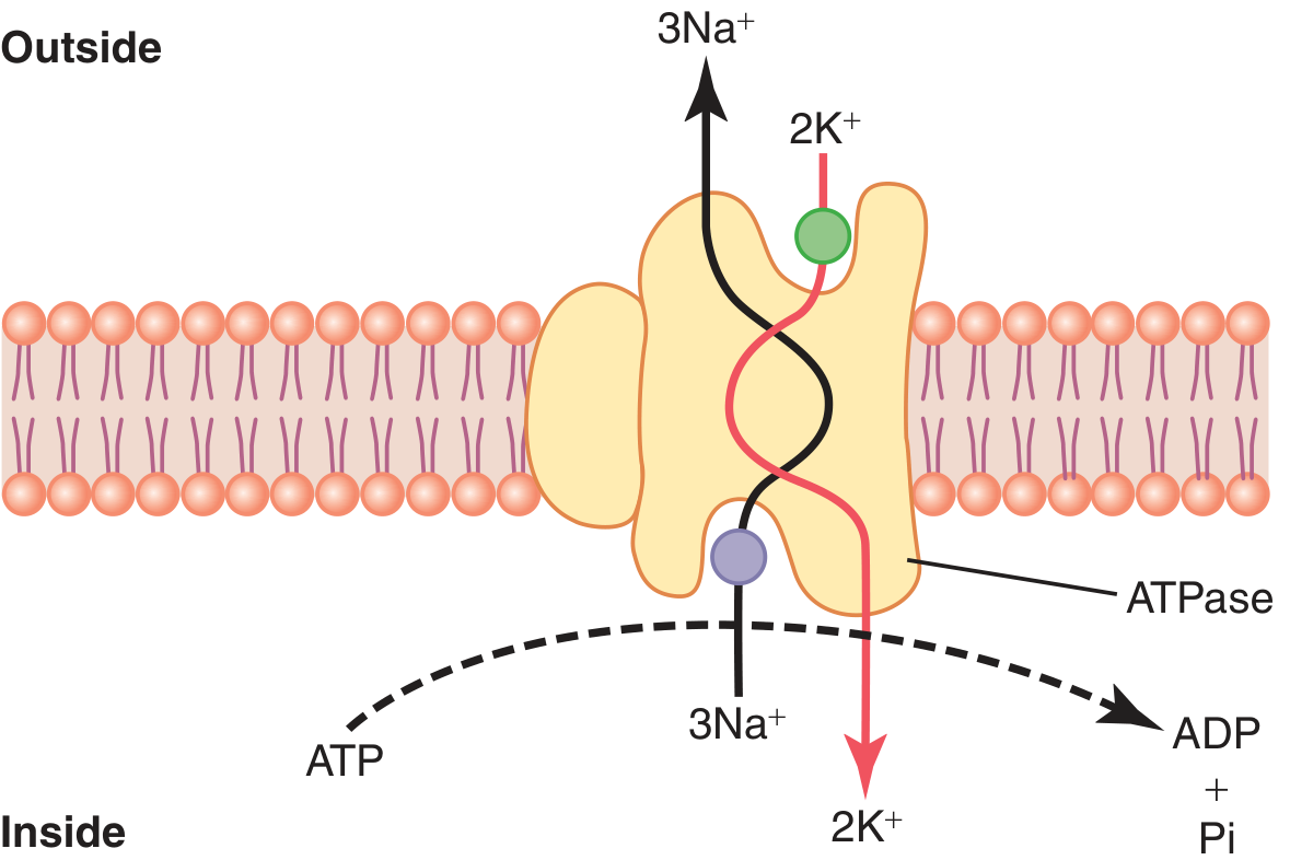
Functions of the Na⁺-K⁺ pump:
- Maintains Na⁺/K⁺ concentration gradients across cell membranes
- Establishes the negative intracellular electrical potential
- Controls cell volume - prevents osmotic swelling and lysis
- Powers nerve signal transmission (60-70% of energy in nerve cells goes to this pump)
B. Secondary Active Transport
Secondary active transport uses the energy stored in an ion gradient (created by primary active transport) rather than directly hydrolyzing ATP. It has two forms:
1. Co-transport (Symport)
Both the driving ion (Na⁺) and the transported substance move in the same direction across the membrane.
Example - Sodium-glucose co-transport (SGLT):
- High extracellular Na⁺ (maintained by the Na⁺-K⁺ pump) provides the driving force
- A carrier protein has two binding sites on the exterior: one for Na⁺, one for glucose
- When both bind simultaneously, a conformational change occurs transporting both into the cell
- This moves glucose against its concentration gradient using Na⁺'s downhill gradient
- This is critical in the intestinal epithelium and renal tubules for glucose absorption
- A similar mechanism exists for amino acid absorption (at least 5 types of amino acid co-transporters)
2. Counter-transport (Antiport)
The driving ion (Na⁺) and the transported substance move in opposite directions.
Example - Na⁺/Ca²⁺ exchanger:
- Na⁺ moves inward down its gradient; Ca²⁺ is simultaneously transported outward
- Critically regulates intracellular Ca²⁺ concentration in cardiac and smooth muscle cells
Example - Na⁺/H⁺ exchanger:
- Na⁺ moves in; H⁺ is extruded outward
- Important in acid-base regulation
Key distinction: In secondary active transport, energy comes indirectly from the Na⁺ gradient established by the primary Na⁺-K⁺ pump. The process is therefore still energy-dependent, just one step removed.
PART 2: FACTORS AFFECTING FACILITATED DIFFUSION
Facilitated diffusion is carrier-mediated passive transport - it moves substances down their concentration gradient (no energy required) using specific membrane carrier proteins. Unlike simple diffusion, it shows saturation kinetics.
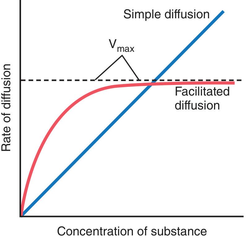
Mechanism of the carrier protein: The substance binds to a receptor inside a pore of the carrier protein. A conformational change opens the pore to the other side of the membrane, and the molecule is released by the weak binding force due to thermal motion. The carrier then reverts to its original conformation.
Factors That Affect the Rate of Facilitated Diffusion
1. Concentration Gradient (most important factor)
- The rate of facilitated diffusion is proportional to the concentration difference (Co - Ci) between outside and inside the cell
- Net diffusion ∝ (Co - Ci)
- A larger gradient → faster net transport rate
- However, unlike simple diffusion, this relationship is not linear - it approaches a maximum (V_max) as the carrier becomes saturated
2. Number and Availability of Carrier Proteins (Transport Maximum / V_max)
- Each carrier protein can only flip conformation at a finite rate
- When all carriers are occupied by substrate molecules, the maximum transport rate (V_max) is reached - this is called saturation or the transport maximum (Tm)
- Increasing the number of carrier proteins in the membrane increases V_max
- Example: Insulin stimulates the translocation of GLUT4 glucose transporters from intracellular vesicles to the plasma membrane, increasing membrane carrier density and raising V_max for glucose uptake in skeletal muscle, adipose tissue, and heart by 10- to 20-fold
3. Specificity of the Carrier Protein
- Each carrier is specific for a particular molecule or group of structurally similar molecules
- For example, GLUT transporters handle glucose, galactose, and fructose (structurally similar monosaccharides) but not other sugars
- Competitive inhibition can occur when two similar substances compete for the same carrier
4. Temperature
- Higher temperature increases the kinetic (thermal) energy of molecules
- This increases both the rate of molecular collisions with the carrier and the speed of conformational changes in the carrier protein
- Within physiological range, increased temperature generally increases the rate of facilitated diffusion
5. Electrical Potential Across the Membrane (for ions)
- For charged particles, the electrochemical gradient (not just concentration) determines net movement
- An electrical potential difference can either enhance or oppose diffusion of ions through their carriers
- At the Nernst potential, electrical and concentration forces exactly balance, producing no net diffusion
6. Pressure Difference
- Increased hydrostatic pressure on one side of the membrane increases the force with which molecules strike the membrane
- This slightly increases the rate of diffusion (including facilitated diffusion) toward the low-pressure side
Summary Table
| Feature | Active Transport | Facilitated Diffusion |
|---|---|---|
| Direction | Against gradient | Down gradient |
| Energy required | Yes (ATP or ion gradient) | No |
| Carrier protein | Yes | Yes |
| Saturation (Vmax) | Yes | Yes |
| Specificity | Yes | Yes |
| Regulation | ATPase activity, hormones | Hormone-mediated carrier insertion (e.g., insulin/GLUT4) |
| Example | Na⁺-K⁺ pump; SGLT1 (glucose reabsorption) | GLUT transporters, amino acid carriers |
References: Guyton and Hall Textbook of Medical Physiology, Chapter 4 (Transport of Substances Through Cell Membranes), pp. 67-74.
Can you do the same by preparing 10 mark answers for Erythropoiesis, Anemia, Immunity, Blood Coagulation and Blood Groups
Searching all 5 topics in parallel right now.
Finding Sources
Finding Sources
Finding Sources
Finding Sources
Finding Sources
Good. Now let me do targeted heading searches for coagulation and blood groups (better terms), and read the key sections simultaneously.
Finding Sources
Finding Sources
Reading File
Reading File
Reading File
Now let me read the coagulation and blood groups sections, plus more on erythropoiesis from Guyton & Hall.
Reading File
Reading File
Finding Sources
Finding Sources
Let me now read the coagulation cascade and blood transfusion/blood groups sections together.
Reading File
Reading File
I now have rich material from authoritative textbooks for all 5 topics. Let me also quickly read the coagulation cascade continuation and the Rh system content.
Reading File
Reading File
I now have excellent content for all 5 topics. Let me read the Rh antigen continuation and the coagulation extrinsic/intrinsic pathway details quickly.
Reading File
Reading File
I now have thorough content from authoritative textbooks for all 5 topics. Let me compile and write all five 10-mark answers.Here are all five complete 10-mark answers:
ANSWER 1: ERYTHROPOIESIS
Definition
Erythropoiesis is the process of red blood cell (erythrocyte) production, which occurs primarily in the red bone marrow of adults. It is regulated by the hormone erythropoietin (EPO) in response to tissue hypoxia.
Regulation by Erythropoietin
The kidneys are the primary sensors of tissue oxygenation. When oxygen delivery falls (due to anemia, high altitude, or cardiorespiratory disease), the kidney releases erythropoietin, which stimulates the multiplication and maturation of erythroid progenitors in the bone marrow.
Stages of Erythropoiesis (Developmental Sequence)
The progression follows a hierarchical stem cell pathway:
Pluripotent Stem Cell
        ↓
CFU-GEMM (Colony-Forming Unit - Granulocyte, Erythroid, Monocyte, Megakaryocyte)
        ↓
BFU-E (Burst-Forming Unit - Erythroid)
        ↓
CFU-E (Colony-Forming Unit - Erythroid)
        ↓
Normoblast (Pronormoblast → Basophilic → Polychromatic → Orthochromatic)
        ↓
Reticulocyte (released into blood)
        ↓
Mature Erythrocyte
Key events at each stage:
-
Pronormoblast (Rubriblast): Largest recognizable red cell precursor; large nucleus; basophilic cytoplasm due to abundant ribosomes for hemoglobin synthesis.
-
Basophilic Normoblast: Active hemoglobin synthesis begins; nucleus shrinks; cytoplasm intensely basophilic.
-
Polychromatic Normoblast: Mixed staining (basophilic + eosinophilic) as both RNA and hemoglobin are present.
-
Orthochromatic Normoblast (Acidophilic): Pyknotic (dense, condensed) nucleus; cytoplasm nearly full of hemoglobin. At this stage, the nucleus is extruded.
-
Reticulocyte: Anucleate; retains ribosomes and mRNA; still capable of synthesizing hemoglobin. Released from bone marrow, circulates for 1-2 days, and matures in the spleen where ribosomes and mRNA are lost. Reticulocyte count in normal blood: ~0.5-1.5%.
-
Mature Erythrocyte: Biconcave disc, ~7-8 µm; no nucleus or organelles; lifespan ~120 days; destroyed by macrophages in the spleen, liver, and bone marrow (reticuloendothelial system).
Requirements for Erythropoiesis
| Nutrient/Factor | Role |
|---|---|
| Iron | Required for heme synthesis (incorporated into hemoglobin) |
| Vitamin B12 (Cobalamin) | Required for DNA synthesis; deficiency → macrocytic anemia |
| Folic Acid | Required for DNA synthesis (thymidylate synthesis) |
| Erythropoietin | Stimulates proliferation and differentiation of erythroid progenitors |
| Vitamin C | Enhances iron absorption |
| Proteins | Globin chain synthesis |
| Copper | Cofactor for iron metabolism |
Each person produces approximately 10¹² red blood cells per day. Any nutritional deficiency in iron, B12, or folate impairs this high-turnover process and causes anemia.
Clinical Significance
- Polycythemia: Excess EPO production (e.g., from renal cell carcinoma or high altitude) → excess RBC production
- Anemia of chronic kidney disease: Reduced EPO secretion → insufficient erythropoiesis (treated with recombinant EPO - erythropoiesis-stimulating agents)
- JAK/STAT signaling disruption is associated with myeloid/lymphoid leukemias and Fanconi anemia (bone marrow failure)
Source: Basic Medical Biochemistry, 6e; Guyton and Hall Textbook of Medical Physiology
ANSWER 2: ANEMIA
Definition
Anemia is defined as a reduction in the concentration of hemoglobin or the number of red blood cells below the normal range for a given age and sex. It leads to decreased oxygen-carrying capacity of the blood.
Normal values:
- Males: Hb < 13.5 g/dL
- Females: Hb < 12.0 g/dL
- Pregnant women: Hb < 11.0 g/dL
Classification
A. By Red Cell Size (MCV-Based Morphological Classification)
1. Microcytic Anemia (MCV < 80 fL)
Caused by any process that impairs hemoglobin production (less hemoglobin → smaller cells):
- Iron deficiency anemia - most common globally; inability to make heme
- Thalassemia - defects in globin protein synthesis (α or β chains)
- Anemia of chronic disease/inflammation - cytokine-mediated inhibition of iron reutilization
- Sideroblastic anemia - defects of heme synthesis
2. Macrocytic Anemia (MCV > 100 fL)
Due to defects in DNA synthesis. Two subtypes:
- Oval macrocytes (megaloblastic): Vitamin B12 deficiency, Folate deficiency, medications (chemotherapy, antiseizure drugs), myelodysplasia
- Round macrocytes (non-megaloblastic): Alcohol use, liver disease, hypothyroidism, reticulocytosis, dysproteinemia
3. Normocytic Anemia (MCV 80-100 fL)
Broad differential - includes aplastic anemia, renal disease, endocrinopathies, bone marrow invasion, multiple myeloma, pure red cell aplasia
B. By Mechanism (Pathophysiological Classification)
1. Decreased Production (Hypoproliferative / Decreased Reticulocyte Count)
- Bone marrow failure (aplastic anemia)
- Nutritional deficiencies (iron, B12, folate)
- Chronic kidney disease (low EPO)
- Myelophthisic anemia (marrow replacement by tumor/fibrosis)
2. Increased Destruction (Hemolytic - Elevated Reticulocyte Count)
- Acquired: Autoimmune hemolytic anemia (warm or cold antibody), mechanical hemolysis (prosthetic valves), microangiopathic (TTP/HUS), infections (malaria)
- Congenital: Hemoglobinopathies (sickle cell disease - HbS polymerization causes vaso-occlusion and hemolysis), enzyme defects (G6PD deficiency), membrane defects (hereditary spherocytosis)
3. Blood Loss
- Acute blood loss → normocytic then regenerative (elevated reticulocytes)
- Chronic blood loss → iron deficiency → microcytic anemia
Key Diagnostic Indicator
The absolute reticulocyte count is the first indicator of mechanism:
- Elevated reticulocytes → hemolysis or blood loss (marrow responding)
- Low reticulocytes → inadequate production (marrow failure or nutritional deficiency)
Clinical Features
- Fatigue, pallor, dyspnea on exertion, palpitations
- Severe anemia: high-output cardiac failure, angina
- Sickle cell disease note: Intravascular hemolysis increases unconjugated bilirubin → exceeds hepatic conjugation capacity → hyperbilirubinemia → pigmented (calcium bilirubinate) gallstones
Treatment Principles
- Iron deficiency: Oral/IV iron supplementation
- B12 deficiency: Cyanocobalamin injection (IM, especially if pernicious anemia)
- Folate deficiency: Folic acid 5 mg/day
- Hemolytic anemia: Treat underlying cause; steroids for autoimmune types
- Severe/symptomatic: Blood transfusion
Source: Harrison's Principles of Internal Medicine, 22e; Robbins & Cotran Pathologic Basis of Disease; Basic Medical Biochemistry, 6e
ANSWER 3: IMMUNITY
Definition
Immunity is the ability of the body to resist or eliminate abnormal cells and foreign organisms (bacteria, viruses, parasites, fungi) and their products. It is divided into two broad categories: Innate (non-specific) and Adaptive (specific) immunity.
I. Innate Immunity (Natural / Non-specific)
Innate immunity is the first line of defense, present before any infection occurs. It responds rapidly within minutes to hours and does NOT improve with repeated exposure to the same pathogen.
Features:
| Feature | Innate Immunity |
|---|---|
| Specificity | Recognizes molecules shared by groups of microbes (PAMPs) |
| Diversity | Low - encoded by germline genes |
| Memory | Absent (or very limited) |
| Response time | Minutes to hours |
Components:
1. Physical and Chemical Barriers
- Skin (intact epithelium)
- Mucosal epithelia (respiratory, GI, urogenital tracts)
- Antimicrobial molecules (defensins, lysozyme in saliva/tears, gastric acid)
- Ciliary action in respiratory tract
2. Cellular Components
- Phagocytes:
- Neutrophils - first responders; engulf and kill bacteria via reactive oxygen species and lysosomal enzymes
- Macrophages - tissue-resident phagocytes; present antigens to adaptive immune cells; produce cytokines
- Dendritic cells - bridge between innate and adaptive immunity; the most potent antigen-presenting cells (APCs)
- Natural Killer (NK) cells - destroy virally infected cells and tumor cells without prior sensitization; recognize absence of MHC class I ("missing self")
- Mast cells - release histamine and other mediators; important in allergy and parasite defense
- Innate Lymphoid Cells (ILCs) - include NK cells; diverse functions
3. Secreted Proteins (Humoral Innate)
- Complement system - cascade of proteins that opsonize bacteria, lyse pathogens (membrane attack complex), and recruit inflammatory cells
- Lectins (mannose-binding lectin - recognizes microbial surface sugars)
- Acute phase proteins (CRP, fibrinogen)
- Cytokines/Interferons (type I IFNs limit viral spread)
4. Pattern Recognition
Innate immunity detects Pathogen-Associated Molecular Patterns (PAMPs) via Pattern Recognition Receptors (PRRs) like Toll-Like Receptors (TLRs). Examples: LPS from gram-negative bacteria, peptidoglycan, double-stranded RNA.
II. Adaptive Immunity (Specific / Acquired)
Adaptive immunity develops in response to specific antigens and improves with each exposure due to immunological memory. It takes days to weeks to mount a primary response but is faster and stronger on re-exposure.
Features:
| Feature | Adaptive Immunity |
|---|---|
| Specificity | For specific antigens (microbial and non-microbial) |
| Diversity | Very high - generated by somatic recombination of gene segments in lymphocytes |
| Memory | Yes - immunological memory |
| Response time | Days to weeks (primary); hours to days (secondary) |
Two Arms of Adaptive Immunity:
1. Humoral Immunity (B Cells / Antibodies)
- B lymphocytes produce antigen-specific antibodies (immunoglobulins)
- Activated by antigen + T helper cell signals (CD4+ T cells)
- Differentiate into plasma cells (antibody factories) and memory B cells
- Antibody functions: neutralization, opsonization, complement activation, ADCC (antibody-dependent cell-mediated cytotoxicity)
- Immunoglobulin classes: IgM (primary response), IgG (secondary response, crosses placenta), IgA (mucosal), IgE (allergy/parasites), IgD (B cell activation)
2. Cell-Mediated Immunity (T Cells)
- CD4+ T helper cells:
- Recognize antigens on MHC class II molecules (on APCs)
- Th1 cells: activate macrophages and CD8+ cells (intracellular pathogens)
- Th2 cells: help B cells; direct response to extracellular parasites
- Th17 cells: defense against fungi and extracellular bacteria
- CD8+ Cytotoxic T lymphocytes (CTLs):
- Recognize antigens on MHC class I (on all nucleated cells)
- Kill virally infected cells and tumor cells via perforin/granzymes and Fas-FasL pathway
- Regulatory T cells (Tregs): Suppress immune responses; prevent autoimmunity
Interaction Between Innate and Adaptive Immunity
- Innate immunity activates adaptive responses - dendritic cells present antigens to T cells in lymph nodes
- Adaptive immunity enhances innate mechanisms (e.g., IgG opsonizes bacteria for phagocytosis)
- This cross-talk is fundamental to effective host defense
Source: Cellular and Molecular Immunology (Abbas, Lichtman & Pillai); Junqueira's Basic Histology, 17e
ANSWER 4: BLOOD COAGULATION
Definition
Blood coagulation (clotting) is the process by which blood forms a solid clot (thrombus) to stop bleeding from a damaged vessel. It is part of hemostasis - the overall process of arresting blood loss.
Events of Hemostasis (Overview)
When a blood vessel is severed or ruptured, hemostasis is achieved in four stages:
- Vascular constriction - immediate reflex
- Platelet plug formation - primary hemostasis
- Coagulation cascade - formation of fibrin clot (secondary hemostasis)
- Fibrous tissue repair - permanent sealing
Stage 1: Vascular Spasm
Trauma to the vessel wall causes smooth muscle contraction via:
- Local myogenic spasm
- Autacoid factors from traumatized tissues
- Nervous reflexes (pain impulses from damaged area)
- Thromboxane A2 released by activated platelets
The spasm can last minutes to hours, buying time for platelet plug and clot formation.
Stage 2: Platelet Plug Formation (Primary Hemostasis)
Platelet characteristics:
- Minute discs, 1-4 µm in diameter
- Formed from megakaryocytes in bone marrow
- Normal count: 150,000-450,000/µL
- Half-life: 8-12 days
- Contain: actin/myosin, thrombostenin, prostaglandin-synthesizing enzymes, fibrin-stabilizing factor, ADP/ATP stores, platelet-derived growth factor (PDGF)
Sequence of platelet plug formation:
- Adhesion: Exposed subendothelial collagen (after endothelial damage) activates platelets via glycoprotein surface receptors. von Willebrand Factor (vWF) acts as a bridge between collagen and platelet receptor GPIb.
- Activation: Platelets become activated → release ADP, thromboxane A2, and serotonin from their granules (degranulation)
- Aggregation: Released ADP causes nearby platelets to swell and aggregate → platelet plug seals small cuts and vascular holes
Stage 3: Blood Coagulation (Coagulation Cascade)
The coagulation cascade involves sequential activation of clotting factors (mostly inactive proteolytic enzymes - zymogens). The cascade leads to:
Prothrombin activator → converts Prothrombin → Thrombin → converts Fibrinogen → Fibrin clot
The cascade has two initiating pathways:
A. Extrinsic Pathway (Tissue Factor Pathway)
Triggered by tissue trauma - faster, takes seconds:
- Tissue Factor (Factor III / Thromboplastin) released from traumatized endothelial cells/tissues
- Tissue factor + Factor VII + Ca²⁺ → activates Factor X (Xa)
- Factor Xa + Factor Va + Ca²⁺ + phospholipids → Prothrombin activator (Prothrombinase complex)
- Prothrombin activator + Ca²⁺ → Prothrombin (II) → Thrombin (IIa)
B. Intrinsic Pathway (Contact Activation Pathway)
Triggered by blood trauma or contact with collagen - slower, takes minutes:
- Factor XII is activated by contact with exposed collagen or foreign surfaces → Factor XIIa
- XIIa activates Factor XI → XIa
- XIa + Ca²⁺ activates Factor IX → IXa
- IXa + Factor VIIIa + Ca²⁺ + phospholipids → activates Factor X → Xa
- Factor Xa + Va → Prothrombin activator → same final pathway as above
C. Common Pathway
Thrombin acts on fibrinogen (Factor I) → cleaves fibrinopeptides → fibrin monomers → polymerize → fibrin mesh. Factor XIIIa (activated by thrombin) cross-links fibrin polymers, creating a stable, insoluble clot.
Clotting Factors Summary:
| Factor | Name |
|---|---|
| I | Fibrinogen |
| II | Prothrombin |
| III | Tissue factor (Thromboplastin) |
| IV | Calcium ions |
| V | Labile factor (Proaccelerin) |
| VII | Proconvertin |
| VIII | Anti-hemophilic factor A |
| IX | Anti-hemophilic factor B (Christmas factor) |
| X | Stuart-Prower factor |
| XI | Plasma thromboplastin antecedent |
| XII | Hageman factor (contact factor) |
| XIII | Fibrin-stabilizing factor |
Role of the Liver and Vitamin K
- Prothrombin, fibrinogen, and most other clotting factors are synthesized in the liver
- Vitamin K is required for hepatic activation of Factors II, VII, IX, X (and Protein C, S)
- Liver disease or vitamin K deficiency → reduced clotting factor synthesis → bleeding tendency
Positive Feedback of Clotting
Once thrombin is formed, it accelerates the process by:
- Acting proteolytically on Factors VIII, IX, X, XI, XII
- Converting more prothrombin to thrombin
- Stimulating platelet aggregation
Prevention of Abnormal Clotting (Anticoagulation)
- Intact endothelium: Produces prostacyclin (PGI2) and nitric oxide - inhibit platelet aggregation; thrombomodulin activates Protein C
- Antithrombin III: Neutralizes thrombin and other proteases
- Heparin: Activates antithrombin III (used clinically)
- Protein C and S: Inactivate Factors Va and VIIIa
- Tissue Plasminogen Activator (t-PA): Activates plasmin → fibrinolysis (clot dissolution)
Source: Guyton and Hall Textbook of Medical Physiology, Chapter 37
ANSWER 5: BLOOD GROUPS
Introduction
Blood groups are systems for classifying blood based on the presence or absence of antigens (agglutininogens) on the surface of red blood cells (and other cells). There are over 30 blood group systems recognized, but the ABO and Rh systems are the most clinically significant.
I. ABO Blood Group System
Antigens
ABO antigens are carbohydrates (not proteins) linked to cell surface proteins and lipids, synthesized by polymorphic glycosyltransferase enzymes. They are present not only on RBCs but also on endothelial cells and some epithelial cells.
Biosynthesis:
- A common core glycan is produced in all individuals
- Most individuals express a fucosyltransferase that converts this to the H antigen
- A gene on chromosome 9 encodes a glycosyltransferase that further modifies H antigen:
- A allele: enzyme adds N-acetylgalactosamine → forms A antigen
- B allele: enzyme adds galactose → forms B antigen
- O allele: gene product has no enzymatic activity → only H antigen expressed
Inheritance (Codominant):
- Blood type O: OO genotype (only H antigen)
- Blood type A: AA or AO genotype
- Blood type B: BB or BO genotype
- Blood type AB: AB genotype (both A and B antigens)
- Bombay phenotype: Rare mutation in fucosyltransferase → cannot make H antigen → no A, B, or H antigens; produces anti-H, anti-A, and anti-B antibodies
Antibodies (Agglutinins)
Individuals produce natural IgM antibodies against blood group antigens they do NOT express. These antibodies likely arise against cross-reacting glycolipids of intestinal bacteria.
| Blood Group | Antigen on RBC | Antibody in Plasma |
|---|---|---|
| A | A antigen | Anti-B IgM |
| B | B antigen | Anti-A IgM |
| AB | A and B antigens | Neither (universal recipient) |
| O | Neither (H antigen only) | Anti-A and Anti-B IgM (universal donor) |
Transfusion Compatibility:
| Donor | Compatible Recipients |
|---|---|
| O (universal donor) | O, A, B, AB |
| A | A, AB |
| B | B, AB |
| AB | AB only |
Transfusion Reaction (ABO Incompatibility)
If incompatible blood is transfused:
- Preexisting IgM antibodies bind donor RBCs
- Complement activation → intravascular hemolysis
- Hemoglobin released → acute renal tubular necrosis and kidney failure
- Massive cytokine release (TNF, IL-1) → high fever, shock
- Disseminated intravascular coagulation (DIC) → paradoxical bleeding (clotting factors consumed faster than synthesized)
ABO antigens are also expressed on endothelial cells, so ABO compatibility is essential not just for blood transfusion but also for organ transplantation (to prevent hyperacute rejection).
II. Rh Blood Group System
Antigens
- Rh antigens are non-glycosylated, hydrophobic membrane proteins (unlike ABO carbohydrate antigens)
- Encoded by two tightly linked, highly homologous genes; the clinically relevant one is RhD
- Rh positive: Expresses RhD antigen (~85% of population)
- Rh negative: ~15% of population has deletion or alteration of the RhD allele
Antibodies
- Unlike ABO, Rh-negative individuals do NOT have pre-formed anti-Rh antibodies
- Anti-Rh antibodies are produced only after exposure to Rh-positive blood (sensitization)
- Being a protein antigen, it stimulates class-switched IgG antibodies (not IgM)
- Critical difference: IgG can cross the placenta; IgM cannot
Hemolytic Disease of the Fetus and Newborn (HDFN / Erythroblastosis Fetalis)
Mechanism:
- Rh-negative mother carries an Rh-positive fetus (if father is Rh-positive)
- During first delivery, fetal RBCs enter maternal circulation → mother gets sensitized → produces anti-RhD IgG
- In a subsequent Rh-positive pregnancy, maternal anti-RhD IgG crosses the placenta
- Maternal IgG destroys fetal RBCs → severe fetal anemia → compensatory extramedullary hematopoiesis → erythroblasts in circulation → erythroblastosis fetalis
- Consequences: hydrops fetalis, kernicterus (bilirubin brain damage), stillbirth
Prevention:
- Administer anti-RhD immunoglobulin (Rh immune globulin / RhoGAM) to the mother within 72 hours of delivery of first Rh-positive baby
- Mechanism: Passive antibodies clear fetal RhD-positive RBCs from maternal circulation before they can trigger an immune response (possibly via Fc receptor-mediated B cell inhibition)
- Also given at 28 weeks gestation as prophylaxis
III. Other Blood Group Systems (Brief)
| System | Antigen Type | Clinical Significance |
|---|---|---|
| Lewis | Carbohydrate (related to ABO) | Weak transfusion reactions; role in leukocyte adhesion (selectin ligands) |
| Kell | Protein | Can cause hemolytic transfusion reactions |
| Duffy | Protein | Receptor for Plasmodium vivax malaria; Duffy-negative individuals (common in West Africa) are resistant |
| Kidd | Protein | Hemolytic transfusion reactions |
| MNS | Glycoprotein | Usually clinically insignificant |
Summary Table: ABO vs. Rh
| Feature | ABO | Rh |
|---|---|---|
| Antigen type | Carbohydrate | Protein (non-glycosylated) |
| Pre-formed antibodies | Yes (IgM) | No |
| Antibody class | IgM | IgG (after sensitization) |
| Crosses placenta | No (IgM) | Yes (IgG) |
| Causes HDFN | Mild (ABO incompatibility) | Severe (anti-D) |
| Universal donor | Blood group O | Rh negative |
| Gene location | Chromosome 9 | Chromosome 1 |
Source: Cellular and Molecular Immunology (Abbas, Lichtman & Pillai), 10e; Bailey and Love's Short Practice of Surgery, 28e; Guyton and Hall Textbook of Medical Physiology
---All five 10-mark answers are complete. Here is a quick guide to what each answer covers:
What's in each answer:
| Topic | Key Content Covered |
|---|---|
| Erythropoiesis | EPO regulation, full developmental sequence (CFU-GEMM → mature RBC), normoblast stages, reticulocyte maturation, nutritional requirements, clinical significance |
| Anemia | Definition, morphological classification (micro/macro/normocytic with causes), pathophysiological classification (production/destruction/loss), reticulocyte index as diagnostic tool, clinical features, treatment |
| Immunity | Innate vs. adaptive comparison table, all innate components (barriers, phagocytes, NK cells, complement, TLRs/PAMPs), adaptive humoral (B cells, antibody classes) and cell-mediated (CD4+ Th1/Th2/Th17, CD8+ CTLs, Tregs), innate-adaptive cross-talk |
| Blood Coagulation | Four stages of hemostasis, platelet structure and plug formation, extrinsic pathway (tissue factor/Factor VII), intrinsic pathway (contact activation/XII→XI→IX→X), common pathway (prothrombin→thrombin→fibrinogen→fibrin), all clotting factors listed, Vitamin K role, anticoagulant mechanisms |
| Blood Groups | ABO antigens (carbohydrate biochemistry, chromosome 9, H antigen, Bombay phenotype), antibodies and compatibility table, transfusion reaction mechanism (DIC, renal failure), Rh system (protein antigen, sensitization, HDFN/erythroblastosis fetalis, RhoGAM prophylaxis), minor blood groups |
Each answer is structured for exam writing - definition first, then detailed content with mechanisms, examples, and clinical relevance, plus summary tables where helpful.
Immunity - i want cell mediated and humoral immunity
Finding Sources
Finding Sources
Finding Sources
Reading File
Reading File
Finding Sources
Finding Sources
Reading File
Reading File
Reading File
Reading File
Reading File
I now have rich material from both Guyton & Hall and Cellular & Molecular Immunology. Let me also fetch one key diagram and get the immunoglobulin classes content.
Finding Sources
Loading Image
I have all the material needed. Here is the complete, focused 10-mark answer:
IMMUNITY: CELL-MEDIATED AND HUMORAL IMMUNITY
(Both are branches of Adaptive / Acquired Immunity, which is antigen-specific, develops after exposure, and possesses immunological memory.)
PART 1: CELL-MEDIATED IMMUNITY (CMI)
Definition
Cell-mediated immunity is the arm of adaptive immunity carried out by T lymphocytes (T cells) that directly attack and destroy cells harboring intracellular pathogens, cancer cells, and foreign grafts. It does NOT involve antibodies - the T cell itself is the effector.
Origin and Maturation of T Cells
- T cells originate from pluripotent stem cells in the bone marrow
- They migrate to the thymus for maturation and education
- In the thymus, T cells undergo:
- Positive selection - T cells that can recognise self-MHC are kept
- Negative selection (clonal deletion) - autoreactive T cells (those that react to self-antigens) are eliminated → this creates self-tolerance and prevents autoimmunity
- Mature T cells leave the thymus and circulate through blood and lymphoid tissues
Antigen Recognition - Role of MHC
T cells cannot recognize free antigen directly. They only respond to antigen presented by Antigen-Presenting Cells (APCs) - macrophages, dendritic cells, and B cells - in the form of peptide fragments bound to MHC (Major Histocompatibility Complex) proteins.
- MHC Class I proteins (on all nucleated cells): present endogenous antigens (e.g., viral peptides, tumor antigens) → recognized by CD8+ T cells
- MHC Class II proteins (on APCs only): present exogenous antigens (e.g., extracellular bacteria) → recognized by CD4+ T cells
Each T cell bears up to 100,000 antigen-specific receptor sites (TCRs) on its surface.
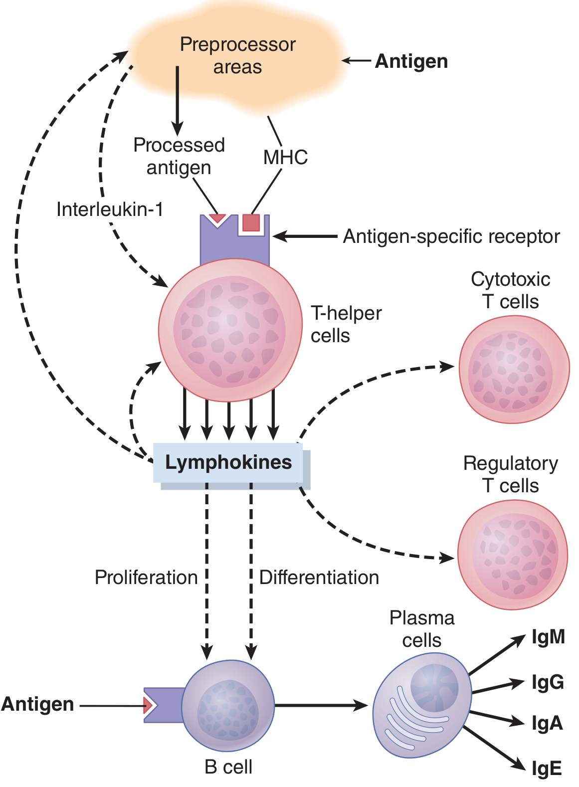
Types of T Cells and Their Functions
1. T-Helper Cells (CD4+ / Th cells) - Most Numerous (~75% of T cells)
These are the master regulators of all immune responses. They act by secreting protein mediators called lymphokines (cytokines).
Three major subsets, differentiated by the cytokine environment:
| Subset | Inducing Cytokines | Cytokines Produced | Main Functions |
|---|---|---|---|
| Th1 | IFN-γ, IL-12 | IFN-γ, IL-2, TNF-α, GM-CSF | Macrophage activation; stimulate IgG production; fight intracellular pathogens |
| Th2 | IL-4 | IL-4, IL-5, IL-6, IL-10, IL-13 | Stimulate IgE; activate mast cells and eosinophils; anti-parasite responses; allergy |
| Th17 | TGF-β, IL-1, IL-6, IL-23 | IL-17, IL-22 | Recruit neutrophils and monocytes; defend against fungi and extracellular bacteria |
Key lymphokine functions:
- IL-2 (Interleukin-2): amplifies the T-helper response (positive feedback); stimulates proliferation of cytotoxic T cells and regulatory T cells
- IFN-γ: activates macrophages to become more efficient killers
- Lymphokines stimulate B-cell growth and differentiation into plasma cells and antibodies
- Lymphokines slow macrophage migration (concentrating them at the site of infection)
HIV/AIDS destroys CD4+ T-helper cells, leaving the immune system almost completely paralyzed - this is why AIDS patients suffer from opportunistic infections.
2. Cytotoxic T Cells (CD8+ / CTLs - Killer Cells)
- Recognize antigen presented on MHC Class I (on infected/tumor cells)
- Directly attack and kill target cells
- Killing mechanism:
- CTL binds tightly to the target cell via antigen-specific TCR
- Releases perforins - hole-forming proteins that punch channels in the target cell membrane
- Releases granzymes (serine proteases) that enter via perforin pores and activate apoptosis
- Also activates Fas-FasL pathway → programmed cell death (apoptosis)
- The CTL detaches and can kill multiple target cells sequentially
- Targets: Virally infected cells, tumor cells, transplanted foreign cells
- After killing, CTLs can persist in tissues for months
3. Regulatory T Cells (CD4+ Tregs)
- Suppress the activity of cytotoxic T cells and T-helper cells
- Prevent autoimmunity and excessive tissue damage
- Maintain immune tolerance to self-antigens
- Secrete inhibitory cytokines (IL-10, TGF-β)
- Also generated by the thymus to suppress any autoreactive T cells that escape negative selection
- Clinical significance: Tregs may suppress anti-tumor immunity → research into downregulating Tregs in cancer immunotherapy (checkpoint inhibitors); upregulating Tregs in autoimmune diseases
Memory T Cells
On first exposure (primary response), activated T cell clones generate memory T cells that:
- Are preserved in lymphoid tissue throughout the body
- Respond far more rapidly and powerfully on subsequent exposure to the same antigen (secondary response)
- Can last for months to years
Examples of Cell-Mediated Immunity in Action
- Defense against intracellular pathogens (Mycobacterium tuberculosis, Listeria, viruses)
- Killing of virus-infected cells (CTLs recognize viral peptides on MHC I)
- Rejection of organ transplants (CTLs attack foreign MHC molecules)
- Tumor surveillance (CTLs destroy cancer cells)
- Delayed-type hypersensitivity (Type IV) - e.g., tuberculin skin test, contact dermatitis
PART 2: HUMORAL IMMUNITY
Definition
Humoral immunity is the arm of adaptive immunity mediated by B lymphocytes and their secreted products - antibodies (immunoglobulins). It primarily defends against extracellular pathogens (bacteria, toxins, viruses in body fluids). The term "humoral" comes from "humor" (body fluid) because antibodies act in blood and secretions.
Origin and Maturation of B Cells
- B cells originate and mature in the bone marrow (where they undergo negative selection to eliminate autoreactive B cells)
- Mature naive B cells express surface IgM and IgD as B cell receptors (BCR)
- They circulate and home to the B cell zones (follicles) of secondary lymphoid organs (lymph nodes, spleen)
Antigen Recognition by B Cells
Unlike T cells, B cells recognize intact, unprocessed antigen in its native conformation via the BCR. The BCR complex consists of:
- Membrane immunoglobulin (binds antigen)
- Igα and Igβ signaling proteins (transmit activation signal into the cell)
Antigen is delivered to B cells in lymph nodes via:
- Afferent lymphatics (small soluble antigens via conduits)
- Subcapsular sinus macrophages (large antigens)
- Follicular dendritic cells (FDCs) - display antigen for prolonged periods (days to weeks) to sustain B cell activation
B Cell Activation
T-Dependent Responses (Protein Antigens - Most Common)
Protein antigens require T-helper cell assistance:
- Antigen binds BCR → B cell internalizes and processes antigen → presents peptide fragments on MHC Class II to a CD4+ Th cell
- CD40L (on T cell) binds CD40 (on B cell) - this costimulatory signal is essential for full B cell activation
- T helper cytokines (IL-2, IL-4, IL-5, IL-6, IL-21) drive B cell proliferation and differentiation
Germinal Center Reaction (in lymphoid follicles):
- Activated B cells move into follicles → form germinal centers
- In germinal centers, two critical processes occur:
- Affinity maturation: Somatic hypermutation of immunoglobulin genes → B cells with higher-affinity receptors are selected (by FDC-displayed antigen) → progressively higher-affinity antibodies are produced as the response matures
- Heavy chain isotype (class) switching: Initial IgM response switches to IgG, IgA, or IgE depending on the cytokine environment (e.g., IL-4 → IgE; TGF-β → IgA)
- B cells differentiate into:
- Plasma cells - antibody-secreting factories; a single B cell generates up to 5000 plasma cells, each secreting ~2000 antibody molecules per second
- Memory B cells - long-lived cells that mediate rapid secondary responses
T-Independent Responses (Polysaccharide/Lipopolysaccharide Antigens)
- Some antigens (e.g., bacterial polysaccharides) can activate B cells without T-helper cell help
- Produce mainly IgM antibodies (no class switching)
- Little affinity maturation or memory generated
- Important in defense against encapsulated bacteria (e.g., Streptococcus pneumoniae, Haemophilus influenzae)
Antibodies (Immunoglobulins) - Structure and Classes
Basic structure: Two heavy chains + two light chains linked by disulfide bonds → Y-shaped molecule
- Variable region (Fab): Binds antigen (unique for each antibody specificity)
- Constant region (Fc): Determines antibody class and effector function (complement activation, Fc receptor binding)
| Class | Main Location | Key Properties |
|---|---|---|
| IgM | Blood (pentamer) | First antibody produced in primary response; excellent complement activator; largest Ig |
| IgG | Blood & tissues | Most abundant; secondary response; crosses placenta (passive immunity to fetus); opsonizes bacteria; activates complement |
| IgA | Secretions (dimer) - saliva, tears, breast milk, mucus | Mucosal immunity; defends GI, respiratory, urogenital tracts; transferred to infant via breast milk |
| IgE | Mast cells & basophils | Mediates allergic reactions (Type I hypersensitivity) and anti-parasite defense |
| IgD | B cell surface | Co-receptor with IgM on naive B cells; signals B cell activation |
Mechanisms by Which Antibodies Eliminate Pathogens
-
Neutralization: Antibodies bind to toxins or viral surface proteins → block their ability to bind host cells (e.g., anti-tetanus toxin IgG)
-
Opsonization: IgG coats bacteria → phagocytes (macrophages, neutrophils) recognize Fc region → enhanced phagocytosis
-
Complement Activation: IgM and IgG activate the classical complement pathway → MAC (membrane attack complex) lyses bacteria; C3b opsonizes; C5a recruits neutrophils
-
Antibody-Dependent Cell-Mediated Cytotoxicity (ADCC): NK cells and macrophages bind Fc portion of IgG on target cells → kill the target cell
-
Agglutination / Precipitation: IgM agglutinates bacteria (clumps them) → easier phagocytosis
Primary vs. Secondary Antibody Response
| Feature | Primary Response | Secondary Response |
|---|---|---|
| Timing | 5-10 days after first exposure | 1-3 days after re-exposure |
| Antibody level | Low | Very high |
| Main antibody | IgM | IgG (mainly) |
| Affinity | Lower | Higher (affinity maturation) |
| Duration | Short | Long-lasting |
| Mediator | Naive B cells | Memory B cells |
Comparison: Cell-Mediated vs. Humoral Immunity
| Feature | Cell-Mediated Immunity | Humoral Immunity |
|---|---|---|
| Key cells | T lymphocytes (CD4+, CD8+) | B lymphocytes → Plasma cells |
| Effector molecule | Cytokines, perforins, granzymes | Antibodies (immunoglobulins) |
| Antigen recognition | Processed peptide + MHC | Intact antigen on BCR |
| Primary target | Intracellular pathogens, tumor cells, grafts | Extracellular pathogens, toxins, free viruses |
| Preprocessing organ | Thymus | Bone marrow |
| Memory | T memory cells | Memory B cells |
| Site of action | Within tissues (direct cell contact) | Blood, lymph, secretions |
| Example | TB, viral infections, transplant rejection | Bacterial infections, vaccine antibodies |
Source: Guyton and Hall Textbook of Medical Physiology, Chapter 35; Cellular and Molecular Immunology (Abbas, Lichtman & Pillai), 10e
This is a shared conversation. Sign in to Orris to start your own chat.