
Invertogram in anorectal malformation
invertogram anorectal malformation imperforate anus X-ray
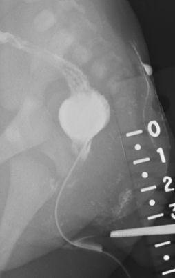
This lateral pelvic x-ray (radiography) demonstrates a contrast-enhanced fistulogram in a neonate with an anorectal malformation (imperforate anus). The diagnostic image highlights a distended, contrast-filled distal rectal pouch positioned superior to the expected level of the anal opening. A long, narrow, radiopaque fistulous tract is visible, originating from the perineal skin surface (vestibular region) and ascending superiorly to communicate with the rectal pouch. A radiopaque ruler is placed externally along the posterior-inferior margin to measure the distance between the skin surface and the terminal end of the rectum, a critical measurement for classifying the malformation as high, intermediate, or low. The vertebral bodies and sacrum are visible, providing anatomical landmarks for evaluating the height of the rectal pouch relative to the pelvic floor. This study is essential for surgical planning, specifically for determining the approach for a posterior sagittal anorectoplasty (PSARP).
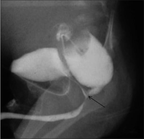
This diagnostic X-ray image is a lateral contrast cologram of a pediatric male patient, illustrating high-type anorectal malformation. The study reveals anorectal agenesis, characterized by a blind-ending distal rectum that fails to reach the perineum. A clear fistulous communication (rectourethral fistula) is demonstrated, where contrast medium flows from the distal rectum into the bulbar segment of the urethra, indicated by a black arrow. The radiograph shows simultaneous opacification of the rectal pouch, the fistulous tract, and the lower urinary tract, including the urethra. This finding is clinically significant for surgical planning in cases of imperforate anus where meconium may be observed in the urine. The image serves as an educational example of congenital urogenital and gastrointestinal anomalies, specifically highlighting the anatomical relationship between the rectum and the bulbar urethra in intermediate to high-level anorectal malformations.
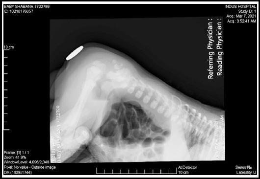
This diagnostic image is a lateral invertogram (Wangensteen-Rice view) of a neonate, a specialized pediatric radiograph used to evaluate anorectal malformations. The infant is positioned in a prone, head-down (inverted) posture to allow bowel gas to rise toward the perineum. A radiopaque marker is placed on the skin surface at the expected site of the anus. The radiograph reveals significant bowel gas distension with multiple dilated intestinal loops, consistent with a distal bowel obstruction. The distance between the most distal rectal gas bubble and the external perineal marker is used to classify the malformation as a 'high' or 'low' type defect. Anatomical landmarks visible include the infant's lumbar and sacral spine, pelvis, and femur, showing age-appropriate incomplete ossification. This imaging modality is critical in pediatric surgery for planning the initial management and surgical approach for congenital imperforate anus.
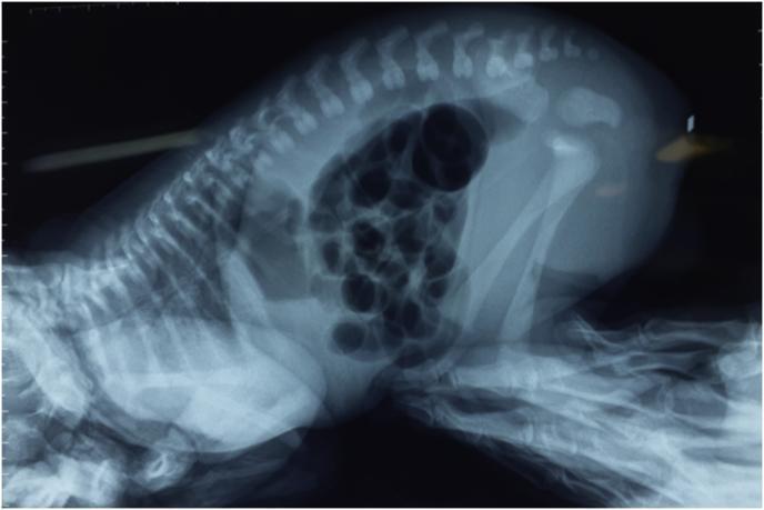
This diagnostic image is a cross-table lateral X-ray of a newborn infant in a prone position, often referred to as an invertogram or Wangensteen-Rice view. The imaging primary focus is the evaluation of anorectal malformations (ARM). The skeletal system is clearly visible, showing the vertebral column with individual vertebrae and spinous processes, the rib cage, and the pelvic girdle comprising the ilium, ischium, and pubis. The femur and tibia of the lower extremities are flexed. The abdomen contains multiple radiolucent (dark) air-filled bowel loops. A key diagnostic feature is the distance between the most distal gas bubble in the rectum and the perineal skin (often marked with a radiopaque marker), used to determine the level of the atresia (high, intermediate, or low). This clinical imaging modality is essential for surgical planning in neonates presenting with imperforate anus to differentiate between various types of anorectal anomalies and associated fistulas.
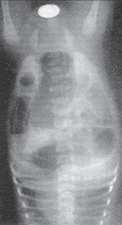
This diagnostic image is a lateral abdominal and pelvic X-ray of a neonate, positioned in an inverted or 'head-down' orientation for the Wangensteen-Rice technique. The radiograph shows gas-filled bowel loops, specifically outlining the distal rectum as the air rises to the highest anatomical point in this position. A radiopaque metallic marker is placed externally on the perineal dimple to serve as a reference point. The primary educational focus is the assessment of an anorectal malformation (imperforate anus). The image allows for the measurement of the distance between the distal rectal gas bubble and the perineal marker, which is critical for classifying the defect as a 'high' or 'low' lesion and determining the appropriate surgical approach. This clinical application is most valid after 14–24 hours of life to ensure bowel gas has reached the terminal rectum. The imaging highlights key findings in pediatric radiology and neonatal gastrointestinal surgery.
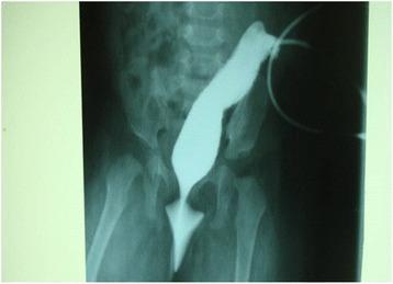
This diagnostic image is a contrast-enhanced colostogram (X-ray radiography) of a pediatric patient, likely an infant, focused on the lower abdomen and pelvic region. The imaging shows a gastrografin contrast medium introduced through a sigmoid colostomy site, filling the distal colon. The radiopaque contrast clearly delineates a blind-ending rectal pouch, characteristic of anorectal malformation (imperforate anus). Crucially, a thin, fistulous tract is visible extending inferiorly from the rectal pouch and terminating at the vulval vestibule, confirming a rectovestibular fistula. The surrounding skeletal anatomy reveals the lumbar spine, pelvic bones, and proximal femurs, which serve as landmarks to assess the level of the rectal pouch relative to the puborectalis muscle and pelvic floor. This image is medically significant for pediatric surgery and gastroenterology, as it demonstrates the morphology of high/intermediate anorectal malformations and guides posterior sagittal anorectoplasty (PSARP) planning.
Invertogram in Anorectal Malformation
What Is an Invertogram?
- Mulholland & Greenfield's Surgery (7e), p. 5492
- Sabiston Textbook of Surgery, p. 2689
Technique
- Wait 12-24 hours after birth - this ensures swallowed air has migrated to the terminal rectum
- Place the infant in the inverted prone (head-down) position for 3-5 minutes before the film
- Place a radiopaque marker (coin, lead shot, or metallic tape) on the anal dimple (perineal skin)
- Take a lateral view of the pelvis
- Measure the distance from the most distal gas bubble to the marker
Classification Based on Invertogram
| Distance from marker | Classification | Surgical Implication |
|---|---|---|
| Gas bubble below the I-line (<1 cm from skin) | Low lesion | Perineal anoplasty without colostomy |
| Gas bubble between PC and I-line | Intermediate lesion | PSARP, may need colostomy |
| Gas bubble above the PC line (>1.5-2 cm from skin) | High lesion | Defunctioning colostomy first, then PSARP |
Why It Matters
-
Low lesions: primary perineal anoplasty in the neonatal period, no colostomy needed
-
High/intermediate lesions: staged repair - diverting colostomy first, then posterior sagittal anorectoplasty (PSARP) at 3-6 months of age
-
Mulholland & Greenfield's Surgery (7e), p. 5492
Limitations and Why It Has Been Replaced
- False high result if gas has not yet reached the terminal rectum (before 18-24h of life)
- Mucus/meconium can plug the distal pouch, preventing gas from reaching it - leading to overestimation of the level (falsely suggesting a "high" lesion)
- Bowel wall thickness and incomplete gaseous distension introduce error
- Cannot demonstrate fistula anatomy
- Poor reproducibility; positioning artifacts are common
-
Perineal/transperineal ultrasound - real-time, no radiation, accurately measures pouch-to-skin distance (now first-line investigation)
-
Distal colostogram (after colostomy is formed) - provides the most reliable anatomy including fistula location, essential for PSARP planning
-
MRI pelvis - best for evaluating the striated muscle complex, puborectalis, and spinal cord
-
CT pelvis - useful in complex/cloacal malformations
-
Mulholland & Greenfield's Surgery (7e), p. 5492
-
Sabiston Textbook of Surgery, p. 2689
Associated Investigations
- Spinal X-ray (sacral ratio - indicator of prognosis for continence; sacral agenesis = poor prognosis)
- Renal USS (up to 50% have urological anomalies)
- Echocardiography (VACTERL association)
- Spinal MRI (tethered cord)
- Karyotype if rectal atresia (association with trisomy 21)
Summary
- Neonatal bowel gas physics (gas rises in inverted position)
- PC line / I-line landmarks for high/low classification
- Timing (must wait 18-24h for reliable results)
- The clinical examination remains paramount - in most cases, perineal inspection alone classifies the lesion, and ultrasound confirms the level when needed.
Treatment
posterior sagittal anorectoplasty PSARP anorectal malformation surgery
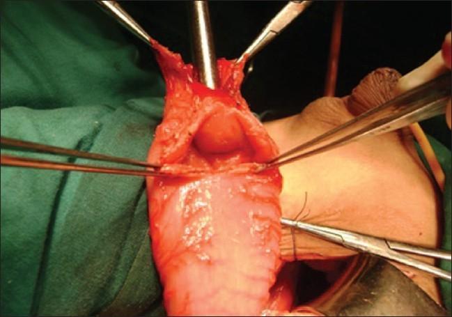
This clinical photograph captures an intraoperative view of a posterior sagittal anorectoplasty (PSARP) for the correction of an anorectal malformation. The image demonstrates a surgically opened, dilated rectal pouch being held apart by multiple surgical forceps. Within the lumen of the reddish-pink rectal tissue, a rare anatomical finding known as a rectal diaphragm is visible. The diaphragm appears as a raised, circular, obstructive shelf of tissue located approximately 2 cm proximal to the distal end of the pouch. A surgical metal probe or sound is positioned within the lumen to highlight the anatomical relationship between the distal blind pouch and the proximal obstruction. The visual evidence illustrates a complex variant of anorectal atresia where internal septation complicates the identification of the proximal bowel lumen during corrective surgery. This photograph is intended for pediatric surgery education, specifically regarding the identification of atypical internal obstructions in neonates and children with anorectal malformations.
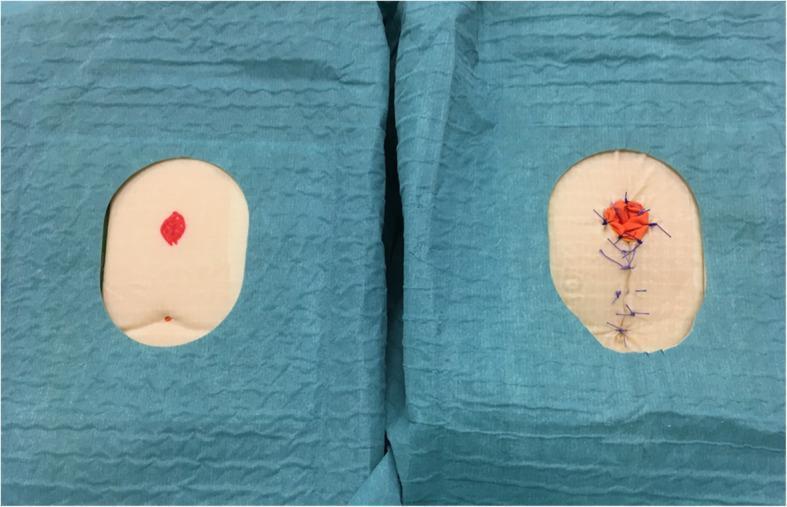
A top-down comparison photograph featuring two synthetic perineal sponge models used for surgical simulation training of the Posterior Sagittal Anorectoplasty (PSARP) procedure. Both models are oval-shaped, light peach in color, and framed by blue sterile surgical drapes. The left model represents the preoperative state, showing a smooth surface with a small, central red marking indicating the planned site for the anorectal malformation (ARM) repair. The right model represents the postoperative state, demonstrating the results of surgical intervention. Key features include an orange material at the original marking site to simulate the transposed rectum and a vertical line of blue surgical sutures extending inferiorly, representing the closure of the sagittal incision. This comparison illustrates the anatomical changes and surgical steps involved in pediatric colorectal reconstructive surgery, including sagittal incision, dissection, and anoplasty, for educational and procedural validation purposes.
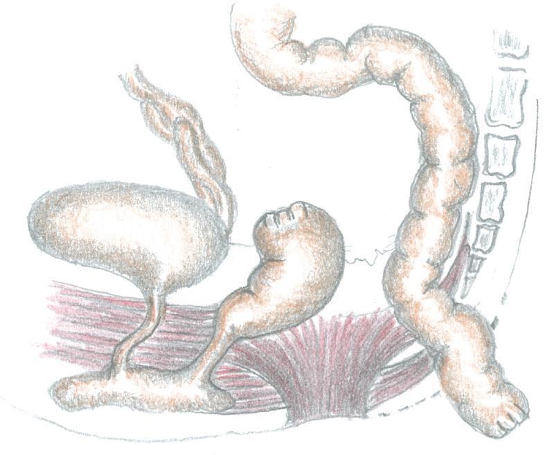
This medical illustration depicts a surgical stage of a posterior sagittal anorectoplasty (PSARP) for the management of a congenital anorectal malformation. The diagram shows the pelvic anatomy from a lateral-posterior perspective. Central to the image is the mobilized sigmoid colon, shown as a long, haustrated tubular structure being transitioned toward the posterior sagittal space. To the left, the urinary bladder is visualized with its connection to the lower urinary tract. Adjacent to it is a blind-ending distal rectal pouch, which has been surgically separated from the proximal bowel. The pelvic floor musculature, including the levator ani and muscle complex, is rendered in red, illustrating the tunnel created for the pull-through procedure. On the right, the sacrum and vertebral column serve as anatomical landmarks, highlighting the retrorectal pathway used for the sigmoid mobilization. This educational illustration demonstrates the surgical technique of bypassing a dilated rectum to utilize the healthy sigmoid colon for neo-anus construction.
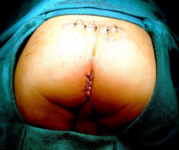
This clinical photograph shows the perineal and sacral region of a pediatric patient immediately following a Sagittal Sacroperineal Anorectoplasty (SSARP) for the correction of an anorectal malformation. The patient is in a prone position, surrounded by green surgical drapes. Two distinct surgical closure sites are visible. Superiorly, a horizontal (transverse) incision located just above the coccygeal region is closed with approximately four dark-colored, non-absorbable interrupted sutures. Inferiorly, a vertical incision at the newly created anal site is closed with a continuous suture pattern using similar dark material. Notably, the posterior midline skin between these two incisions remains entirely intact and spared, a characteristic feature of the SSARP technique that distinguishes it from the Posterior Sagittal Anorectoplasty (PSARP). The surrounding skin appears healthy with no signs of excessive inflammation, significant edema, or bruising. This image serves as a clinical example of postoperative anatomy in pediatric colorectal surgery, illustrating successful wound closure with preservation of midline skin integrity.
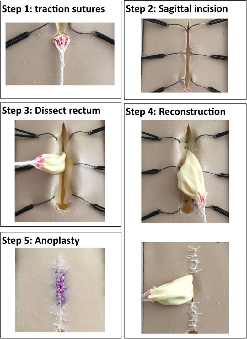
This surgical simulation infographic details the sequential steps of a Posterior Sagittal Anorectoplasty (PSARP) for Anorectal Malformation (ARM) using a synthetic training model. The procedure is shown in five distinct intraoperative stages. Step 1 (Traction Sutures) demonstrates the placement of silk and colored sutures at the ectopic anal site to provide orientation and traction. Step 2 (Sagittal Incision) shows a vertical midline perineal incision with the skin edges retracted by stay sutures to expose deep tissues. Step 3 (Dissect Rectum) depicts the mobilization of the rectum, represented by a beige balloon-like structure, through the sagittal opening. Step 4 (Reconstruction) shows the anatomical repositioning and tapering of the rectum within the muscle complex. Step 5 (Anoplasty) illustrates the final closure, where the rectum is sutured to the skin to create a new anal opening, featuring visible blue and white interrupted sutures. The series serves as an educational tool for pediatric surgical trainees to visualize surgical landmarks, tissue manipulation, and suture techniques required for repairing anorectal defects.
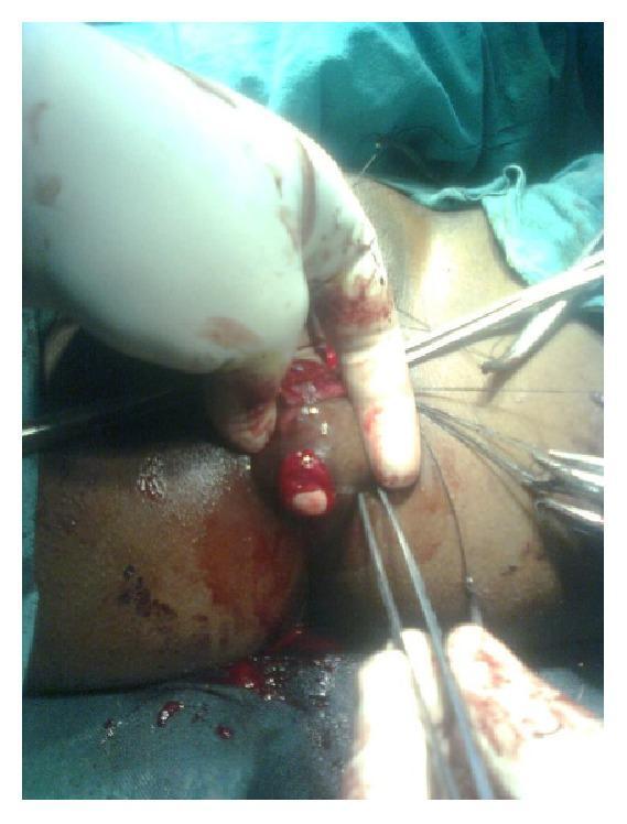
An intraoperative clinical photograph documenting a posterior sagittal anorectoplasty (PSARP) or similar anal transposition procedure for an anorectal malformation. The surgical field focuses on the perineum of a patient in the lithotomy position. A new anal opening has been surgically created through the identified striated muscle complex (sphincter). A gloved hand and surgical instruments, including fine-tipped forceps and hemostats, are visible manipulating the raw, vascularized perineal tissues. Stay sutures are in place to provide traction and exposure of the operative site. The image shows the newly formed ovoid opening with some internal mucosal or rectal tissue visible at the margins. Visible blood and tissue inflammation are consistent with an active surgical dissection. This visual illustrates the surgical management of imperforate anus or rectovestibular fistula in an adult patient, emphasizing the creation of a neo-anus within the sphincteric muscle complex.
Treatment of Anorectal Malformation
Step 1 - Initial Neonatal Assessment (All Patients)
- Place orogastric tube; IV access, NPO
- Observe for 12-24 hours - a visible perineal/vestibular fistula may only appear once meconium descends distally
- Look for meconium in urine (suggests rectourethral fistula = high lesion in males)
- Investigations for associated anomalies:
- Renal USS (urological anomaly in ~50%)
- Echocardiogram (cardiac anomaly)
- Spinal X-ray + spinal USS (sacral ratio; tethered cord)
- Sacral X-ray - sacral agenesis = poor prognosis for continence
- Imperforate anus alone is not immediately life-threatening; there is time to properly classify before rushing to theatre
Step 2 - Surgical Management by Lesion Type
LOW LESIONS (pouch <1 cm from perineum)
| Lesion | Procedure |
|---|---|
| Thin anal membrane / covered anus | Simple dilatation or unroofing |
| Anterior perineal fistula / anal stenosis | Cutback anoplasty - incise anterior fistula posteriorly to the sphincter |
| Female with anterior perineal/vestibular fistula | Transposition anoplasty - circumferential mobilization + transposition to center of external sphincter + perineal body reconstruction |
INTERMEDIATE AND HIGH LESIONS (pouch above I-line or PC line)
Stage 1 - Neonatal Diverting Colostomy
- Performed in the neonatal period (within first few days)
- Divided loop colostomy (preferred over loop colostomy) in the sigmoid colon - provides complete fecal diversion from the downstream rectourethral/vestibular fistula
- Must leave adequate length and mobility of distal colon for future pull-through
- After colostomy is formed, a distal colostogram (contrast study through distal limb) defines the fistula anatomy and the level of the rectal pouch - this is the most reliable pre-repair anatomical study
Stage 2 - Anorectoplasty (at 8-12 months of age)
- Posterior midline sagittal incision along the gluteal crease
- Muscle stimulator used to identify and map the striated muscle complex (external sphincter + levator ani) - ensures symmetric midline dissection
- Levator ani and external sphincter complex are divided posteriorly in the midline
- Rectal pouch is exposed and mobilized
- Rectourinary fistula is identified and closed directly (divided and oversewn)
- Mobilized rectum is placed centrally within the striated muscle complex
- Striated muscle complex is reconstructed circumferentially around the rectum
- Anoplasty - neoanus centred within the external sphincter; mucosa sutured to perineum
- Colostomy maintained until anorectoplasty is fully healed
- (a) Relieve rectal obstruction
- (b) Create a new anus
- (c) Position rectum centrally within the striated muscle complex
- (d) Divide and close the rectourinary fistula
- (e) Preserve surrounding structures (prostate, urethra, seminal vesicles, vaginal wall)
Stage 3 - Colostomy Closure
- Performed electively once the anorectoplasty is healed
- Followed by anal dilatation programme starting 2-3 weeks post-PSARP to prevent stricture
SPECIAL CASE - Persistent Cloaca (Females)
- Careful urological evaluation at the time of colostomy (vesicostomy may also be needed if bladder drainage is impaired)
- Definitive reconstruction is a complex combined urogenital and rectal repair, timing and approach depending on the length of the common channel (>3 cm = "long cloaca" = more complex)
Other Surgical Approaches (Historical / Alternative)
| Approach | Description |
|---|---|
| Sacroperineal (Stephens) | Involves sacral incision; blind pull-through without direct muscle visualization |
| Endorectal (Rehbein) | Endorectal dissection component; also a blind pull-through |
| Laparoscopic-assisted PSARP | Useful for high recto-bladder neck fistulas; avoids laparotomy |
- Mulholland & Greenfield's Surgery (7e), p. 5495
- Schwartz's Principles of Surgery (11e), p. 1764
Postoperative Management
- Anal dilatation started 2-3 weeks post-PSARP; continued for weeks to months to prevent stricture (stricture/leak in 5-10% after tapering rectoplasty)
- Bowel management programme - enemas, cathartics to prevent fecal impaction and secondary motility dysfunction
- In refractory incontinence: antegrade enemas via appendicostomy (Malone MACE) or cecostomy
- Rarely, a permanent diverting colostomy may be required for socially unacceptable fecal incontinence
Long-term Outcomes and Prognosis
| Lesion Level | Continence Outcome |
|---|---|
| Low lesions | Generally good - normal rectal descent within muscle complex |
| High lesions | Less predictable - higher incidence of fecal incontinence |
| All patients | ~75% have voluntary bowel movements; ~40% achieve full continence (Peña series of 1192 patients) |
- Degree of rectal descent within the muscle complex
- Integrity of the striated muscle complex
- Sacral innervation (sacral ratio on X-ray - good predictor)
- Low lesions tend toward constipation; high lesions toward incontinence
- All children should be followed until at least 4 years of age as continence cannot be accurately predicted earlier
- Mulholland & Greenfield's Surgery (7e), p. 5494-5496
- Schwartz's Principles of Surgery (11e), p. 1764