

Answer them for my university exam
Pathology Paper I - Complete Exam Answers
SECTION: Injury, Cellular Adaptations and Cellular Aging
SN 1. Types of Calcification; Dystrophic vs Metastatic Calcification
Types of Pathologic Calcification
- Dystrophic calcification
- Metastatic calcification
Dystrophic Calcification
- Definition: Deposition of calcium salts in dead or dying (necrotic) tissues, despite normal serum calcium levels and no derangement in calcium metabolism.
- Mechanism: The underlying disease damages cell membranes, leading to calcium influx. Phospholipid-rich cell membranes in necrotic cells act as nucleation sites. Membrane-bound vesicles concentrate calcium and phosphate, initiating crystalline hydroxyapatite formation.
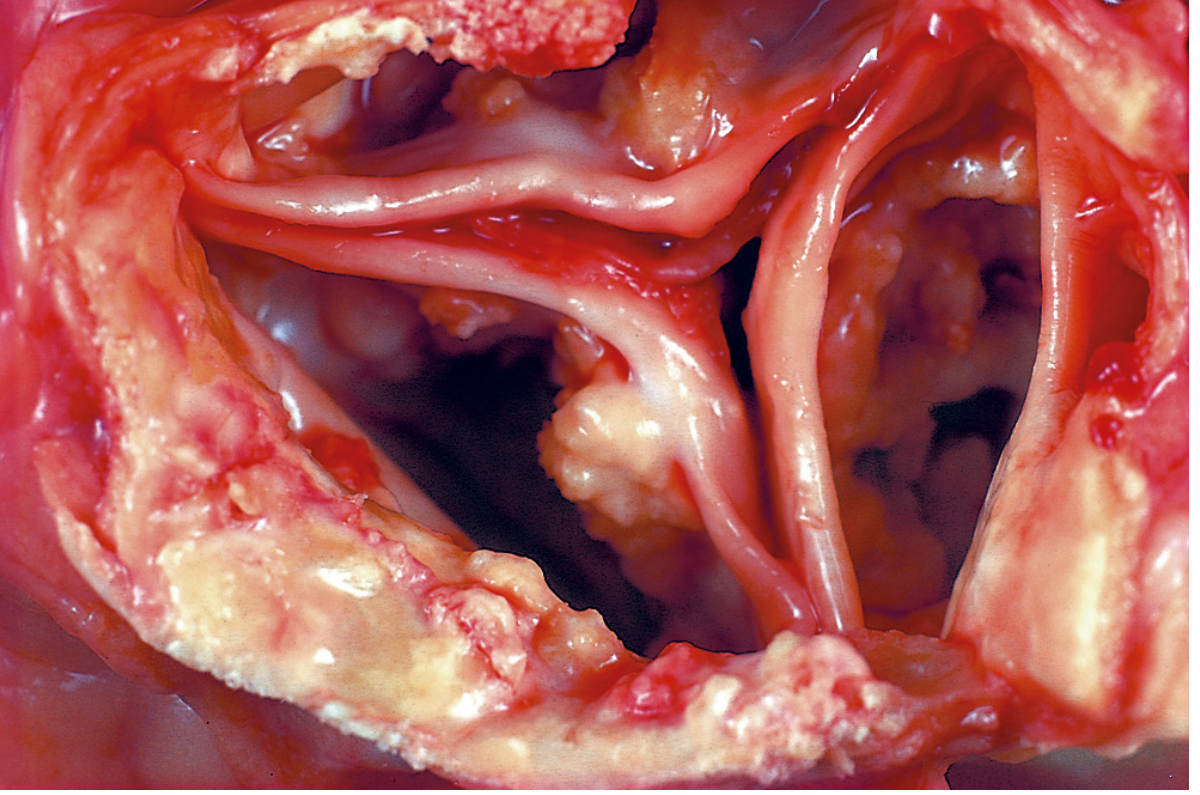
- Sites: Areas of coagulative, caseous, or liquefactive necrosis; atheromas of advanced atherosclerosis; aging or damaged heart valves (calcific aortic stenosis); tuberculous lymph nodes (may convert to stone); fat necrosis.
Metastatic Calcification
- Definition: Deposition of calcium salts in normal, otherwise healthy tissues, almost always resulting from hypercalcemia due to a disturbance in calcium metabolism.
- Causes of hypercalcemia:
- Hyperparathyroidism (primary or secondary)
- Destruction of bone (metastases, multiple myeloma, Paget disease)
- Vitamin D-related disorders (intoxication, sarcoidosis)
- Renal failure
- Milk-alkali syndrome
- Sites: Interstitial tissues of gastric mucosa, kidneys (nephrocalcinosis), lungs, systemic arteries, pulmonary veins, and cornea (all sites that lose acid, allowing pH to rise, which favors calcium precipitation).
Differences: Dystrophic vs Metastatic Calcification
| Feature | Dystrophic | Metastatic |
|---|---|---|
| Site of deposition | Necrotic/dead tissue | Normal viable tissue |
| Serum calcium | Normal | Elevated (hypercalcemia) |
| Calcium metabolism | Normal | Disturbed |
| Underlying cause | Local tissue necrosis/injury | Systemic calcium disorder |
| Examples | TB lymph node, atheroma, damaged valves | Hyperparathyroidism, renal failure |
| Clinical significance | Marker of prior injury/necrosis | Indicates systemic metabolic disease |
| Serum phosphate | Normal | Often elevated |
SN 2. Apoptosis - Definition, Examples, Morphological Changes, Mechanisms, Physiological and Pathological Causes
Definition
Morphological Changes
- Cell shrinkage - the cell is smaller; cytoplasm is dense; organelles are more tightly packed
- Chromatin condensation (pyknosis) - chromatin aggregates peripherally under the nuclear membrane into dense masses
- Nuclear fragmentation (karyorrhexis) - the nucleus breaks up into two or more fragments
- Cytoplasmic blebs - the cell surface shows irregular budding
- Apoptotic body formation - blebs pinch off to form membrane-bound apoptotic bodies, which may contain fragments of nucleus and/or cytoplasm
- Phagocytosis - apoptotic bodies are rapidly eaten by macrophages or neighboring cells. No inflammation occurs because membrane integrity is preserved.
Mechanisms (Pathways)
- Triggered by: DNA damage, oxidative stress, ER stress, growth factor withdrawal
- Key players: BCL2 family proteins
- Anti-apoptotic: BCL2, BCL-XL, MCL1 (keep mitochondrial outer membrane impermeable)
- Pro-apoptotic: BAX, BAK (permeabilize the membrane)
- BH3-only sensors: BAD, BIM, BID, PUMA, NOXA (detect stress, activate BAX/BAK)
- When stress signals dominate, BAX/BAK oligomerize in the outer mitochondrial membrane → cytochrome c leaks into cytosol
- Cytochrome c + APAF-1 → forms the apoptosome → activates caspase-9 (initiator) → activates caspase-3 (executioner) → cell death
- Triggered by: Fas ligand binding to Fas (CD95), TNF binding to TNFR1
- Fas-FasL interaction → recruitment of FADD (Fas-associated death domain) → activates caspase-8 (initiator) → activates caspase-3 → cell death
- Cross-talk: Caspase-8 cleaves BID (a BH3-only protein) → truncated BID (tBID) activates the mitochondrial pathway, amplifying the signal
Physiological Causes (Normal Events)
- Programmed cell death during embryogenesis (organogenesis, limb development - interdigital web removal)
- Involution of hormone-dependent tissues after hormone withdrawal (e.g., endometrial shedding during menstrual cycle; post-lactation breast involution)
- Cell deletion in proliferating cell populations to maintain constant cell numbers (e.g., intestinal crypt cells)
- Elimination of self-reactive lymphocytes in the thymus and periphery (negative selection)
- Death of immune effector cells (cytotoxic T lymphocytes) after serving their protective role
Pathological Causes (Disease States)
- DNA damage - radiation, cytotoxic drugs causing unrepaired double-strand DNA breaks
- Viral infections - hepatitis B and C (apoptosis of infected hepatocytes); also used by some viruses to spread
- Accumulation of misfolded proteins - ER stress (e.g., in neurodegenerative diseases like Alzheimer, Parkinson)
- Obstruction of ducts - pancreatic duct, salivary duct obstruction leads to apoptosis of gland cells
- Cell injury in diseases - myocardial infarction, neurodegenerative diseases
- Tumor regression after chemotherapy/radiotherapy
SN 3. Metaplasia, Hypertrophy, Hyperplasia, and Atrophy - Definitions with Examples
Atrophy
- Physiologic: Normal aging (brain atrophy after age 60)
- Pathologic: Denervation atrophy, pressure atrophy, disuse atrophy, inadequate nutrition, loss of endocrine stimulation, ischemia
Hypertrophy
- Physiologic: Pregnant uterus (smooth muscle hypertrophy due to estrogen); skeletal muscle in bodybuilders
- Pathologic: Cardiac hypertrophy in hypertension or aortic stenosis (left ventricular hypertrophy)
Hyperplasia
- Physiologic: Hormonal (breast enlargement during pregnancy); compensatory (liver regeneration after partial hepatectomy)
- Pathologic: Endometrial hyperplasia (excess estrogen); BPH (benign prostatic hyperplasia due to DHT); psoriasis (epidermal hyperplasia)
Metaplasia
- Columnar to squamous (most common): Respiratory epithelium in smokers; cervical ectropion with squamocolumnar junction transformation; stones in ducts (salivary, pancreatic, bile ducts)
- Squamous to columnar: Barrett esophagus - esophageal squamous epithelium replaced by intestinal columnar cells due to chronic GERD (risk: adenocarcinoma)
- Connective tissue: Myositis ossificans (bone formation in muscle after intramuscular hemorrhage)
SN 4. Free Radicals and Free Radical-Induced Cell Injury
Definition
Generation of Free Radicals
- Normal mitochondrial respiration - reduction of O2 generates small amounts of O2•-, H2O2, and •OH as byproducts
- Ionizing radiation - hydrolyzes water into •OH and H• radicals
- Activated leukocytes during inflammation - NADPH oxidase generates O2•- (respiratory burst)
- Enzymatic metabolism of drugs/chemicals - e.g., CCl4 → •CCl3 (toxic radical)
- Transition metals (iron, copper) - Fenton reaction: H2O2 + Fe2+ → Fe3+ + •OH + OH- (most damaging radical)
- Reperfusion of ischemic tissue - burst of ROS generation
Removal / Antioxidant Defense
- Superoxide dismutase (SOD): converts O2•- to H2O2
- Catalase: breaks down H2O2 to H2O + O2
- Glutathione peroxidase: reduces H2O2 and lipid peroxides
- Vitamins E, A, and C - antioxidants that scavenge free radicals
- Ceruloplasmin, transferrin - sequester iron/copper to prevent Fenton reaction
Mechanisms of Free Radical-Induced Cell Injury
- •OH attacks double bonds in polyunsaturated fatty acids of cell membranes
- Creates lipid peroxides and new free radicals (autocatalytic chain reaction)
- Results in: increased membrane permeability, loss of membrane potential, organelle dysfunction
- Oxidation of amino acid side chains
- Covalent cross-linking of proteins (disulfide bonds)
- Oxidation of protein backbone → enzyme active site disruption
- Enhanced proteasomal degradation of misfolded proteins
- Single- and double-strand breaks in DNA
- Cross-linking of DNA strands, adduct formation
- Implicated in: cell aging, malignant transformation
- Free radicals can trigger the intrinsic mitochondrial apoptotic pathway (at lower doses)
- At higher doses: necrosis predominates
Pathologic Conditions Caused by Free Radical Injury
- Ischemia-reperfusion injury (MI, stroke)
- Chemical and drug toxicity (CCl4 hepatotoxicity)
- Radiation injury
- Oxygen toxicity (in premature neonates - retrolental fibroplasia, BPD)
- Aging
- Atherosclerosis (oxidized LDL)
- Carcinogenesis
LAQ 1. Necrosis - Definition, Types, Caseous Morphology, Coagulative vs Liquefactive Necrosis
Clinical Scenario Link:
- Diabetic patient with pale, dry leg ulcer = coagulative necrosis (ischemic gangrene)
- TB patient with cheesy lung lesion = caseous necrosis
- Stroke patient with brain liquefaction = liquefactive necrosis
Definition of Necrosis
Morphological Features Common to All Necrotic Cells
- Cytoplasm: Eosinophilia (due to denatured cytoplasmic proteins binding eosin; loss of RNA)
- Nuclear changes (3 patterns):
- Karyolysis - basophilia fades due to DNase activity
- Pyknosis - nuclear shrinkage and increased basophilia (chromatin condenses)
- Karyorrhexis - fragmentation of pyknotic nucleus
- Over 1-2 days, nucleus totally disappears
- Cytoplasm: Vacuolated, moth-eaten appearance; myelin figures (phospholipid precipitates) may form
Types of Necrosis
| Type | Key Feature | Example |
|---|---|---|
| Coagulative | Architecture preserved; firm, eosinophilic "ghost" cells | Kidney/heart/spleen infarct |
| Liquefactive | Complete dissolution; fluid-filled cavity | Brain infarct; pyogenic abscess |
| Caseous | Cheese-like; granuloma with central necrosis | Tuberculosis |
| Fat | Chalky white deposits (saponification) | Acute pancreatitis; breast trauma |
| Fibrinoid | Pink fibrin-like material; immune complex deposition | Vasculitis, malignant hypertension |
| Gangrenous | Clinical term; dry = coagulative; wet = liquefactive | Diabetic foot |
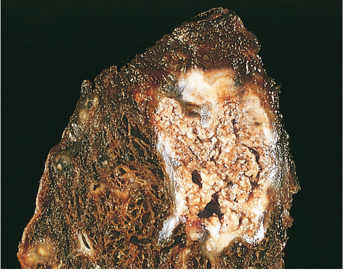
Morphology of Caseous Necrosis
- Central area: acellular, granular, eosinophilic debris with loss of all architectural tissue details (unlike coagulative necrosis, ghost outlines are NOT preserved)
- Surrounding zone: epithelioid macrophages (activated macrophages with abundant pale cytoplasm)
- Langhans giant cells (multinucleated cells formed by fusion of macrophages, with nuclei arranged in a horseshoe/peripheral pattern)
- Peripheral lymphocyte infiltration
- Occasional plasma cells
- Over time: dystrophic calcification may occur in the necrotic center
Coagulative vs Liquefactive Necrosis (in Detail)
- Mechanism: Denaturation of both structural proteins AND enzymes (proteolytic enzymes are denatured, so they cannot digest the dead cells)
- Result: Cell architecture is preserved for days - "ghost outlines" or "tombstone cells" visible microscopically (cell shape preserved but nucleus gone)
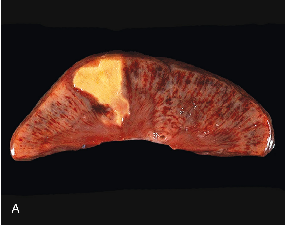
- Gross: Firm, pale, dry, well-demarcated area. In the heart: yellow-white area. In kidney: wedge-shaped infarct.
- Microscopy: Intensely eosinophilic cells with indistinct or absent nuclei; preserved cellular outlines; inflammatory infiltrate at periphery
- Cause: Ischemia (obstruction of blood supply to all organs except the brain)
- Clinical example: Myocardial infarction, renal infarct, splenic infarct, pale infarct of any solid organ. The 58-year-old diabetic's ischemic leg ulcer = dry gangrene = coagulative necrosis.
- Mechanism: Enzymatic digestion dominates - dead cells are rapidly digested by their own hydrolases and by leukocyte-derived enzymes. Entire tissue is liquefied.
- Result: Tissue is completely dissolved; the area is converted into a liquid viscous mass, eventually forming a cyst-like cavity
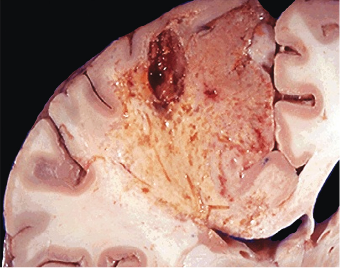
- Gross: Soft, fluid-filled cavity
- Microscopy: Complete loss of cellular architecture; only fluid, debris, and leukocytes visible
- Causes:
- Brain ischemia/infarction - neurons are very rich in hydrolytic enzymes, and the brain has poor structural protein support, so liquefaction always occurs (even with ischemia - unlike other organs)
- Bacterial (pyogenic) infections - neutrophil enzymes liquefy the tissue → pus (the liquid mass of liquefactive necrosis is called "pus" in infections)
- Clinical example: Brain infarct (stroke) - forms a fluid-filled cavity (the 3rd patient); pyogenic abscess (lung abscess, brain abscess)
LAQ 2. Atrophy: Definition, Morphology and Causes of Brown Atrophy of the Heart
Definition of Atrophy
Brown Atrophy of the Heart
Gross Morphology
- Heart is smaller than normal (reduced weight and size)
- The organ has a brown discoloration (instead of normal red-brown)
- "Tigroid" or nutmeg pattern may coexist if there is also chronic venous congestion
- The coronary arteries may appear tortuous and dilated relative to the shrunken myocardium
- Cut surface shows smaller, denser myocardial fibers
Microscopic Morphology
- Cardiomyocytes are reduced in size with smaller, darker nuclei
- Lipofuscin granules are visible as yellow-brown, perinuclear pigment granules within the cytoplasm (best seen with Sudan black or periodic acid-Schiff stain; also autofluorescent under UV light)
- Lipofuscin = residual bodies from incomplete lysosomal digestion of damaged organelles (accumulated over a lifetime; the "wear and tear" pigment)
- Myofibrils are reduced in number
- Interstitial fibrosis may be present
Causes of Brown Atrophy of the Heart
- Advanced age (senile atrophy) - most common cause; the normal aging process leads to gradual cell loss and lipofuscin accumulation
- Cachexia (extreme malnutrition):
- Advanced malignancy (cancer cachexia)
- Severe chronic illness (chronic heart failure, TB, AIDS)
- Starvation
- Prolonged bed rest / immobility - disuse atrophy from reduced cardiac workload
- Loss of endocrine stimulation - e.g., hypopituitarism
- Chronic ischemia with gradual loss of cardiomyocytes
SECTION: Immunopathology Including Amyloidosis
Immunopathology SN 1. Hypersensitivity Reactions - Classification + Type I in Detail
Definition
Classification (Gell and Coombs Classification)
| Type | Name | Mechanism | Mediators | Examples |
|---|---|---|---|---|
| Type I | Immediate (Anaphylactic) | IgE antibody + antigen → mast cell degranulation | Histamine, leukotrienes, prostaglandins | Anaphylaxis, asthma, allergic rhinitis, urticaria |
| Type II | Cytotoxic (Antibody-mediated) | IgG/IgM antibodies against cell-surface or matrix antigens → complement + ADCC | Complement, NK cells, phagocytes | Hemolytic anemia, Goodpasture syndrome, blood transfusion reactions |
| Type III | Immune Complex-mediated | Antigen-antibody complexes (IgG) deposited in tissues → complement + neutrophil activation | Complement, neutrophils | Serum sickness, SLE, post-streptococcal glomerulonephritis, Arthus reaction |
| Type IV | Delayed (Cell-mediated) | Sensitized T lymphocytes (CD4+ or CD8+) react with antigen | Cytokines, cytotoxic T cells | Contact dermatitis, tuberculin test, transplant rejection, Type 1 DM |
Type I Hypersensitivity in Detail
- Sensitization phase (first exposure) - no symptoms
- Effector phase (re-exposure) - immediate reaction within minutes
- First exposure to antigen (allergen)
- Antigen is processed by APCs (dendritic cells) and presented to CD4+ T helper cells
- TH2 cells are activated → produce IL-4, IL-5, IL-13
- IL-4 → induces B cell class switching to IgE
- IgE binds to FcεRI receptors on mast cells and basophils (sensitization) - no symptoms yet
- Re-exposure to the same allergen
- Allergen cross-links IgE molecules on mast cell surface
- Cross-linking triggers mast cell degranulation
- Histamine - most important: vasodilation, increased vascular permeability, smooth muscle contraction, mucus secretion
- Heparin - anticoagulant
- Proteases (tryptase, chymase) - tissue damage, activate complement
- Leukotrienes (LTC4, LTD4, LTE4) - 1000x more potent than histamine in bronchoconstriction; prolonged action (the "slow-reacting substances of anaphylaxis" - SRS-A)
- Prostaglandin D2 - bronchoconstriction, vasodilation
- Platelet-activating factor (PAF) - platelet aggregation, bronchoconstriction
- IL-4, IL-5, IL-13 - promote TH2 response, IgE production, eosinophil recruitment
- TNF-α - leukocyte recruitment
- Recruitment of eosinophils, basophils, neutrophils, TH2 cells
- Eosinophils release: major basic protein (MBP), eosinophil peroxidase, eosinophil cationic protein
- Cause prolonged tissue damage, edema, mucus secretion
- Responsible for the "late-phase" bronchospasm in asthma
| Organ affected | Manifestation |
|---|---|
| Systemic mast cell degranulation | Anaphylaxis (life-threatening) |
| Airways | Asthma (bronchoconstriction) |
| Nasal mucosa | Allergic rhinitis (hay fever) |
| Skin | Urticaria (hives), angioedema |
| GI tract | Nausea, vomiting, diarrhea |
- Epinephrine (anaphylaxis) - reverses vasodilation and bronchospasm
- Antihistamines (H1 blockers) - block histamine effects
- Corticosteroids - reduce late-phase inflammation
- Leukotriene antagonists (montelukast) - block LTD4 receptors
Immunopathology SN 2. Amyloidosis (Clinical Case: RA with Nephrotic Syndrome + Multiple Myeloma)
Clinical Scenario Analysis:
- 58-year-old with rheumatoid arthritis + nephrotic syndrome + hepatosplenomegaly + firm spleen with pale tapioca-like nodules = Secondary (AA) amyloidosis
- Patient with multiple myeloma with restrictive [cardiomyopathy implied] = Primary (AL) amyloidosis
Definition of Amyloidosis
- Resistance to proteolysis
- Ability to bind Congo red dye (with apple-green birefringence under polarized light)
- Positive staining with crystal violet (metachromasia) and thioflavin T/S (fluorescence)
Chemical Nature and Types
| Type | Precursor Protein | Clinical Association |
|---|---|---|
| AL amyloid | Immunoglobulin light chains (lambda > kappa) | Multiple myeloma, primary amyloidosis |
| AA amyloid | Serum amyloid A (SAA) - acute phase reactant | Rheumatoid arthritis, chronic infections (TB, osteomyelitis), chronic inflammatory conditions |
| Aβ amyloid | Beta-amyloid precursor protein (APP) | Alzheimer disease |
| ATTR amyloid | Transthyretin | Senile systemic amyloidosis, familial amyloidosis |
| Aβ2M | Beta-2 microglobulin | Long-term hemodialysis |
Classification
- Associated with plasma cell dyscrasias (multiple myeloma, Waldenström)
- Amyloid derived from immunoglobulin light chains (mostly lambda)
- Deposits in: heart (restrictive cardiomyopathy), kidney, tongue (macroglossia), peripheral nerves, skin
- Associated with chronic inflammatory diseases (RA, IBD, ankylosing spondylitis, chronic infections like TB, osteomyelitis, bronchiectasis)
- Amyloid derived from SAA (serum amyloid A, an acute-phase protein made by the liver)
- Deposits in: kidneys (most common - nephrotic syndrome), liver, spleen, adrenals
- Familial Mediterranean fever (AA type)
- Familial amyloid polyneuropathy (ATTR from mutant transthyretin)
Morphology
- Sago spleen - amyloid deposits in the follicles (white pulp) as small, grey/translucent nodules resembling sago grains (tapioca-like) - matches the case description
- Lardaceous spleen - diffuse red pulp involvement; gives waxy, lard-like appearance, large rubbery spleen
- Amyloid deposits in the glomerular mesangium and GBM → nephrotic syndrome (proteinuria, hypoalbuminemia, edema)
- Kidney enlarged, pale, waxy
- Hepatomegaly; amyloid deposits in the space of Disse (between hepatocytes and sinusoids)
- Restrictive cardiomyopathy; amyloid deposits between myofibers
- "Sparkling" appearance on echocardiogram
Staining and Diagnosis
- Congo red stain - salmon-pink color on routine light microscopy; apple-green birefringence under polarized light (PATHOGNOMONIC)
- Electron microscopy - non-branching fibrils, 7.5-10 nm in diameter
- Crystal violet - metachromasia (amyloid stains red/violet against blue background)
- Thioflavin T or S - fluorescence under UV light
Clinical Features
- Renal: Nephrotic syndrome, progressive renal failure
- Cardiac: Restrictive cardiomyopathy, arrhythmias, heart block
- Hepatic: Hepatomegaly, elevated ALP, rarely liver failure
- Neurologic: Peripheral neuropathy, autonomic neuropathy, carpal tunnel syndrome
- GI: Malabsorption, dysmotility
- Macroglossia (enlarged tongue) - classic sign of AL amyloidosis


Do same for these make flowchart where ever necessary
Pathology Paper I - Complete Exam Answers (Pages 4-7)
IMMUNOPATHOLOGY (Continued from Page 4)
LAQ 1. Autoimmunity - Definition and Pathogenesis
Definition
Self-Tolerance - Normal Mechanisms
- In the thymus (T cells) and bone marrow (B cells), lymphocytes that recognize self-antigens with high affinity are deleted by clonal deletion (apoptosis)
- Remaining weakly self-reactive T cells in the thymus may be diverted to become Tregs (regulatory T cells)
- Clonal anergy: Self-reactive T cells that escape the thymus encounter self-antigen without co-stimulation → become anergic (unresponsive)
- Regulatory T cells (Tregs): CD4+CD25+FoxP3+ Tregs suppress self-reactive effectors via IL-10, TGF-β
- Clonal deletion: Peripheral apoptosis of self-reactive cells
Pathogenesis of Autoimmunity (Flowchart)
GENETIC SUSCEPTIBILITY (HLA genes, non-HLA genes)
+
ENVIRONMENTAL TRIGGERS (infections, drugs, UV radiation, tissue injury)
↓
FAILURE OF SELF-TOLERANCE
↓
┌──────────────────────────────────────────────┐
│ │
CENTRAL TOLERANCE FAILURE PERIPHERAL TOLERANCE FAILURE
(clonal deletion incomplete) (anergy failure, Treg deficiency)
│ │
└─────────────────┬────────────────────────────┘
↓
ACTIVATION OF SELF-REACTIVE LYMPHOCYTES
↓
┌─────────────────┴──────────────────┐
│ │
Self-reactive CD4+ T cells Self-reactive B cells
│ │
↓ ↓
Cytokine release → inflammation Autoantibodies (IgG, IgM)
│ │
└─────────────────┬──────────────────┘
↓
TISSUE DAMAGE (autoimmune disease)
Mechanisms of Autoimmunity
- Mutations in AIRE gene (autoimmune regulator) in thymic medullary epithelial cells → failure to present peripheral self-antigens to developing T cells → self-reactive T cells escape deletion
- AIRE mutations → APS-1 (Autoimmune Polyglandular Syndrome type 1)
- Loss of anergy: If self-antigens are presented with co-stimulatory signals (e.g., during inflammation), anergic self-reactive cells get reactivated
- Treg deficiency/dysfunction: Mutation in FoxP3 gene → IPEX syndrome (immune dysregulation, polyendocrinopathy, enteropathy, X-linked)
- Failure of activation-induced cell death (AICD): Defects in Fas-FasL pathway → lymphoproliferative disease (ALPS)
- Microbial antigens share structural similarity with self-antigens
- Antibodies or T cells generated against the microbe cross-react with self-tissue
- Example: Group A Streptococcus M protein → cross-reacts with cardiac myosin → Rheumatic fever
- Infections cause local inflammation, danger signals (DAMPs), and upregulation of MHC molecules and co-stimulators on APCs
- Self-reactive T cells that had been previously anergic are now activated by the inflammatory milieu without specific antigen recognition
- Tissue injury exposes cryptic self-antigens (normally sequestered, e.g., intracellular proteins)
- These "neoantigens" were never tolerized during thymic development
- Post-translational modifications (citrullination) can alter self-peptides → new epitopes
- Example: Citrullinated peptides in rheumatoid arthritis → anti-CCP antibodies
- HLA associations (strongest genetic risk factor):
- HLA-DR4 → RA
- HLA-DR3/DR4 → Type 1 Diabetes
- HLA-DR2 → SLE, Goodpasture
- HLA-B27 → Ankylosing spondylitis (100-200x risk)
- Non-HLA genes: PTPN22 (T cell signaling), CTLA4, IL-2RA
LAQ 2. AIDS - Etiology, Routes of Transmission, Natural History (Phases), Diagnosis
Definition
Etiology
- Causative agent: HIV-1 (most common worldwide) and HIV-2 (West Africa, less virulent)
- Classification: Retrovirus, family Retroviridae, subfamily Lentivirinae
- Structure:
- Outer lipid envelope with gp120 (binds CD4 and co-receptor) and gp41 (mediates membrane fusion)
- Core: contains 2 copies of single-stranded RNA genome + reverse transcriptase, integrase, protease
- Genome: gag (core proteins), pol (enzymes), env (envelope glycoproteins) + regulatory genes (tat, rev, nef, vif, vpr, vpu)
- gp120 binds CD4 receptor on T helper cells, macrophages, dendritic cells
- Conformational change → gp120 binds co-receptor (CCR5 on macrophages - early infection; CXCR4 on T cells - late infection)
- gp41 mediates membrane fusion
- Viral core enters cell → reverse transcriptase converts RNA → double-stranded DNA
- Viral DNA integrates into host genome as provirus
- Viral replication → new virions bud off → CD4+ T cell death
Routes of Transmission
- Sexual transmission (most common globally):
- Unprotected heterosexual intercourse (main route in Africa/Asia)
- MSM (men who have sex with men) - highest risk per exposure
- Receptive anal intercourse carries highest risk
- Parenteral (blood-borne):
- Intravenous drug users sharing needles
- Blood transfusions and blood products (before screening was implemented)
- Accidental needlestick injuries (healthcare workers - ~0.3% risk per exposure)
- Mother-to-child (vertical) transmission:
- In utero (transplacental, 20-30%)
- During delivery (most common)
- Via breastfeeding (post-natal, 10-15%)
Natural History / Phases of HIV Infection (Flowchart)
HIV EXPOSURE & INFECTION
↓
[PHASE 1] ACUTE HIV SYNDROME (2-4 weeks after exposure)
• "Mononucleosis-like" illness
• Fever, malaise, lymphadenopathy, rash, myalgia
• High viral load (viremia) → virus spreads to lymphoid tissue
• CD4+ count drops transiently
• Seroconversion occurs (anti-HIV antibodies appear)
• Self-limiting (resolves in weeks)
↓
[PHASE 2] CLINICAL LATENCY / CHRONIC ASYMPTOMATIC INFECTION
• Duration: 2-10 years (average ~7-10 years without treatment)
• Patient feels well; may have persistent generalized lymphadenopathy (PGL)
• CD4+ count slowly declines (normal 500-1500/μL; declines ~50 cells/year)
• Continuous HIV replication in lymph nodes; viral load ~10,000-100,000 copies/mL
• At CD4 <500: minor opportunistic infections begin
↓
[PHASE 3] AIDS (CD4+ <200 cells/μL OR AIDS-defining illness)
• Constitutional symptoms: fever, night sweats, weight loss (>10% body weight)
• Severe opportunistic infections (see below)
• AIDS-defining malignancies
• Neurological disease
• Without treatment: death within 2-3 years
| CD4 Count | Opportunistic Infection |
|---|---|
| <500 | Herpes zoster, oral thrush (Candida), TB |
| <200 | PCP (Pneumocystis jirovecii pneumonia), Toxoplasma encephalitis |
| <100 | Cryptosporidium, Cryptococcal meningitis, CMV retinitis |
| <50 | MAC (Mycobacterium avium complex), CMV colitis |
- Kaposi sarcoma (HHV-8; purplish skin/oral lesions)
- Non-Hodgkin lymphoma (especially CNS lymphoma)
- Invasive cervical carcinoma (HPV-related)
Diagnosis
- ELISA (4th generation): Detects both p24 antigen AND anti-HIV antibodies; window period ~18-45 days; high sensitivity (~99.9%)
- Rapid HIV antibody tests (point-of-care)
- Western Blot: Detects antibodies to specific HIV proteins (gp120, gp41, p24); positive if bands for ≥2 of: gp24, gp41, gp120/160
- HIV RNA PCR (viral load): Detects HIV RNA directly; used for diagnosis in early infection (before seroconversion), newborns (maternal antibodies confound serology), and for monitoring treatment response
- CD4+ T cell count - monitors immune status; <200/μL = AIDS
- HIV RNA viral load - monitors response to antiretroviral therapy (ART); goal is undetectable (<50 copies/mL)
- CBC, chemistry panel - baseline and monitoring for ART toxicity
- Resistance genotyping - before starting ART
SECTION: Derangements of Homeostasis and Haemodynamics
SN 1. Infarct - Definition and Types
Definition
Types of Infarcts
| Feature | Red (Hemorrhagic) Infarct | White (Anemic/Pale) Infarct |
|---|---|---|
| Color | Red due to blood extravasation | Pale/white due to lack of blood |
| Tissue type | Loose/spongy texture (lung); dual circulation (lung, small intestine) | Solid organs with end-arteries |
| Mechanism | Venous occlusion; dual blood supply; reperfusion | Arterial occlusion in solid organs |
| Examples | Lung infarct, bowel infarct, testicular torsion | Renal infarct, splenic infarct, cardiac infarct |
| Shape | Irregular | Wedge-shaped (base toward surface, apex toward hilum) |
- Bland (aseptic) infarct - no bacterial infection (most infarcts)
- Septic infarct - bacterial emboli seed the infarct; can form abscesses; e.g., infective endocarditis
- Arterial infarct - most common; arterial thrombosis or embolism
- Venous infarct - venous occlusion; uncommon; more likely in organs with single efferent vein (testis, ovary)
- Adequacy of collateral circulation (e.g., dual supply of lung)
- Rate of development of occlusion (gradual = collaterals develop)
- Tissue vulnerability to ischemia: neurons (4-5 min) > myocardium (20-30 min) > fibroblasts (hours)
- Oxygen content of blood (anaemia worsens outcome)
SN 2. Gross and Microscopic Features of Liver and Spleen in Right-Sided Heart Failure
Right-Sided Heart Failure (RHF)
LIVER (Nutmeg Liver / Chronic Passive Congestion)
- Elevated CVP → hepatic veins dilate → centrilobular sinusoids become engorged with blood → centrilobular hepatocyte necrosis (zone 3 = most vulnerable, least oxygen) → fibrosis over time (cardiac cirrhosis)
- Liver is enlarged (hepatomegaly), heavy, firm, tense
- Cut surface shows a characteristic "nutmeg" pattern: alternating dark red (congested centrilobular areas) and yellow-tan (fatty change in periportal hepatocytes) areas - resembles the cut surface of a nutmeg
- In chronic cases: cardiac cirrhosis - fine fibrous strands radiating from central veins (reversed lobulation pattern)
- Centrilobular (zone 3) sinusoidal dilation - sinusoids engorged with red blood cells
- Centrilobular hepatocyte necrosis - hepatocytes in zone 3 show cell death
- Fatty change (steatosis) in periportal (zone 1) hepatocytes - contrast creates the nutmeg appearance
- In chronic/severe cases: centrilobular fibrosis → bridges between central veins (cardiac cirrhosis / "reversed lobulation")
- Over time: atrophy and loss of hepatocytes in centrilobular zones
SPLEEN (Congestive Splenomegaly)
- Enlarged (splenomegaly) - may reach 250-300g (normal ~150g)
- Capsule is tense and smooth
- Cut surface: dark red/bluish-red, firm, congested
- Dilated, engorged sinusoids in red pulp
- Fibrosis of sinusoidal walls (Gamna-Gandy bodies - hemosiderin-laden macrophages with calcium deposits)
- Red pulp is expanded; white pulp is compressed and atrophied
- Occasional foci of old hemorrhage with hemosiderin deposition
- In chronic cases: thickening of sinusoidal walls (fibrocongestive splenomegaly)
SN 3. Virchow's Triad - Role in Thrombus Formation
Virchow's Triad
┌─────────────────────────────────────────┐
│ VIRCHOW'S TRIAD │
│ │
│ ENDOTHELIAL INJURY │
│ ↕ (most important) │
│ ABNORMAL BLOOD FLOW ←→ HYPERCOAGULABILITY│
│ (stasis or turbulence) │
└─────────────────────────────────────────┘
These three may promote thrombosis independently or together
- Normal endothelium is anti-thrombotic: produces prostacyclin (PGI2), NO, thrombomodulin, heparin-like molecules, t-PA
- Injured/activated endothelium becomes PRO-thrombotic:
- Downregulates thrombomodulin, protein C, t-PA
- Exposes subendothelial collagen and von Willebrand factor (vWF) → platelet adhesion
- Synthesizes tissue factor → activates extrinsic coagulation cascade
- Releases PAI-1 (inhibits fibrinolysis)
- Causes: atherosclerosis, hypertension, hypercholesterolaemia, bacterial toxins, radiation, smoking
- Stasis: Slow-moving blood allows accumulation of activated clotting factors; prevents their dilution; prevents mixing with natural anticoagulants; allows platelets to contact endothelium
- Causes of stasis: atrial fibrillation, dilated cardiomyopathy, deep vein thrombosis in immobilized patients, varicose veins
- Turbulence: Disrupts laminar flow; causes endothelial injury; creates downstream eddies that promote platelet contact with vessel wall
- Causes of turbulence: atherosclerotic plaques, vessel bifurcations, aneurysms, prosthetic heart valves
- Primary (hereditary): Factor V Leiden mutation (most common - activated protein C resistance), Prothrombin gene G20210A mutation, Antithrombin III deficiency, Protein C or S deficiency
- Secondary (acquired): Prolonged bed rest, cancer (Trousseau syndrome - migratory thrombophlebitis), pregnancy, oral contraceptives, antiphospholipid antibody syndrome (lupus anticoagulant), HIT (heparin-induced thrombocytopenia)
SN 4. Air Embolism
Definition
Causes
- Trauma to large veins (neck veins, subclavian veins): penetrating injuries; central venous catheter insertion/removal (especially if patient is upright and inspires)
- Obstetric procedures: during delivery, abortion, or insufflation procedures (uterine veins torn; air sucked in by negative uterine pressure) 3.Decompression sickness (Caisson disease): Rapid ascent from high-pressure environments (deep-sea diving). Nitrogen dissolved under high pressure forms bubbles in blood and tissues as pressure rapidly drops.
- Laparoscopic surgery: CO2 insufflation may accidentally enter a vessel
- Cardiopulmonary bypass and open-heart surgery
Pathogenesis and Effects
- Air enters a vein → travels to right heart → frothy air/blood mixture → right ventricular outflow obstruction
- Frothy blood cannot be pumped efficiently by the right ventricle → acute right heart failure
- Large air emboli can occlude the pulmonary outflow tract → sudden death ("air-lock")
- Air enters arterial system (through pulmonary AVMs, cardiac defects, or iatrogenic) → coronary arteries → MI; cerebral arteries → stroke
- Gas emboli in joints → joint pain ("the bends")
- Pulmonary emboli → "the chokes" (dyspnoea, coughing)
- Cerebral emboli → neurological symptoms ("the staggers")
- Chronic form: caisson disease - aseptic bone necrosis (femoral head most common) due to ischemic necrosis from persistent nitrogen bubbles in bone
SN 5. Pulmonary Thromboembolism
Definition
Pathogenesis
DVT (deep veins of leg/pelvis)
↓
Thrombus detaches (embolus)
↓
Travels through IVC → right atrium → right ventricle
↓
Enters pulmonary arterial circulation
↓
Lodges in pulmonary artery or branch
↓
Depends on SIZE:
├─ MASSIVE (>60% pulmonary circulation occluded)
│ → Acute cor pulmonale → sudden death
├─ MAJOR/SUBMASSIVE (medium-sized arteries)
│ → Pulmonary infarction (red hemorrhagic infarct)
│ → Pleuritic chest pain, haemoptysis, dyspnoea
└─ MINOR/SMALL (arterioles, small branches)
→ Often asymptomatic
→ Multiple episodes → pulmonary hypertension
Morphological Appearances
- Wedge-shaped, with base toward the pleural surface and apex pointing toward the hilum
- Red/hemorrhagic (hemorrhagic infarct) - because the lung has dual circulation
- Located in lower lobes (preferential blood flow)
- Well-demarcated, hemorrhagic, firm
- Overlying pleura shows fibrinous pleuritis
- Alveolar walls filled with red blood cells (hemorrhagic necrosis)
- Necrosis of alveolar septa
- Over time: organization with fibrous scar formation
Clinical Features
| Type | Features |
|---|---|
| Massive PE | Sudden onset dyspnoea, hypotension, shock, cyanosis, syncope, death |
| Major PE | Pleuritic chest pain, haemoptysis, dyspnoea, friction rub, pleural effusion |
| Small/Multiple PE | Dyspnoea on exertion, pulmonary hypertension, right heart failure |
LAQ 1. Oedema - Definition, Types, Pathogenesis, Pulmonary Oedema, Transudate vs Exudate
Definition
- Hydrothorax (pleural cavity)
- Hydropericardium (pericardial cavity)
- Hydroperitoneum/Ascites (peritoneal cavity)
- Anasarca = generalized, massive oedema of the whole body
Pathogenesis - Starling Forces (Flowchart)
NORMAL FLUID BALANCE:
Capillary hydrostatic pressure pushes fluid OUT
Plasma oncotic (colloid osmotic) pressure holds fluid IN
Net filtration = Net reabsorption (lymphatics drain excess)
OEDEMA WHEN:
┌──────────────────────────────────────────────────┐
│1. ↑ Hydrostatic pressure → excess filtration out │
│ (Heart failure, venous obstruction, cirrhosis) │
├──────────────────────────────────────────────────┤
│2. ↓ Plasma oncotic pressure → less reabsorption │
│ (Hypoalbuminaemia: nephrotic, malnutrition, CLD)│
├──────────────────────────────────────────────────┤
│3. ↑ Vascular permeability → protein leaks out │
│ (Inflammation, burns, allergy, sepsis) │
├──────────────────────────────────────────────────┤
│4. Lymphatic obstruction → fluid not drained │
│ (Filariasis, tumour, post-mastectomy lymphoedema)│
├──────────────────────────────────────────────────┤
│5. Sodium and water retention │
│ (Renal failure, CHF → RAAS activation) │
└──────────────────────────────────────────────────┘
Types of Oedema
| Type | Mechanism | Examples |
|---|---|---|
| Cardiac oedema | ↑ venous hydrostatic pressure + Na+ retention | CHF: bilateral dependent, pitting oedema; ascites |
| Renal oedema | ↓ oncotic pressure (proteinuria) + Na+ retention | Nephrotic syndrome: periorbital oedema first, then generalised |
| Hepatic oedema | ↓ albumin synthesis + portal hypertension | Cirrhosis: ascites predominantly |
| Inflammatory oedema | ↑ vascular permeability (exudate) | Infections, allergy, burns |
| Lymphoedema | Lymphatic obstruction | Filariasis (elephantiasis), post-mastectomy |
| Nutritional | ↓ oncotic pressure (hypoalbuminaemia) | Kwashiorkor, starvation |
Pulmonary Oedema (Primary Left Heart Failure - LAQ sub-question)
Left ventricular failure
↓
↑ Left ventricular end-diastolic pressure (LVEDP)
↓
↑ Left atrial pressure
↓
↑ Pulmonary venous pressure
↓
↑ Pulmonary capillary hydrostatic pressure
(exceeds oncotic pressure of ~25 mmHg)
↓
Fluid leaks from pulmonary capillaries into:
1. Perivascular and peribronchial space (first)
2. Interstitial space (alveolar walls thicken)
3. Alveolar space (frothy pink sputum)
↓
Impaired gas exchange → hypoxia → dyspnoea
- Lungs are heavy, wet (2-3x normal weight; normal ~400g per lung)
- Cut surface oozes frothy, blood-tinged fluid (mixture of air + oedema fluid + extravasated RBCs)
- Congested, deep red/dark colour
- Dilated and engorged capillaries in alveolar walls (hyperaemia)
- Alveolar septa thickened by oedema fluid
- Alveolar spaces filled with:
- Proteinaceous eosinophilic fluid
- Red blood cells (diapedesis)
- Macrophages with engulfed haemosiderin = "heart failure cells" (siderophages - brown granular cytoplasm, positive with Prussian blue stain)
- In chronic cases: haemosiderosis (widespread haemosiderin deposition)
Transudate vs Exudate
| Feature | Transudate | Exudate |
|---|---|---|
| Protein content | Low (<3 g/dL) | High (>3 g/dL) |
| Specific gravity | <1.012 | >1.020 |
| Cells | Few (mainly mesothelial) | Many (neutrophils, macrophages) |
| LDH | Low | High |
| Appearance | Clear, straw-coloured, watery | Cloudy, turbid, may be purulent |
| Light's criteria | Does not meet criteria | Meets ≥1 Light criterion |
| Fibrin/Clot | Does not clot | May clot (fibrinogen present) |
| Mechanism | Increased hydrostatic pressure or decreased oncotic pressure; intact capillary wall | Increased vascular permeability; protein leaks through damaged capillary wall |
| Causes | CHF, nephrotic syndrome, cirrhosis, hypoalbuminaemia | Infections, malignancy, pancreatitis, TB, rheumatoid pleuritis |
- Pleural protein/serum protein >0.5
- Pleural LDH/serum LDH >0.6
- Pleural LDH >2/3 upper limit of normal serum LDH
LAQ 2. Embolism - Definition, Types, Pathogenesis, Morphology, Fate
Definition
Types of Emboli
| Type | Source | Destination |
|---|---|---|
| Thromboembolism | DVT of leg veins (most common) | Pulmonary arteries |
| Fat embolism | Bone marrow fat; traumatic adipose | Lung, brain, kidney |
| Air embolism | Veins (during surgery, trauma, decompression) | Right heart, lung |
| Amniotic fluid embolism | Uterine veins during delivery | Lung (fatal) |
| Tumour embolism | Malignant cells | Any organ (basis of haematogenous metastasis) |
| Cholesterol embolism | Atheromatous plaque rupture | Lower extremity arteries, kidneys |
| Septic embolism | Infected thrombus (IE, sepsis) | Any organ |
| Paradoxical embolism | Venous thrombus crosses ASD/VSD/PFO → arterial circulation | Brain, kidneys, extremities |
Fat Embolism Syndrome
- Fractures of long bones (femur, tibia) - bone marrow fat released
- Severe soft tissue trauma (crush injuries)
- Orthopaedic procedures
Bone/soft tissue injury
↓
Fat globules enter disrupted venous sinusoids/vessels
↓
Travel to pulmonary microcirculation
↓
Physical obstruction of capillaries
+
Hydrolysis of neutral fat → free fatty acids (toxic to endothelium)
↓
Pulmonary oedema, haemorrhage, ARDS
↓
Fat crosses through pulmonary AV anastomoses OR through patent foramen ovale
↓
Systemic (cerebral, renal) fat emboli
- Respiratory failure (dyspnoea, hypoxia)
- Neurological features (confusion, restlessness, coma)
- Petechial rash (on chest, axillae, conjunctivae) - pathognomonic
Fate of Emboli (Pulmonary Thromboemboli specifically)
PULMONARY EMBOLUS
↓
┌────┴────────────────────────────────────────┐
│ │
IMMEDIATE DEATH LODGES IN PULMONARY ARTERY
(massive saddle embolus) ↓
┌──────────┴───────────────┐
│ │
FIBRINOLYSIS ORGANIZATION &
(clot dissolves, INCORPORATION
vessel reopens) into vessel wall
↓ (eventual recanalisation)
Complete recovery
┌────────────────────┐
│ IF INFARCTION occurs│
│ (depends on adequacy│
│ of collateral circ) │
↓ │
Red wedge-shaped │
haemorrhagic infarct │
↓ │
Fibrous scar (white) │
└────────────────────┘
MULTIPLE SMALL EMBOLI OVER TIME
↓
Pulmonary hypertension
↓
Right heart failure (cor pulmonale)
LAQ 3. Thrombus - Definition, Pathogenesis, Types, Fate, Complications
Definition
Pathogenesis (Virchow's Triad - detailed above)
ENDOTHELIAL INJURY / STASIS / HYPERCOAGULABILITY
↓
Platelet adhesion to exposed collagen (via vWF)
↓
Platelet activation → shape change → granule release
(ADP, TXA2 = amplify platelet activation)
↓
Platelet aggregation (platelet plug formation)
↓
Coagulation cascade activation
(Tissue factor → extrinsic pathway;
exposed collagen activates factor XII → intrinsic pathway)
↓
Thrombin generated → fibrinogen → FIBRIN
↓
Fibrin mesh entraps platelets, RBCs, WBCs
↓
THROMBUS formed
Lines of Zahn
Types of Thrombi
| Feature | Arterial Thrombus | Venous Thrombus | Cardiac Mural Thrombus |
|---|---|---|---|
| Colour | White/pale (platelet-rich) | Red (RBC-rich) | Mixed |
| Structure | Lines of Zahn visible | Homogeneous red clot | Lines of Zahn if in ventricle |
| Cause | Atherosclerosis, turbulence | Stasis, hypercoagulability | MI, AF, cardiomyopathy |
| Attachment | Firmly attached to wall | May be free at one end | Attached to myocardium/valve |
| Tendency | Embolise to brain, kidney | Embolise to lungs | Embolise systemically or to lungs |
| Location | Coronary, cerebral, femoral arteries | Deep leg veins, pelvic veins | Left ventricle, atria |
Fate of Thrombus (4R + 2C)
THROMBUS
├─→ 1. RESOLUTION (fibrinolysis)
│ Small, fresh thrombus dissolved by plasminogen/t-PA system
│ → Complete vascular recanalization
│
├─→ 2. ORGANISATION & RECANALISATION
│ Thrombus invaded by fibroblasts, smooth muscle cells, endothelial cells
│ → Fibrosed thrombus incorporated into vessel wall
│ → New capillary channels form through it (recanalisation)
│
├─→ 3. PROPAGATION
│ Thrombus enlarges (adds more clot) → greater risk of embolism
│
├─→ 4. CALCIFICATION
│ Old organized thrombi may calcify
│ → "Phleboliths" in veins; calcified plaques in arteries
│
├─→ 5. EMBOLISATION (complication)
│ Thrombus fragments detach → travel to distant sites
│
└─→ 6. INFECTION (complication)
Bacteria seed the thrombus → mycotic aneurysm, septic emboli
Complications of Thrombosis
- Embolism - most serious; DVT → pulmonary embolism; cardiac mural thrombus → stroke, renal infarct
- Infarction - downstream ischemic necrosis (MI, stroke)
- Venous congestion - DVT causing oedema, pain, skin ulceration
- Post-thrombotic syndrome - chronic venous insufficiency after DVT
- DIC (disseminated intravascular coagulation) - widespread microvascular thrombosis → consumption of clotting factors → bleeding
- Paradoxical embolism - VTE crossing to arterial side via PFO
SECTION: Inflammation and Healing
SN 1. Chemotaxis and Phagocytosis
Chemotaxis
- Bacterial products: N-formyl-methionyl peptides (fMLP) - the most potent chemotaxin
- Complement components: C5a (most important endogenous chemotaxin)
- Arachidonic acid metabolites: Leukotriene B4 (LTB4)
- Cytokines: IL-8 (CXCL8), MCP-1 (for monocytes)
- Chemotaxin binds to G-protein-coupled receptor on neutrophil
- Activates PLC → IP3 (Ca2+ release) + DAG (PKC activation)
- Cytoskeletal reorganisation: F-actin polymerises at leading edge (lamellipodia form)
- Cell polarises and migrates toward the gradient
Phagocytosis
RECOGNITION AND ATTACHMENT
(Opsonization enhances this step)
Opsonins: IgG (Fc receptor), C3b (CR1 receptor), MBL
↓
ENGULFMENT
Phagocyte extends pseudopods around the particle
Pseudopods fuse → PHAGOSOME forms
↓
FUSION WITH LYSOSOMES
Phagosome + lysosome → PHAGOLYSOSOME
↓
KILLING AND DEGRADATION
├─ OXYGEN-DEPENDENT (most important)
│ ├─ NADPH oxidase → superoxide (O2•-) → H2O2 → HOCl (hypochlorite) - MPO system
│ └─ Myeloperoxidase (MPO) + H2O2 + Cl- → hypochlorite (HOCL) - most potent bactericide
│
└─ OXYGEN-INDEPENDENT
├─ Lysozyme (attacks bacterial cell wall)
├─ Lactoferrin (chelates iron)
├─ Defensins (membrane-disruptive peptides)
├─ Major basic protein (eosinophils - parasites)
└─ Cathepsin G, elastase (proteolytic)
↓
DEAD MICROBE DEGRADED BY LYSOSOMAL ENZYMES
(proteases, lipases, nucleases)
SN 2. Common Sites of Primary Tuberculosis and Pathogenesis of TB
Primary Tuberculosis
- Intestine (especially ileocaecal region) - ingested bovine TB
- Tonsillar/pharyngeal - via ingestion or inhalation
- Skin (primary cutaneous TB) - rare
Pathogenesis of TB (Flowchart)
INHALATION of M. tuberculosis (droplet nuclei, <5 μm)
↓
Bacteria reach alveoli
↓
Phagocytosed by ALVEOLAR MACROPHAGES
(but TB survives by inhibiting phagolysosome fusion)
↓
Bacteria replicate within macrophages → spread to
regional lymph nodes (hilar nodes)
↓
GHON FOCUS forms (subpleural lung lesion)
+ hilar lymph node involvement
= GHON COMPLEX (PRIMARY COMPLEX)
↓
┌────┴────────────────────────┐
│ │
T-cell-mediated immunity Bacteria persist
develops after 4-8 weeks in granulomas
│
↓
CELL-MEDIATED IMMUNE RESPONSE
(Th1 cells, CD4+, IFN-γ)
│
↓
Macrophage activation by IFN-γ
│
↓
GRANULOMA FORMATION
(Epithelioid macrophages + Langhans giant cells + lymphocytes)
│
↓
Central CASEOUS NECROSIS
│
↓
├─ HEALING: calcification, fibrosis (most patients)
│
└─ PROGRESSION (if immunity fails):
↓
SECONDARY/POST-PRIMARY TB
(reactivation or reinfection)
↓
Cavitation, haematogenous spread, miliary TB
SN 3 & 4. Morphological Features of Pulmonary Tuberculosis and Ghon's Complex
Ghon's Complex (Primary Complex)
- Ghon focus (Ghon lesion): Subpleural area of pneumonia (consolidation) in the lower lobe or upper lobe (mid-zone), 1-1.5 cm. Initially shows non-specific exudative inflammation, then develops into a caseating granuloma.
- Lymphangitis: Lymphatic spread from the Ghon focus toward the hilum
- Hilar/mediastinal lymph node enlargement: Caseating granulomas in the draining lymph nodes
- Small (1-1.5 cm), firm, yellowish-white, subpleural lesion
- Cheese-like (caseous) centre
- Surrounding fibrous capsule
- Central caseous necrosis (acellular, eosinophilic, granular debris)
- Surrounding rim of epithelioid macrophages (activated macrophages with pale eosinophilic cytoplasm and vesicular nuclei)
- Langhans giant cells (nuclei arranged in horseshoe/peripheral pattern)
- Peripheral lymphocytes and plasma cells
- ± fibrosis and calcification
GHON COMPLEX
├─ HEALING (majority - immunity intact):
│ Caseous necrosis → inspissation → CALCIFICATION (dystrophic)
│ → Fibrosis and dense fibrous scar
│ Calcified Ghon complex on CXR = "Ranke complex"
│
├─ PROGRESSIVE PRIMARY TB (young children, immunocompromised):
│ Ghon focus enlarges → lobar pneumonia
│ Hilar node softens → erodes into bronchus → endobronchial TB
│ Haematogenous spread → Miliary TB
│
└─ LATENT INFECTION:
Bacteria remain dormant in calcified foci
Reactivation years later = Post-primary/Secondary TB
Morphological Features of Pulmonary Tuberculosis (Post-primary/Secondary TB)
- Consolidation (exudative) with caseous centre
- Cavitation - caseous material liquefied and drains into bronchus → cavity (hallmark of secondary TB); cavity wall lined by caseous material
- Fibrocaseous lesions - older lesions with fibrosis + caseous centres
- Satellite lesions around main foci
LAQ 1. Inflammation - Cardinal Signs, Acute Inflammation (Vascular + Cellular + Chemical Mediators), Acute vs Chronic
Definition of Inflammation
- Rubor (Redness) - vasodilation
- Calor (Heat) - increased blood flow
- Tumor (Swelling) - increased vascular permeability + exudate
- Dolor (Pain) - prostaglandins, bradykinin stimulate nociceptors
- Functio laesa (Loss of function) - added by Virchow
Acute Inflammation - VASCULAR EVENTS
INJURY
↓
TRANSIENT VASOCONSTRICTION (seconds)
↓
VASODILATION (arterioles first, then capillaries)
- Mediators: HISTAMINE (immediate), NITRIC OXIDE (sustained)
- Result: increased blood flow → heat and redness
↓
INCREASED VASCULAR PERMEABILITY
- Endothelial cells contract → intercellular gaps form
- Mediators: Histamine, serotonin, C3a/C5a (anaphylatoxins), bradykinin, leukotrienes
- Protein-rich fluid (exudate) leaks into interstitium → OEDEMA
↓
STASIS OF BLOOD FLOW
- Increased viscosity (fluid lost; concentrated RBCs)
- Leukocytes accumulate along vessel margin
↓
MARGINATION of leukocytes (pavementing)
Acute Inflammation - CELLULAR EVENTS (Leukocyte Recruitment)
MARGINATION
(Leukocytes move to periphery of blood vessel)
↓
ROLLING
(selectin-mediated - loose, transient adhesion)
- Endothelial: E-selectin, P-selectin (expressed after IL-1, TNF, histamine)
- Leukocyte: sialyl-Lewis X ligand (carbohydrate)
↓
ADHESION (firm)
(integrin-mediated)
- Endothelial: ICAM-1, VCAM-1 (upregulated by IL-1, TNF)
- Leukocyte: CD11/CD18 (LFA-1, MAC-1) integrins
- Leukocyte integrins activated by chemokines (IL-8)
↓
TRANSMIGRATION (DIAPEDESIS)
- Leukocyte squeezes between endothelial cells
- PECAM-1 (CD31) on both endothelial cells and leukocytes mediates diapedesis
- Then traverses basement membrane (collagenases help degrade)
↓
CHEMOTAXIS
(Leukocyte migrates toward site of injury)
↓
PHAGOCYTOSIS AND KILLING
(See SN 1 above)
- First 6-24 hours: Neutrophils predominate (fast, first responders)
- After 24-48 hours: Monocytes/macrophages predominate (longer-lived, arrive later)
CHEMICAL MEDIATORS of Inflammation
| Mediator | Source | Effects |
|---|---|---|
| Histamine | Mast cells, basophils | Vasodilation, ↑ permeability, smooth muscle spasm |
| Serotonin (5-HT) | Platelets, enterochromaffin cells | Vasodilation, ↑ permeability |
| Lysosomal enzymes | Neutrophils, macrophages | Tissue destruction, bactericidal |
| Mediator | Source | Effects |
|---|---|---|
| PGI2 (prostacyclin) | Endothelium | Vasodilation, inhibits platelet aggregation |
| PGE2 | Many cells | Vasodilation, fever, pain sensitisation |
| TXA2 | Platelets | Vasoconstriction, platelet aggregation |
| LTB4 | Neutrophils | Potent chemotaxis for neutrophils |
| LTC4/D4/E4 | Mast cells | Bronchoconstriction, ↑ permeability (SRS-A) |
| PAF | Many cells | Platelet aggregation, bronchoconstriction |
| System | Key Mediator | Effects |
|---|---|---|
| Complement | C3a, C5a (anaphylatoxins) | Mast cell degranulation, chemotaxis (C5a), opsonisation (C3b) |
| Kinin | Bradykinin | Pain, vasodilation, ↑ permeability |
| Coagulation | Thrombin, fibrin | Endothelial activation, fibrin deposition |
| Fibrinolysis | Plasmin | Complement activation, fibrin degradation |
- IL-1, TNF-α: Principal mediators of systemic effects (fever, acute phase response, endothelial activation)
- IL-6: Acute phase protein synthesis
- IL-8 (CXCL8): Chemotaxis for neutrophils
- IFN-γ: Macrophage activation
- Produced by iNOS in macrophages (large amounts - microbicidal)
- Produced by eNOS in endothelium (small amounts - vasodilation, anti-platelet)
Acute vs Chronic Inflammation
| Feature | Acute Inflammation | Chronic Inflammation |
|---|---|---|
| Duration | Hours to days | Weeks to years |
| Onset | Rapid | Gradual (or follows acute) |
| Predominant cell | Neutrophils | Mononuclear cells (macrophages, lymphocytes, plasma cells) |
| Tissue injury | Mild (often reversible) | More severe, ongoing tissue destruction |
| Exudate | Rich (protein, cells) | Less prominent |
| Vascular changes | Prominent | Less prominent |
| Angiogenesis | Not typical | Present (granulation tissue) |
| Fibrosis | Absent | Present (progressive) |
| Outcome | Resolution, repair, or chronicity | Fibrosis, granuloma, tissue destruction |
| Examples | Acute appendicitis, acute lobar pneumonia, acute abscess | TB, RA, Crohn disease, silicosis |
| Special feature | - | Granuloma formation (in specific chronic inflammations) |
LAQ 2. Wound Healing - Regeneration/Repair, Factors, Complications, Fracture Healing, Primary/Secondary Intention
Regeneration vs Repair
| Regeneration | Repair (Scar Formation) | |
|---|---|---|
| Definition | Replacement of injured cells by cells of same type | Replacement by fibrous connective tissue (scar) |
| Result | Full restoration of structure and function | Scar with loss of specialised function |
| Occurs in | Labile and stable cells | All cells; predominates in permanent cells |
| Example | Liver regeneration, skin epidermis | Myocardial infarction scar, skin dermis |
- Labile cells (continuously dividing): skin epidermis, GI mucosa, haematopoietic cells → excellent regeneration
- Stable cells (quiescent but can divide): hepatocytes, renal tubular cells, fibroblasts, smooth muscle → good regeneration
- Permanent cells (cannot divide): neurons (CNS), cardiac myocytes, skeletal muscle → NO regeneration; repair by scar
Steps in Wound Repair by Scar Formation (Flowchart)
INJURY
↓
[Hours] Haemostasis: platelet aggregation, fibrin clot
↓
[Hours-Days] Acute INFLAMMATION: neutrophil influx, cleansing of debris
↓
[Days] MACROPHAGE PHASE: macrophages replace neutrophils;
phagocytosis of debris; release of growth factors (EGF, PDGF, TGF-β, FGF, VEGF)
↓
[Days-Weeks] GRANULATION TISSUE FORMATION:
- Angiogenesis (VEGF, FGF) → new capillaries
- Fibroblast proliferation (PDGF, TGF-β) → collagen synthesis (Type III initially)
- Pink granular tissue with thin-walled capillaries (histologically)
↓
[Weeks-Months] REMODELLING:
- Type III collagen → Type I collagen (stronger)
- Matrix metalloproteinases (MMPs) remodel the ECM
- Wound contraction (myofibroblasts, α-SMA positive)
- Scar strengthens to ~80% of original tensile strength
↓
MATURE SCAR
(Avascular, pale, firm collagenous tissue)
Wound Healing by Primary vs Secondary Intention
| Feature | Primary Intention (First Intention) | Secondary Intention (Second Intention) |
|---|---|---|
| Type of wound | Clean, surgical incision; well-apposed edges | Large, open wound; irregular/infected edges |
| Inflammation | Minimal | Intense and prolonged |
| Granulation tissue | Small amount | Abundant ("exuberant") |
| Epithelialisation | Bridged in 24-48 hours | Takes longer; must cover large defect |
| Wound contraction | Minimal | Significant (myofibroblasts) |
| Scar | Neat, linear, minimal | Large, irregular, contracted scar |
| Healing time | Days to 1-2 weeks | Weeks to months |
| Example | Sutured surgical wound | Pressure sore, burn, infected wound |
Factors Affecting Wound Healing
- Infection - most important local factor; prolongs inflammation, delays healing
- Blood supply - ischemia severely impairs healing
- Foreign body - perpetuates inflammation
- Size and location - avascular areas (cartilage, tendons) heal poorly
- Mechanical stress - movement over wound delays healing
- Diabetes mellitus - impaired neutrophil function, microangiopathy, neuropathy
- Malnutrition - protein deficiency (impairs collagen synthesis); Vitamin C deficiency (cofactor for collagen hydroxylation → hydroxyprolyl and hydroxylysyl residues) → scurvy; Zinc deficiency (cofactor for MMPs)
- Corticosteroids - suppress inflammation, inhibit fibroblast proliferation and collagen synthesis
- Anaemia and hypoxia - O2 required for collagen synthesis (proline/lysine hydroxylation)
- Age - impaired cellular responses; reduced growth factor production
- Obesity - impaired blood supply to fatty tissue
Complications of Wound Healing
- Wound dehiscence - reopening (especially abdominal wounds; post-operative)
- Incisional hernia - after dehiscence of abdominal wall
- Hypertrophic scar - excessive collagen within wound boundaries; tends to regress
- Keloid - scar extends beyond wound margins; due to excess TGF-β; does NOT regress; more common in dark-skinned individuals and on face, chest, deltoid
- Contracture - excessive wound contraction → restricted movement (serious over joints, digits)
- Wound infection - bacterial contamination
- Ulceration - non-healing wound (due to ischemia, infection, neuropathy)
- Malignant change - rare; Marjolin's ulcer (SCC in chronic scar/burn)
Complications of Bone (Fracture) Healing
- Non-union - failure to heal (due to infection, poor immobilisation, poor blood supply, gap between fragments)
- Delayed union - takes longer than expected
- Malunion - heals in wrong position
- Avascular necrosis - disruption of blood supply (scaphoid, femoral head)
- Myositis ossificans - heterotopic ossification in muscle adjacent to fracture
- Fracture disease - prolonged immobilisation → joint stiffness, muscle atrophy
Fracture Healing (Sequence)
FRACTURE
↓
[Immediate] HAEMATOMA formation at fracture site
(torn vessels, periosteum, endosteum)
↓
[Days 1-5] ACUTE INFLAMMATION
Neutrophils, then macrophages; fibrin clot; growth factor release
↓
[Days 5-14] SOFT CALLUS (procallus) formation
- Fibroblasts and chondroblasts invade clot
- Type II collagen + cartilage bridging fracture gap (enchondral ossification)
- Periosteal cells differentiate → cartilage
- X-ray: no visible callus yet
↓
[Weeks 2-6] HARD CALLUS (bony callus) formation
- Woven bone laid down (by osteoblasts)
- Cartilage calcifies → replaced by woven bone
- X-ray: callus visible at 3-6 weeks
↓
[Months] REMODELLING
- Woven bone → lamellar bone (stronger)
- Medullary cavity restored
- Callus remodels to original bone shape (Wolff's law)
- Can take 1-2 years for complete remodelling
SECTION: Neoplasia
SN 1. Precancerous Lesions
| Condition | Cancer Risk |
|---|---|
| Barrett's oesophagus (intestinal metaplasia) | Oesophageal adenocarcinoma |
| Chronic atrophic gastritis with intestinal metaplasia | Gastric carcinoma |
| Ulcerative colitis (>10 years duration) | Colorectal carcinoma |
| Cervical CIN III / Severe dysplasia (HPV) | Cervical squamous cell carcinoma |
| Leukoplakia of oral mucosa | Oral SCC |
| Bowen's disease (carcinoma in situ of skin) | Skin SCC |
| Actinic (solar) keratosis | Skin SCC |
| Adenomatous polyps of colon | Colorectal carcinoma |
| Xeroderma pigmentosum (DNA repair defect) | Skin cancers, multiple |
| Hepatic cirrhosis | Hepatocellular carcinoma |
| Paget's disease of bone | Osteosarcoma |
| Cryptorchidism (undescended testis) | Testicular germ cell tumour |
| Li-Fraumeni syndrome (TP53 mutation) | Multiple cancers |
| Dysplastic naevus syndrome | Malignant melanoma |
SN 2. Role of Tumour Markers in Diagnosis
| Marker | Cancer | Normal Range | Notes |
|---|---|---|---|
| PSA (Prostate-Specific Antigen) | Prostate carcinoma | <4 ng/mL | Can be elevated in BPH; screening controversial |
| AFP (Alpha-fetoprotein) | Hepatocellular carcinoma; Non-seminomatous germ cell tumours (NSGCT) | <10 ng/mL | Elevated in liver disease, pregnancy |
| β-hCG | Choriocarcinoma; NSGCT | Minimal in males | Also made by placenta |
| CEA (Carcinoembryonic antigen) | Colorectal, pancreatic, gastric, lung cancer | <5 ng/mL | Non-specific; used for monitoring |
| CA-125 | Ovarian carcinoma | <35 U/mL | Elevated in endometriosis, pregnancy |
| CA 19-9 | Pancreatic, biliary carcinoma | <37 U/mL | Useful for monitoring |
| CA 15-3 | Breast cancer | Monitoring only | |
| LDH | Lymphoma, testicular germ cell tumours | General | Non-specific |
| Calcitonin | Medullary thyroid carcinoma | <10 pg/mL | Screening in MEN2 families |
| Thyroglobulin | Differentiated thyroid carcinoma | Post-thyroidectomy monitoring | |
| S-100 protein | Melanoma, schwannoma | Neural crest origin | |
| Chromogranin A | Neuroendocrine tumours (carcinoid, pheochromocytoma) | Monitoring |
- Diagnosis: AFP in hepatocellular carcinoma; hCG in choriocarcinoma; calcitonin in medullary thyroid carcinoma
- Monitoring response to treatment (most common use): PSA after prostatectomy; AFP/hCG after testicular tumour chemotherapy; CEA after colorectal surgery
- Detecting recurrence - rising PSA after prostatectomy = recurrence
- Prognosis - higher AFP = worse prognosis in HCC
SN 3. Paraneoplastic Syndromes
| Syndrome | Manifestation | Tumour | Mediator |
|---|---|---|---|
| Hypercalcaemia | Most common paraneoplastic syndrome | SCC lung, breast, renal, myeloma | PTHrP (parathyroid hormone-related peptide) |
| SIADH | Hyponatraemia, water retention | Small cell lung cancer | Ectopic ADH |
| Cushing syndrome | Hypertension, hyperglycaemia, obesity | Small cell lung cancer, carcinoid, pheochromocytoma | Ectopic ACTH |
| Polycythaemia | ↑ RBCs, plethora | Renal cell carcinoma, hepatocellular, cerebellar haemangioblastoma | Ectopic erythropoietin |
| Hypoglycaemia | ↓ Blood glucose | Hepatocellular, fibrosarcoma, retroperitoneal tumours | IGF-2 or insulin-like peptides |
| Carcinoid syndrome | Flushing, diarrhoea, bronchoconstriction, right heart disease | Carcinoid tumour (with liver metastases) | Serotonin (5-HT), bradykinin |
| Trousseau syndrome | Migratory thrombophlebitis | Pancreatic, lung cancer | Mucin activates coagulation |
| Eaton-Lambert syndrome | Proximal muscle weakness (unlike myasthenia) | Small cell lung cancer | Anti-VGCC antibodies |
| Acanthosis nigricans | Velvety hyperpigmented skin folds | Gastric, GI, lung cancer | EGF receptor activation |
| Hypertrophic osteoarthropathy | Periosteal new bone, clubbing, arthritis | Lung cancer | Unknown |
| Cerebellar degeneration | Ataxia, dysarthria | Lung, breast, ovary | Anti-Yo, anti-Hu antibodies |
| Dermatomyositis | Proximal muscle weakness + rash | Lung, GI, breast | Immune-mediated |
| Nephrotic syndrome | Proteinuria, oedema | Hodgkin lymphoma (membranous nephropathy) | Immune complex |
LAQ 1. Laboratory Diagnosis of Cancer + Chemical Carcinogenesis
Laboratory Diagnosis of Cancer
- Incisional biopsy: Sample from large lesion
- Excisional biopsy: Complete removal of small lesion
- Core needle biopsy: Percutaneous (breast, liver, prostate)
- Fine-Needle Aspiration Cytology (FNAC): Aspirate cells only (no architecture); rapid, minimal invasive; good for thyroid, breast, lymph node
- Frozen sections - intraoperative rapid diagnosis
- Exfoliative cytology: Shed cells in secretions/washings
- Pap smear (cervical cancer screening)
- Sputum cytology (lung cancer)
- Urine cytology (bladder cancer)
- CSF cytology (CNS tumours)
- Antibodies to specific antigens identify cell of origin in undifferentiated tumours
- Key markers: Cytokeratins (carcinomas), Vimentin (sarcomas), LCA/CD45 (lymphomas), S-100 (melanoma, neural), Desmin (muscle), CD30 (Hodgkin/ALCL), ER/PR/HER2 (breast cancer - guides treatment)
- Immunophenotyping of haematologic malignancies
- Cell cycle analysis (DNA ploidy - aneuploidy suggests malignancy)
- PCR: Detect specific mutations (KRAS, EGFR, BRAF), gene rearrangements (BCR-ABL in CML)
- FISH (Fluorescence in situ hybridisation): Detect gene amplifications (HER2/neu in breast cancer) and translocations (t(9;22) in CML)
- Comparative Genomic Hybridisation (CGH) / Next-generation sequencing (NGS): Comprehensive genomic profiling
- BRCA1/2 mutation testing (hereditary breast/ovarian)
Chemical Carcinogenesis
INITIATION → PROMOTION → PROGRESSION
INITIATION:
Chemical carcinogen enters cell
↓
Metabolic activation (mainly by P450 enzymes in liver)
↓
ULTIMATE CARCINOGEN (electrophilic, reactive)
↓
Binds covalently to DNA → ADDUCTS
↓
If not repaired → MUTATION in proto-oncogenes or tumour suppressor genes
↓
Initiated cell (permanent, irreversible change)
(cell itself appears normal; does not proliferate abnormally yet)
PROMOTION:
Initiated cell exposed to PROMOTER
(not carcinogenic alone; not mutagenic; reversible)
↓
Clonal expansion of initiated cell
↓
Benign tumour or dysplastic lesion
PROGRESSION:
Additional mutations accumulate
↓
Malignant conversion
↓
Invasive cancer
| Class | Examples | Cancer |
|---|---|---|
| Polycyclic aromatic hydrocarbons | Benzo[a]pyrene (cigarette smoke, coal tar) | Lung, skin |
| Aromatic amines | 2-Naphthylamine (dye industry) | Bladder cancer |
| Alkylating agents | Nitrogen mustards, cyclophosphamide (therapeutic) | Secondary leukaemias |
| Nitrosamines | N-nitrosodiethylamine (processed meats, tobacco smoke) | Gastric, oesophageal cancer |
| Aflatoxin B1 | Aspergillus flavus (contaminated grain/peanuts) | Hepatocellular carcinoma (+ HBV synergy) |
| Azo dyes | β-Naphthylamine | Bladder cancer |
| Vinyl chloride | PVC manufacturing | Angiosarcoma of liver |
| Benzene | Industrial solvent | AML |
| Asbestos | Construction, shipbuilding | Mesothelioma (with crocidolite), lung cancer |
| Arsenic | Pesticides, contaminated water | Skin, lung, liver angiosarcoma |
LAQ 2. Benign vs Malignant Neoplasms, Routes of Spread, Classification
Definition of Neoplasia
Classification of Neoplasms
| Tissue | Benign | Malignant |
|---|---|---|
| Epithelium (squamous) | Squamous papilloma | Squamous cell carcinoma |
| Epithelium (glandular) | Adenoma | Adenocarcinoma |
| Fibrous tissue | Fibroma | Fibrosarcoma |
| Adipose tissue | Lipoma | Liposarcoma |
| Smooth muscle | Leiomyoma | Leiomyosarcoma |
| Cartilage | Chondroma | Chondrosarcoma |
| Bone | Osteoma | Osteosarcoma |
| Blood vessels | Haemangioma | Angiosarcoma |
| Lymphoid tissue | - | Lymphoma |
| Plasma cells | - | Multiple myeloma |
| Melanocytes | Naevus (mole) | Melanoma |
| Nerve sheath | Neurofibroma | Malignant peripheral nerve sheath tumour |
- Teratoma - contains elements from all 3 germ layers (can be benign or malignant)
- Hamartoma - disorganised but mature tissue indigenous to that site
- Choristoma - normal tissue in an abnormal location
Differences: Benign vs Malignant Neoplasms
| Feature | Benign | Malignant |
|---|---|---|
| Growth rate | Slow | Rapid (variable) |
| Border | Well-defined, encapsulated | Irregular, infiltrative, no capsule |
| Differentiation | Well-differentiated (resembles parent tissue) | Poorly differentiated to anaplastic |
| Mitoses | Rare; normal | Frequent; atypical (tripolar, multipolar) |
| Nuclear features | Normal N:C ratio | ↑ N:C ratio; hyperchromatic; pleomorphic nuclei |
| Metastasis | ABSENT (most important criterion) | PRESENT |
| Local invasion | No (compresses but does not invade) | Yes (invades and destroys adjacent tissue) |
| Recurrence | Rare after excision | Common |
| Effect on host | Usually local pressure | Local invasion + systemic effects |
| Necrosis/Haemorrhage | Rare | Common |
| Vascularisation | Adequate | Inadequate (central necrosis common) |
| Examples | Lipoma, uterine fibroid, thyroid adenoma | Carcinoma, sarcoma, lymphoma |
Routes of Spread of Malignant Neoplasms (LAQ 3 + 4)
- Tumour cells invade adjacent tissues by proteolytic degradation of ECM (MMPs, cathepsins)
- Loss of E-cadherin (loss of cell-cell adhesion) → epithelial-mesenchymal transition (EMT)
- Example: Rectal carcinoma invading bladder; breast carcinoma invading chest wall
- Tumour cells enter lymphatic channels → regional lymph nodes
- Lymph node involvement = metastatic lymphadenopathy (hard, matted, non-tender)
- Sentinel lymph node biopsy - first lymph node draining a tumour (if negative = node negative disease)
- Example: Breast carcinoma → axillary nodes; Lung carcinoma → hilar/mediastinal nodes
- Tumour cells enter veins (thin walls, easier than arteries)
- Portal system → liver (GI cancers - liver most common visceral metastatic site)
- Systemic veins → lungs (second most common metastatic site)
- Vertebral (Batson's) venous plexus → spine, pelvis (prostate, breast, thyroid cancers)
- Target organ tropism:
- Liver: GI cancers, pancreas
- Lung: Nearly any cancer (very vascular)
- Bone: Prostate (osteoblastic/sclerotic), breast, lung, kidney, thyroid (lytic/mixed) - mnemonic: PB LKT
- Brain: Lung, breast, melanoma, kidney, colon ("Let Me Buy Coca-Cola")
- Tumours penetrating peritoneum, pleura, pericardium, subarachnoid space
- Spread along surfaces → implants throughout the cavity
- Example: Krukenberg tumour (gastric signet ring carcinoma metastases to both ovaries via transcoelomic spread); pseudomyxoma peritonei (mucin-secreting ovarian/appendiceal tumour seeds peritoneum); malignant ascites
- Tumour grows along nerve sheaths
- Example: Prostate cancer, head and neck cancers
LAQ 3 + 4. Metastasis - Definition, Mechanism, and Modes
Definition of Metastasis
Mechanism of Metastasis (The Metastatic Cascade - Flowchart)
PRIMARY TUMOUR (established)
↓
[Step 1] LOCAL INVASION
- Loss of E-cadherin → cells detach from neighbours
- Metalloproteinases (MMPs) degrade basement membrane & ECM
- Epithelial-Mesenchymal Transition (EMT): tumour cells acquire
mesenchymal phenotype (spindle shape, motile, invasive)
↓
[Step 2] INTRAVASATION
- Tumour cells enter blood vessels or lymphatics
- Facilitated by VEGF (tumour angiogenesis) + leaky tumour vessels
↓
[Step 3] SURVIVAL IN CIRCULATION
- Circulating Tumour Cells (CTCs) must evade:
→ NK cell killing
→ Shear forces in blood
→ Anoikis (apoptosis from loss of matrix contact)
- May be protected by platelet coating
↓
[Step 4] ARREST IN DISTANT ORGAN
- CTCs arrest in capillaries (size restriction or receptor-ligand binding)
- Organ tropism determined by:
→ "Seed and Soil" hypothesis (Paget, 1889): metastatic cells (seeds) will only
colonise organs (soil) that provide a favourable microenvironment
→ Expression of chemokine receptors (CXCR4 on breast cancer cells → CXCL12 rich in bone)
↓
[Step 5] EXTRAVASATION
- Tumour cells exit capillaries into target organ
↓
[Step 6] FORMATION OF MICROMETASTASIS
- Tumour cells survive and proliferate in new site
- May remain dormant for years (cancer dormancy)
↓
[Step 7] ANGIOGENESIS & COLONISATION
- Tumour induces VEGF → new blood vessels
- Establishes MACROMETASTASIS (clinically detectable secondary)
SECTION: Genetic and Paediatric Diseases
SN 1 + 2. Down's Syndrome / Trisomy 21
Definition
Karyotypes
| Type | Mechanism | Proportion |
|---|---|---|
| Trisomy 21 (classic) | Non-disjunction during meiosis I or II (mostly maternal) | ~95% |
| Translocation Down's | Robertsonian translocation: extra chr. 21 fused to chr. 14 (or 13, 22) | ~4% - FAMILIAL; not related to maternal age |
| Mosaic Down's | Post-fertilisation non-disjunction → some cells normal, some trisomy 21 | ~1% - milder phenotype |
Clinical Features
- Flat facial profile (flat nasal bridge)
- Upward-slanting palpebral fissures (mongoloid slant)
- Epicanthal folds (bilateral)
- Brushfield spots (white/grey spots on iris periphery)
- Protruding tongue (macroglossia + small oral cavity) and small, open mouth
- Small, low-set ears
- Short, broad neck with excess nuchal skin
- Short, stubby fingers (brachydactyly)
- Simian crease (single transverse palmar crease) - in 50%
- Clinodactyly (incurved 5th finger)
- Short stature and hypotonia (floppy baby at birth)
- Wide gap between 1st and 2nd toes
- Intellectual disability (IQ usually 40-60; mild to moderate)
- Hypotonia at birth
- Alzheimer disease in virtually ALL Down's patients by age 40 (APP gene on chr. 21 → excess amyloid precursor protein → Aβ plaques)
- Congenital heart defects in 40-50%: most commonly ASD + VSD (atrioventricular septal defect / endocardial cushion defect, AVSD)
- Increased risk of leukaemia: 10-20x higher risk; ALL (children), AML particularly (transient myeloproliferative disorder in newborns)
- Duodenal atresia (double-bubble sign on X-ray)
- Hirschsprung's disease
- Hypothyroidism (increased susceptibility)
- Increased susceptibility to infections (especially respiratory)
- Prenatal: NIPT (non-invasive prenatal testing) - maternal blood cfDNA; NT measurement; triple/quadruple serum screen (↓AFP, ↓uE3, ↑hCG, ↑inhibin A)
- Diagnostic: Karyotype (amniocentesis or CVS)
- Postnatal: Karyotype from peripheral blood lymphocytes
SN 3. Klinefelter's Syndrome
Definition
Karyotypes
| Karyotype | Proportion | Severity |
|---|---|---|
| 47,XXY (classic) | ~80% | Standard features |
| 48,XXXY | Rare | More severe |
| 48,XXYY | Rare | Tall, aggressive |
| Mosaic 46,XY/47,XXY | ~10% | Milder; may be fertile |
Pathogenesis
- Extra X chromosome: X-inactivation (Barr body formation) is incomplete → one inactive Barr body visible in 47,XXY cells
- Hypogonadism: Small testes → ↓ testosterone → clinical features
- Number of Barr bodies = number of X chromosomes minus 1 (so 47,XXY has 1 Barr body)
Clinical Features
47,XXY (Klinefelter's)
↓
SMALL, FIRM TESTES (most constant finding; <2 mL)
↓ Testosterone ↓ Inhibin B
↓ ↓
↑ FSH, ↑ LH (hypergonadotropic) Sertoli cell dysfunction
↓ ↓
HYPOGONADISM AZOOSPERMIA (infertility - major presenting complaint)
↓
TALL STATURE (due to delayed epiphyseal closure from low oestrogen)
Long legs (eunuchoid proportions): arm span > height; lower segment > upper segment
↓
GYNAECOMASTIA (in ~50%; due to ↑ oestrogen:testosterone ratio)
↓
SPARSE FACIAL, AXILLARY, PUBIC HAIR
↓
MILD INTELLECTUAL DISABILITY (IQ ~10-15 below siblings; mainly language-based)
↓
BEHAVIOURAL: Shy, poor social skills, impulsivity; increased risk of anxiety/depression
- Increased risk of breast cancer (20-50x higher than normal males due to gynaecomastia)
- Increased risk of extragonadal germ cell tumours
- Mitral valve prolapse
- Systemic lupus erythematosus
- Metabolic syndrome
- Karyotype (diagnostic)
- Hormones: ↑ FSH, ↑ LH, ↓ testosterone, ↑ oestradiol
- Semen analysis: azoospermia
- Testicular biopsy: hyalinisation and fibrosis of seminiferous tubules + Sertoli cell-only pattern; Leydig cell hyperplasia
- Testosterone replacement (from adolescence onward) - develops male secondary sexual characteristics, prevents osteoporosis
- Infertility: Testicular sperm extraction (TESE) + ICSI in some cases
- Psychological support
Give me revised answer with flow charts for the very 1st image I sent
- SN 1-4: Calcification, Apoptosis, Cellular Adaptations, Free Radicals
- LAQ 1: Necrosis types + Caseous/Coagulative/Liquefactive necrosis
- LAQ 2: Brown atrophy of heart
- Immunopathology SN 1: Hypersensitivity reactions + Type I
- Immunopathology SN 2: Amyloidosis
Pathology Paper I - Revised with Flowcharts
SECTION: Injury, Cellular Adaptations and Cellular Aging
SN 1. Types of Calcification | Dystrophic vs Metastatic
Types of Pathologic Calcification
PATHOLOGIC CALCIFICATION
│
┌────┴────────────────────┐
│ │
DYSTROPHIC METASTATIC
(dead tissue) (normal tissue)
Normal serum Ca²⁺ Elevated serum Ca²⁺
Dystrophic Calcification - Mechanism (Flowchart)
CELL NECROSIS / INJURY
↓
Membrane damage → uncontrolled Ca²⁺ influx into cell
↓
Phospholipid-rich membrane vesicles released
↓
Vesicles concentrate Ca²⁺ and phosphate
↓
Initial crystalline nucleation (hydroxyapatite)
↓
Propagation → calcium deposits grow
↓
DYSTROPHIC CALCIFICATION
(despite normal serum Ca²⁺ levels)
Metastatic Calcification - Mechanism (Flowchart)
SYSTEMIC HYPERCALCAEMIA
(hyperparathyroidism / bone destruction / vit D excess / renal failure)
↓
Elevated serum Ca²⁺ × phosphate product
↓
Supersaturation of interstitial fluids
↓
Precipitation of calcium salts in NORMAL tissues
(preferentially in tissues that lose acid → ↑ local pH)
↓
METASTATIC CALCIFICATION
Dystrophic vs Metastatic - Comparison Table
| Feature | Dystrophic | Metastatic |
|---|---|---|
| Serum calcium | Normal | Elevated |
| Tissue involved | Dead / necrotic | Normal / viable |
| Calcium metabolism | Normal | Disturbed |
| Cause | Local injury/necrosis | Systemic hypercalcaemia |
| Examples | TB lymph node, atheroma, damaged valves | Hyperparathyroidism, renal failure, bony metastases |
| Clinical significance | Marker of prior necrosis | Indicates systemic metabolic disease |
SN 2. Apoptosis - Definition, Examples, Morphological Changes, Mechanisms, Physiological and Pathological Causes
Definition
Morphological Changes (Sequence)
NORMAL CELL
↓
1. CELL SHRINKAGE
(cytoplasm condenses; organelles tightly packed)
↓
2. CHROMATIN CONDENSATION (PYKNOSIS)
(chromatin aggregates peripherally under nuclear membrane)
↓
3. NUCLEAR FRAGMENTATION (KARYORRHEXIS)
(nucleus breaks into fragments)
↓
4. CYTOPLASMIC BLEBBING
(irregular budding of plasma membrane)
↓
5. APOPTOTIC BODY FORMATION
(membrane-bound fragments containing organelles ± nuclear debris)
↓
6. PHAGOCYTOSIS by macrophages / neighbouring cells
(no leakage of contents → NO INFLAMMATION)
Mechanisms / Pathways of Apoptosis
INTRINSIC (Mitochondrial) Pathway
STRESS SIGNALS
(DNA damage, oxidative stress, ER stress, growth factor withdrawal)
↓
BH3-only proteins activated (BAD, BIM, BID, PUMA, NOXA)
↓
BH3-only proteins → inhibit anti-apoptotic BCL2/BCL-XL
→ directly activate pro-apoptotic BAX/BAK
↓
BAX/BAK oligomerise in outer mitochondrial membrane
↓
CYTOCHROME C leaks into cytosol
↓
Cytochrome c + APAF-1 → APOPTOSOME
↓
Activates CASPASE-9 (initiator caspase)
↓
Activates CASPASE-3 (executioner caspase)
↓
APOPTOSIS (DNA fragmentation, membrane blebbing, etc.)
BCL2 family roles:
Anti-apoptotic: BCL2, BCL-XL, MCL1 → block cytochrome c release
Pro-apoptotic: BAX, BAK → pore formation
BH3-only sensors: BAD, BIM, BID, PUMA, NOXA → activate BAX/BAK
EXTRINSIC (Death Receptor) Pathway
FAS LIGAND (FasL) binds FAS (CD95) receptor
OR TNF binds TNFR1
↓
Receptor trimerisation → recruitment of FADD
(Fas-Associated Death Domain protein)
↓
Activates CASPASE-8 (initiator)
↓
┌──┴──────────────────────┐
↓ ↓
Direct activation Cleavage of BID (BH3-only)
of CASPASE-3 ↓
tBID activates BAX/BAK
→ cross-talks with intrinsic pathway
↓
Caspase-9 → Caspase-3
↓
APOPTOSIS
Physiological Causes (Normal Programmed Cell Death)
| Situation | Example |
|---|---|
| Embryogenesis | Interdigital web removal; organ morphogenesis |
| Hormone withdrawal | Endometrial shedding (menstruation); breast involution post-lactation |
| Immune regulation | Deletion of self-reactive lymphocytes in thymus (negative selection) |
| Maintaining cell number | Intestinal crypt cell turnover |
| Post-immune response | Death of effector T and B lymphocytes after infection is cleared |
Pathological Causes
| Condition | Mechanism |
|---|---|
| DNA damage (radiation, chemotherapy) | p53 activates BH3-only proteins → intrinsic pathway |
| Viral infections | Hepatitis B/C → hepatocyte apoptosis |
| Neurodegenerative diseases | ER stress from misfolded proteins → UPR → apoptosis |
| Duct obstruction | Pancreatic/salivary duct obstruction → gland cell apoptosis |
| Myocardial ischaemia | Apoptosis at border zone of infarct |
| Cytotoxic T cell killing | Extrinsic pathway (perforin-granzyme and Fas-FasL) |
SN 3. Metaplasia, Hypertrophy, Hyperplasia, and Atrophy
Cellular Adaptations Overview (Flowchart)
CELL UNDER STRESS
│
┌────┴────────────────────────────────────────┐
│ │ │ │
ATROPHY HYPERTROPHY HYPERPLASIA METAPLASIA
(↓ size) (↑ size) (↑ number) (change in type)
│ │ │ │
Reduced Increased Cell One adult
workload/ demand, division cell type →
nutrition/ hormones stimulated another adult
denervation stimulate by growth cell type
protein factors/
synthesis hormones
ATROPHY
ATROPHIC STIMULUS
(disuse, denervation, ischaemia, malnutrition, ageing)
↓
┌───┴────────────────────┐
↓ ↓
↓ Protein synthesis ↑ Protein degradation
(↓ IGF-1, ↓ nutrients) (Ubiquitin-proteasome pathway)
└───────────┬────────────┘
↓
AUTOPHAGY
(cell digests own organelles via autophagosomes)
↓
Accumulation of lipofuscin (residual bodies)
↓
SMALLER CELL / ORGAN (atrophy)
- Physiologic: brain atrophy in old age; uterus after menopause
- Pathologic: limb muscle in plaster cast (disuse); muscle after nerve section (denervation); thyroid after loss of TSH; kidney with reduced blood supply; cancer cachexia
HYPERTROPHY
INCREASED WORKLOAD (hypertension, valve stenosis)
↓
Mechanical sensors in cardiomyocyte detect load
↓
Activation of signalling pathways:
- PI3K/AKT pathway (physiologic - exercise)
- G-protein-coupled receptors (pathologic - angiotensin II, ET-1, α-adrenergic)
↓
Transcription factors activated: GATA4, NFAT, MEF2
↓
↑ Contractile protein synthesis
Switch: α-myosin → β-myosin heavy chain (slower, economical)
↑ Atrial natriuretic peptide (↓ blood volume/pressure)
↓
LARGER CARDIOMYOCYTES → LVH
(Compensated initially; eventually decompensation → heart failure)
- Physiologic: gravid uterus (smooth muscle + hypertrophy); bodybuilder's biceps; cardiac hypertrophy in trained athletes
- Pathologic: LVH in hypertension; right ventricular hypertrophy in pulmonary hypertension
HYPERPLASIA
GROWTH FACTOR / HORMONAL STIMULUS
(EGF, HGF, oestrogen, TSH, ACTH, DHT)
↓
Binding to cell surface receptors
↓
Signal transduction (RAS-MAPK, PI3K-AKT)
↓
G1 → S phase transition (cyclin D/CDK4 activated)
↓
Stem cell proliferation + daughter cell differentiation
↓
INCREASED CELL NUMBER (Hyperplasia)
- Physiologic: Breast glandular hyperplasia during pregnancy; liver regeneration after partial hepatectomy (compensatory); thyroid hyperplasia in iodine deficiency (goitre)
- Pathologic: Endometrial hyperplasia (excess oestrogen); BPH (benign prostatic hyperplasia, due to DHT); psoriasis; adrenal cortical hyperplasia (excess ACTH)
METAPLASIA
CHRONIC IRRITATION / STRESS
(smoking, reflux, stone, vitamin A deficiency)
↓
Reprogramming of LOCAL TISSUE STEM CELLS
(not transdifferentiation of existing differentiated cells)
↓
Stem cells produce new cell lineage
(driven by altered gene expression: e.g., CDX2 in Barrett's)
↓
METAPLASTIC EPITHELIUM
(better suited to survive the stress; but functional cost)
↓
If stimulus persists → DYSPLASIA → CARCINOMA
| Type | Example | Cancer Risk |
|---|---|---|
| Columnar → Squamous | Smoker's bronchus; cervical ectropion; stones in ducts | SCC (e.g., lung SCC in smokers) |
| Squamous → Columnar | Barrett's oesophagus (GERD → intestinal metaplasia) | Oesophageal adenocarcinoma |
| Connective tissue | Myositis ossificans (bone in muscle after haematoma) | None |
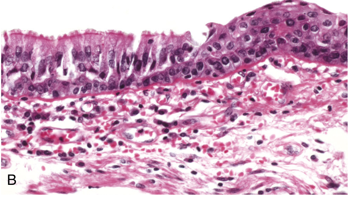
SN 4. Free Radicals and Free Radical-Induced Cell Injury
Definition
Generation of Free Radicals (Flowchart)
SOURCES OF FREE RADICAL GENERATION:
│
┌────┼──────────────────────────────────────┐
↓ ↓ ↓ ↓
Normal Radiation Activated Drug/Chemical
Mitochondrial (UV, X-ray) Leukocytes metabolism
respiration ↓ (NADPH oxidase) (CCl₄ → •CCl₃)
↓ Water → O₂•⁻ ↓
O₂ partially hydrolysis ↓ P450 enzymes
reduced → → •OH H₂O₂ generate radicals
O₂•⁻ ↓
↓ + Fe²⁺ (Fenton reaction):
H₂O₂ H₂O₂ + Fe²⁺ → Fe³⁺ + •OH + OH⁻
↓ (MOST DAMAGING radical)
•OH (via
Fenton rxn)
Antioxidant Defence Systems
FREE RADICALS GENERATED
↓
DEFENCE MECHANISMS:
├─ Superoxide dismutase (SOD): O₂•⁻ + O₂•⁻ → H₂O₂ + O₂
├─ Catalase: H₂O₂ → H₂O + O₂
├─ Glutathione peroxidase: H₂O₂ + 2GSH → GSSG + 2H₂O
├─ Vitamins E (membrane), A, C (aqueous) - scavengers
└─ Transferrin, ceruloplasmin - sequester iron/copper
↓
If defence is OVERWHELMED:
OXIDATIVE STRESS
Mechanisms of Free Radical-Induced Cell Injury
EXCESS FREE RADICALS (Oxidative Stress)
│
┌────┼────────────────────────────────┐
↓ ↓ ↓ ↓
LIPID PROTEIN DNA DAMAGE APOPTOSIS
PEROXIDATION OXIDATION ACTIVATION
↓ ↓ ↓ ↓
•OH attacks Amino acid Single/double Caspase
double bonds side chain strand breaks cascade
in membrane oxidation in DNA activated
polyunsaturated ↓ ↓ (low doses)
fatty acids Enzyme active DNA adducts
↓ site disrupted → mutations
Lipid peroxide Cross-linking → cancer,
↓ of proteins aging
New radicals ↓
generated Proteasomal
(chain rxn) degradation
↓ of misfolded
Membrane proteins
damage:
↑ permeability,
organelle dysfunction,
cell death (NECROSIS
at high doses)
Clinical Conditions Caused by Free Radical Injury
| Condition | Mechanism |
|---|---|
| Ischaemia-reperfusion injury | Burst of ROS on reperfusion; mitochondrial dysfunction |
| Chemical toxicity (CCl₄) | CYP450 converts CCl₄ → •CCl₃ → hepatic necrosis |
| Radiation injury | Ionising radiation → •OH → DNA damage |
| Atherosclerosis | LDL oxidation by ROS → foam cell formation |
| Aging | Cumulative oxidative DNA damage |
| Oxygen toxicity (neonates) | Excess O₂ → ROP, BPD |
| Paraquat poisoning | Generates O₂•⁻ → pulmonary fibrosis |
| Carcinogenesis | Oxidative DNA damage → oncogene activation |
LAQ 1. Necrosis - Clinical Scenario, Definition, Types, Caseous Morphology, Coagulative vs Liquefactive
Clinical Scenario Link
Patient 1: Diabetic, pale dry leg ulcer
→ Ischaemia (arterial occlusion in atherosclerosis)
→ Tissue preserved but dead → COAGULATIVE NECROSIS (dry gangrene)
Patient 2: TB patient, cheesy lung lesion
→ Mycobacterium tuberculosis → granulomatous inflammation
→ CASEOUS NECROSIS
Patient 3: Stroke → brain liquefaction
→ Cerebral ischaemia in neuron-rich tissue
→ LIQUEFACTIVE NECROSIS (brain always liquefies)
Definition of Necrosis
General Morphological Features of Necrotic Cells
- Eosinophilia (↑ eosin binding due to denatured proteins + loss of RNA)
- Vacuolated, moth-eaten cytoplasm
- Myelin figures (whorled phospholipid precipitates from degraded membranes)
NUCLEAR CHANGES IN NECROSIS:
1. KARYOLYSIS: Basophilia fades (DNase degrades DNA)
Nucleus ghost-like → disappears
2. PYKNOSIS: Nuclear shrinkage + increased basophilia
(chromatin condenses into dense mass)
3. KARYORRHEXIS: Pyknotic nucleus fragments
Nuclear debris scattered in cytoplasm
→ After 1-2 days: nucleus COMPLETELY DISAPPEARS
Types of Necrosis - Summary Table
| Type | Key Feature | Gross | Micro | Example |
|---|---|---|---|---|
| Coagulative | Architecture preserved; ghost cells | Firm, pale, dry | Eosinophilic cells with no nuclei; architecture intact | Renal, cardiac, splenic infarct |
| Liquefactive | Complete dissolution; fluid cavity | Soft, fluid, cavity | No architecture; fluid + debris + leukocytes | Brain infarct; pyogenic abscess |
| Caseous | Cheese-like; granuloma | Soft, yellow-white, cheesy | No architecture; acellular granular debris + granuloma | Tuberculosis |
| Fat | Saponification (chalky white) | White chalky deposits | Fat cells replaced by shadowy outlines; calcium soaps | Acute pancreatitis; breast trauma |
| Fibrinoid | Pink homogeneous fibrin-like material | Not grossly distinctive | Vessel wall necrosis; fibrin + immune complexes | Malignant hypertension; vasculitis; PAN |
| Gangrenous | Clinical term (not true histologic type) | Dry = coagulative; Wet = + liquefactive | Depends on type | Diabetic foot (dry/wet gangrene) |
Caseous Necrosis - Morphology in Detail
CASEOUS NECROSIS (Gross):
Soft, friable, CHEESE-LIKE material
Colour: Yellow-white (like dry crumbled cottage cheese)
"Caseous" from Latin: caseus = cheese
Enclosed in FIBROUS CAPSULE (granuloma wall)
Classic site: TB lymph node, TB lung lesion
CASEOUS NECROSIS (Micro):
CENTRAL ZONE:
┌──────────────────────────────────────────────┐
│ ACELLULAR, GRANULAR, EOSINOPHILIC DEBRIS │
│ - Complete loss of all tissue architecture │
│ - No ghost cell outlines (unlike coagulative)│
│ - Amorphous pink material on H&E │
└──────────────────────────────────────────────┘
↑ surrounded by ↑
PERIPHERAL ZONE (GRANULOMA):
┌──────────────────────────────────────────────────┐
│ EPITHELIOID MACROPHAGES │
│ (activated macrophages: pale, abundant cytoplasm;│
│ vesicular nuclei; elongated - look like │
│ epithelial cells) │
│ │
│ LANGHANS GIANT CELLS │
│ (fused macrophages; 10-50 nuclei arranged in │
│ horseshoe/peripheral pattern at cell rim) │
│ ← specific to TB and related granulomas │
│ │
│ LYMPHOCYTES (peripheral cuff) │
│ PLASMA CELLS (occasional) │
│ │
│ Later: FIBROSIS + DYSTROPHIC CALCIFICATION │
└──────────────────────────────────────────────────┘
Coagulative Necrosis - Detail
ISCHAEMIA (arterial occlusion → O₂ deprivation)
↓
ATP depletion → cell swelling → membrane damage
↓
PROTEIN DENATURATION
(both structural proteins AND proteolytic enzymes denatured)
↓
Enzymes cannot digest the dead cells
↓
Cell shape PRESERVED for days
("Ghost cells" / "tombstone cells")
Architecture maintained
↓
Eventually leukocytes arrive (days later) and
lysosomal enzymes degrade the dead cells
↓
Absorbed → SCAR FORMS
Liquefactive Necrosis - Detail
ISCHAEMIA TO BRAIN
OR
BACTERIAL (PYOGENIC) INFECTION
↓
ENZYMATIC DIGESTION dominates over denaturation
(Brain: rich in hydrolytic enzymes + poor structural protein support)
(Bacteria: secrete proteolytic enzymes + recruit neutrophils)
↓
Neutrophils/macrophages release lysosomal enzymes
↓
Complete dissolution of ALL tissue components
- No ghost cells
- No architectural preservation
↓
FLUID-FILLED CAVITY
(cerebral infarct → cystic cavity)
(bacterial infection → PUS = liquefied dead tissue + dead neutrophils)
Coagulative vs Liquefactive - Direct Comparison
| Feature | Coagulative Necrosis | Liquefactive Necrosis |
|---|---|---|
| Tissue architecture | Preserved (ghost cells) | Completely lost |
| Enzymatic digestion | Inhibited (enzymes denatured) | Dominant |
| Consistency | Firm, dry | Soft, fluid |
| Appearance | Pale, firm, dry infarct | Fluid-filled cavity / pus |
| Primary mechanism | Protein denaturation | Enzymatic digestion |
| Sites | All solid organs EXCEPT brain | Brain; pyogenic abscesses |
| Cause | Ischaemia (arterial occlusion) | Brain ischaemia; pyogenic bacteria |
| Examples | Myocardial infarct, renal infarct | Stroke, lung abscess, brain abscess |
| Outcome | Scar after resolution | Cyst / fibrous wall (abscess) |
LAQ 2. Atrophy - Definition, Morphology and Causes of Brown Atrophy of the Heart
Definition
Brown Atrophy of the Heart - Pathogenesis (Flowchart)
ADVANCED AGE / CACHEXIA / CHRONIC ILLNESS
↓
Reduced workload on heart
(↓ cardiac output demand, muscle mass loss)
↓
Autophagy activated in cardiomyocytes
(intracellular organelles digested by lysosomes)
↓
Incomplete lysosomal digestion → LIPOFUSCIN
(oxidised lipid-protein residues accumulate
as indigestible brown granules)
↓
LIPOFUSCIN accumulates in perinuclear cytoplasm
↓
Combined with overall cell shrinkage:
BROWN ATROPHY
(macroscopic brown discolouration + small heart)
Gross Morphology
BROWN ATROPHY HEART (Gross):
Normal heart (350-400g) → REDUCED in weight and size
Colour: BROWN (instead of normal reddish-brown)
↓
Coronary arteries appear TORTUOUS and DILATED
relative to the shrunken myocardium
(vessel length unchanged, heart shrinks → redundant tortuous vessels)
↓
Epicardial fat: may be gelatinous/serous atrophy
(fat replaced by fluid in severe cachexia)
↓
Cut section: Smaller, firmer myocardial fibres
Microscopic Morphology
CARDIOMYOCYTES: Smaller diameter, elongated
NUCLEI: Smaller, darker (hyperchromatic)
LIPOFUSCIN GRANULES:
→ Yellow-brown perinuclear pigment granules
→ "Wear and tear" or "aging" pigment
→ Stains: Sudan black positive, PAS positive
Autofluorescent under UV light
Prussian blue NEGATIVE (not iron)
→ Represents incomplete lysosomal digestion
of oxidised lipids and proteins over lifetime
MYOFIBRILS: Reduced in number
INTERSTITIAL FIBROSIS: May be present
NO INFLAMMATORY INFILTRATE (not inflammatory)
Causes of Brown Atrophy of the Heart
| Cause | Mechanism |
|---|---|
| Advanced age (Senile atrophy) | Most common; normal aging → progressive lipofuscin accumulation + cell loss |
| Cancer cachexia | TNF-α, IL-1, IL-6 from tumour → ↑ protein catabolism; starvation of nutrients |
| Chronic debilitating illness | TB, AIDS, CHF, COPD → generalised wasting |
| Severe starvation/malnutrition | Inadequate caloric intake → protein catabolism |
| Prolonged immobility | Disuse → reduced cardiac demand |
| Hypopituitarism | ↓ growth hormone, ↓ thyroid hormone → reduced anabolic stimulation |
IMMUNOPATHOLOGY
SN 1. Hypersensitivity Reactions - Classification + Type I in Detail
Definition
Classification (Gell & Coombs)
HYPERSENSITIVITY REACTIONS
│
┌────┼──────────────────────────────────────────────┐
↓ ↓ ↓ ↓
TYPE I TYPE II TYPE III TYPE IV
(IgE) (IgG/IgM (Immune (T-cell
Immediate cytotoxic) complex) Delayed)
Mast cell Cell surface/ Soluble Ag-Ab CD4+ or CD8+
degranulation matrix Ag complexes T cells
↓ ↓ ↓ ↓
Minutes Complement Complement + 24-72 hours
+ NK cells neutrophils
↓ ↓ ↓ ↓
Anaphylaxis, Haemolytic Serum sickness, Contact
asthma, anaemia, SLE, post-strep dermatitis,
urticaria Goodpasture GN, Arthus rxn tuberculin
test, Type 1 DM
Type I Hypersensitivity - Detailed Flowchart
Phase 1: SENSITISATION (First Exposure - No Symptoms)
FIRST EXPOSURE TO ALLERGEN (antigen)
↓
Antigen processed by APCs (dendritic cells)
↓
TH2 CD4+ T cells activated
(IL-4 producing environment favours TH2 differentiation)
↓
TH2 cells produce:
├─ IL-4 → B cell class switching to IgE
├─ IL-5 → Eosinophil recruitment/activation
└─ IL-13 → Mucus secretion, smooth muscle contraction
↓
IgE produced by plasma cells
↓
IgE binds to FcεRI receptors on MAST CELLS and BASOPHILS
(HIGH AFFINITY receptor - mast cells coated with IgE)
↓
SENSITISED MAST CELLS (no symptoms yet)
Phase 2: EFFECTOR PHASE (Re-exposure - Immediate Reaction, within minutes)
RE-EXPOSURE TO SAME ALLERGEN
↓
Allergen CROSS-LINKS IgE molecules on mast cell surface
(must bind ≥2 IgE molecules simultaneously)
↓
FcεRI aggregation → signal transduction
↓
MAST CELL DEGRANULATION
│
┌────┴─────────────────────────────────────────────┐
↓ ↓ ↓
PREFORMED NEWLY SYNTHESISED CYTOKINES
MEDIATORS MEDIATORS (over hours)
(released (minutes-hours)
immediately)
↓ ↓ ↓
HISTAMINE LEUKOTRIENES (AA pathway) TNF-α
(most important) LTC4, LTD4, LTE4 IL-4, IL-5
→ vasodilation (SRS-A, 1000x >histamine IL-13
→ ↑ permeability in bronchoconstriction) ↓
→ smooth muscle PGD2 (prostaglandin) Recruit
contraction → bronchoconstriction eosinophils,
→ mucus secretion PAF (platelet activating basophils,
factor) TH2 cells
SEROTONIN → platelet aggregation → LATE PHASE
HEPARIN
TRYPTASE
EOSINOPHIL
CHEMOTACTIC
FACTOR
Phase 3: LATE PHASE REACTION (2-8 hours, persisting 24 hours)
Mast cell cytokines (TNF-α, IL-5) recruit:
EOSINOPHILS, BASOPHILS, NEUTROPHILS, TH2 CELLS
↓
Eosinophils release:
- Major Basic Protein (MBP) → tissue damage
- Eosinophil Peroxidase (EPO)
- Eosinophil Cationic Protein (ECP)
- IL-3, IL-5, GM-CSF (amplify further eosinophil production)
↓
PROLONGED bronchoconstriction, oedema, mucus production
(this phase explains SUSTAINED ASTHMA attacks)
Clinical Manifestations of Type I Hypersensitivity
| Target Organ | Allergen Route | Manifestation |
|---|---|---|
| Systemic | IV or widespread | Anaphylaxis - urticaria, angioedema, bronchospasm, hypotension, shock (life-threatening) |
| Lung | Inhaled | Asthma - bronchoconstriction, wheezing, dyspnoea |
| Nasal mucosa | Inhaled | Allergic rhinitis - sneezing, watery discharge, nasal obstruction |
| Skin | Contact/systemic | Urticaria (hives), Angioedema - wheals, flares |
| GI | Ingested | Nausea, vomiting, diarrhoea, cramping |
| Eye | Inhaled/contact | Allergic conjunctivitis - itching, redness |
SN 2. Amyloidosis - Clinical Scenario, Definition, Classification, Molecular Structure, Special Stains
Clinical Scenario Analysis
Patient 1: RA (long-standing) + nephrotic syndrome + hepatosplenomegaly
+ spleen with "pale tapioca-like nodules"
→ SECONDARY (AA) AMYLOIDOSIS
(chronic inflammatory disease → SAA protein → AA amyloid deposits in kidney, spleen, liver)
Patient 2: Multiple myeloma + restrictive cardiomyopathy
+ Congo red → apple-green birefringence
→ PRIMARY (AL) AMYLOIDOSIS
(plasma cell dyscrasia → light chains → AL amyloid deposits in heart, kidney, tongue)
Definition
Classification (Flowchart)
AMYLOIDOSIS
│
┌────┴─────────────────────────────────────────────┐
↓ ↓ ↓
PRIMARY (AL) SECONDARY (AA) HEREDOFAMILIAL
↓ ↓ ↓
Precursor: Precursor: Precursor:
Immunoglobulin Serum Amyloid A Transthyretin (ATTR)
LIGHT CHAINS (SAA - acute Fibrinogen Aα chain
(λ > κ) phase protein) Others
↓ ↓ ↓
Associated with: Associated with: Associated with:
Multiple myeloma RHEUMATOID ARTHRITIS Familial amyloid
Waldenström's Chronic infections polyneuropathy
monoclonal (TB, bronchiectasis, Familial
gammopathy osteomyelitis) Mediterranean fever
MGUS IBD, Ankylosing (AA type in FMF)
spondylitis Senile systemic
amyloidosis (ATTR
in elderly hearts)
↓ ↓ ↓
Deposits in: Deposits in: Deposits in:
Heart KIDNEY (most Heart (ATTR-CA)
Kidney common → nephrotic) Peripheral nerves
Tongue (macro- LIVER (hepato- Carpal tunnel
glossia) splenomegaly)
GI tract SPLEEN (sago/
Peripheral nerves lardaceous)
Skin (waxy) Adrenals
- Aβ amyloid (APP-derived) → Alzheimer disease (senile plaques)
- β₂-microglobulin amyloid → long-term haemodialysis (carpal tunnel syndrome)
- Calcitonin-derived → Medullary thyroid carcinoma
Molecular Structure of Amyloid
AMYLOID FIBRIL STRUCTURE:
Precursor protein (soluble, native conformation)
↓
MISFOLDING (abnormal processing, overproduction)
↓
BETA-SHEET CONFORMATION adopted
(antiparallel β-pleated sheet structure)
↓
Individual fibrils: NON-BRANCHING, 7.5-10 nm diameter
Composed of: β-pleated sheets running perpendicular to fibril axis
↓
CROSS-BETA PATTERN (X-ray diffraction signature)
↓
Fibrils arranged in bundles → macroscopic deposits
↓
Also contains:
- Serum Amyloid P (SAP) component - all types
- Apolipoprotein E - all types
- Glycosaminoglycans (heparan sulphate) - all types
These non-fibril components stabilise the deposit
RESULT: INSOLUBLE, RESISTANT TO PROTEOLYSIS
→ Deposits accumulate progressively → organ damage
Special Staining for Amyloid
SPECIAL STAINS FOR AMYLOID:
1. CONGO RED (most important / pathognomonic)
└─ Routine light: SALMON PINK / APPLE RED colour
└─ Polarised light: APPLE-GREEN BIREFRINGENCE ← PATHOGNOMONIC
(due to ordered beta-pleated sheet binding Congo red dye
and rotating polarised light)
2. CRYSTAL VIOLET (Metachromatic stain)
└─ Amyloid stains RED/VIOLET against blue background
(metachromasia = colour different from the dye used)
3. THIOFLAVIN T or S
└─ FLUORESCENCE under UV light
└─ Used for small deposits and frozen sections
4. PERIODIC ACID-SCHIFF (PAS)
└─ Weakly positive (glycoprotein component)
5. ELECTRON MICROSCOPY
└─ Fibrils: non-branching, 7.5-10 nm diameter
└─ Beta-sheet confirmed by X-ray diffraction (CROSS-BETA pattern)
Morphology of Amyloid Deposits in Key Organs
SAGO SPLEEN (most common in AA amyloidosis):
- Amyloid in WHITE PULP (follicles)
- Gross: Small, grey/translucent tapioca/sago grain-like
nodules = "tapioca appearance" (matches the case!)
- Histology: Amyloid replacing lymphoid follicles
↓
vs.
LARDACEOUS SPLEEN (advanced/severe):
- Amyloid in RED PULP (diffuse)
- Gross: Large, firm, waxy, pale/greyish, "lard-like"
- Histology: Amyloid diffusely replacing red pulp sinusoids
- Glomerular mesangium + GBM → nephrotic syndrome (massive proteinuria, oedema)
- Tubular basement membrane deposits
- Kidney: enlarged, pale, waxy ("large white kidney")
- Restrictive cardiomyopathy (stiff, non-compliant ventricles)
- Amyloid between myofibrils
- Echo: "sparkling" granular echogenicity of myocardium
- Deposits in space of Disse (between hepatocytes and sinusoids) and portal areas
- Hepatomegaly, elevated ALP, rarely liver failure
Make a colour graded PDF of all the data except for the data of 1st image use data which you created afterwards for 1st image
mkdir -p /tmp/workspace/pathology-pdf && ls /tmp/workspace/
Make pdf
mkdir -p /tmp/workspace/pathology-pdf && ls /tmp/workspace/pathology-pdf/
Make a colour graded PDF of all the data except for the data of 1st image use data which you created afterwards for 1st image
mkdir -p /tmp/workspace/pathology-pdf && python3 -c "from reportlab.platypus import SimpleDocTemplate; print('reportlab ok')"


Answer the question for my university exams and add flow chart where ever necessary
Haematology - Paper II Complete Exam Answers
SECTION: Introduction to Haematopoietic System and Disorders of Erythroid Series
SN 1. Etiopathogenesis of Sickle Cell Anaemia | Laboratory Investigations | PBS Findings
Definition
Genetic Basis
| Genotype | Condition | HbS level |
|---|---|---|
| HbSS (homozygous) | Sickle cell anaemia (disease) | ~100% HbS |
| HbAS (heterozygous) | Sickle cell trait (carrier) | ~40% HbS |
| HbSC | Compound heterozygote | HbS + HbC |
| HbS-β thal | Sickle-thal disease | Variable |
Etiopathogenesis (Flowchart)
POINT MUTATION: β-globin gene (GAG→GTG)
Glutamate → Valine at position 6
↓
HAEMOGLOBIN S (HbS) formed
(α₂β₂S instead of normal α₂β₂A)
↓
DEOXYGENATION (low O₂ tension, acidosis, dehydration)
↓
HbS molecules undergo conformational change
↓
HbS polymers form via intermolecular contacts
(abnormal valine residue creates "sticky patch")
↓
SICKLING of red cells (crescentic/sickle shape)
Initially reversible on reoxygenation
↓
REPEATED SICKLING EPISODES
↓
┌────┴─────────────────────────────────────┐
↓ ↓
Ca²⁺ influx → K⁺/H₂O loss Membrane damage
↓ accumulates
IRREVERSIBLY SICKLED CELLS
↓ ↓
HAEMOLYSIS VASCULAR OCCLUSION
(intravascular (sickled cells are
+ extravascular) rigid, adhere to
↓ endothelium)
CHRONIC HAEMOLYTIC ↓
ANAEMIA VASO-OCCLUSIVE CRISES
(Hb 6-8 g/dL) (pain crises, infarcts)
↓ ↓
Jaundice Organ damage: spleen,
Splenomegaly bone, kidney, brain,
(early then retina, lung (ACS)
autosplenectomy)
- Low PO₂ (hypoxia)
- Acidosis (↓ pH)
- Dehydration (increased MCHC)
- Infection/fever
- Cold exposure
- High altitude
- Slow blood flow (stasis)
- HbF (foetal Hb) - protective; does not polymerise with HbS
- HbA (in sickle trait) - protective
- Hydroxyurea (↑ HbF production)
Clinical Features
- Pallor, fatigue, jaundice (unconjugated hyperbilirubinaemia)
- Splenomegaly (early) → autosplenectomy (repeated infarctions → fibrotic shrunken spleen by age 5)
- Gallstones (pigment stones from chronic haemolysis)
- Aplastic crisis (parvovirus B19 infection)
- Painful crises (most common) - bone pain (back, chest, limbs)
- Acute chest syndrome (ACS) - chest pain, fever, pulmonary infiltrates; most common cause of death
- Stroke (cerebrovascular occlusion) - especially in children
- Dactylitis (hand-foot syndrome) - infants; painful swelling of hands/feet
- Avascular necrosis (femoral head, humeral head)
- Priapism (painful prolonged erection)
- Renal papillary necrosis → haematuria
- Encapsulated organisms: S. pneumoniae, H. influenzae, N. meningitidis
- Salmonella osteomyelitis (distinctive)
- Parvovirus B19 aplastic crisis
Laboratory Investigations
| Test | Result in Sickle Cell Anaemia |
|---|---|
| Haemoglobin | Low: 6-8 g/dL |
| Reticulocyte count | Elevated (5-15%) - compensatory |
| Bilirubin (indirect) | Elevated (haemolysis) |
| LDH | Elevated (haemolysis marker) |
| Haptoglobin | Low/absent (binds free Hb) |
| Urine urobilinogen | Elevated |
| WBC | Mild leucocytosis (baseline) |
| Serum ferritin | Normal or elevated (no iron deficiency; repeated transfusions) |
| Haemoglobin electrophoresis | HbSS: ~100% HbS, absent HbA, slight HbF; GOLD STANDARD |
| Sickling test (metabisulphite) | Positive - cells sickle on deoxygenation |
| Solubility test (Sickledex) | Positive - HbS insoluble in reduced solution |
| HPLC | Quantifies HbS, HbF, HbA accurately |
| Bone marrow | Erythroid hyperplasia |
Peripheral Blood Smear (PBS) Findings
PBS in SICKLE CELL ANAEMIA:
DEFINITIVE FINDING:
→ SICKLE CELLS (drepanocytes) - elongated, crescent/holly-leaf/boat-shaped cells
OTHER CELLS:
→ TARGET CELLS (codocytes) - central and peripheral Hb with clear ring
→ POLYCHROMASIA - reticulocytes (bluish tinge) - compensatory
→ ANISOCYTOSIS & POIKILOCYTOSIS
→ NUCLEATED RED BLOOD CELLS (nRBCs) - stress erythropoiesis
→ HOWELL-JOLLY BODIES - nuclear remnants (sign of hyposplenism/autosplenectomy)
→ ACANTHOCYTES - post-splenectomy
→ BASOPHILIC STIPPLING - occasionally
→ PAPPENHEIMER BODIES - iron granules (post-splenectomy)
WBCs: Neutrophilia during crises
PLATELETs: Normal or increased
LAQ 1. Megaloblastic Anaemia (60-year-old vegetarian, ↓B12, macrocytic anaemia, hypersegmented neutrophils)
Sub-question A: Laboratory Investigations and PBS Findings for Megaloblastic Anaemia
Complete Blood Count (CBC)
| Parameter | Finding |
|---|---|
| Haemoglobin | Low (moderate to severe anaemia) |
| MCV | >100 fL (macrocytosis) - often >110-115 fL in megaloblastic |
| MCH | Elevated |
| MCHC | Normal |
| RBC count | Markedly reduced |
| Reticulocyte count | Low/inappropriately low (ineffective erythropoiesis) |
| WBC | Leucopenia (hypersegmented neutrophils, giant metamyelocytes) |
| Platelets | Thrombocytopenia |
| Pancytopenia | In severe cases |
Biochemical Tests
| Test | Finding | Significance |
|---|---|---|
| Serum Vitamin B12 | < 200 pg/mL (normal 200-900) | Primary deficiency |
| Serum Folate | Low if folate deficiency | Alternative cause |
| RBC folate | More reliable than serum | Better indicator of tissue stores |
| Serum homocysteine | Elevated | Elevated in BOTH B12 AND folate deficiency |
| Serum methylmalonic acid (MMA) | Elevated | Specific for B12 deficiency (DIFFERENTIATES from folate deficiency) |
| Serum bilirubin (indirect) | Elevated (ineffective erythropoiesis → intramedullary haemolysis) | |
| LDH | Markedly elevated (massive cell destruction in marrow) | |
| Serum iron/ferritin | Normal or elevated (no iron deficiency) | |
| Schilling test | Identifies cause (pernicious anaemia vs dietary) - less used now | |
| Anti-intrinsic factor antibodies | Positive in pernicious anaemia (>50% sensitivity; high specificity) | |
| Anti-parietal cell antibodies | Positive in ~90% of pernicious anaemia (less specific) |
Peripheral Blood Smear (PBS) Findings
PBS in MEGALOBLASTIC ANAEMIA:
HALLMARK FINDINGS:
→ MACRO-OVALOCYTES (macroovalocytes) - large, oval-shaped RBCs
(oval macrocytes are more specific than round macrocytes)
→ HYPERSEGMENTED NEUTROPHILS ← PATHOGNOMONIC
(5+ lobes in >5% of neutrophils; OR any neutrophil with 6+ lobes)
Mechanism: Impaired DNA synthesis → neutrophil cannot divide properly
→ nuclear segments keep forming without cell division
OTHER FINDINGS:
→ ANISOCYTOSIS (varied cell sizes) - often marked (high RDW)
→ POIKILOCYTOSIS (varied cell shapes)
→ TEAR-DROP CELLS (dacrocytes) - occasionally
→ NUCLEATED RBCs (megaloblasts escape from marrow in severe cases)
→ BASOPHILIC STIPPLING
→ CABOT RINGS (remnants of mitotic spindle in RBCs) - rare
WBCs:
→ Leucopenia
→ Hypersegmented neutrophils (most specific finding)
→ Giant band forms, giant metamyelocytes
PLATELETS:
→ Thrombocytopenia in severe disease
→ Giant platelets occasionally
OVERALL PICTURE: Pancytopenia with macro-ovalocytes and hypersegmented neutrophils
Sub-question B: Bone Marrow Findings in Megaloblastic Anaemia
BONE MARROW in MEGALOBLASTIC ANAEMIA:
HYPERCELLULAR marrow (despite peripheral pancytopenia)
→ This is INEFFECTIVE ERYTHROPOIESIS
(cells are made but destroyed in marrow before release)
ERYTHROID SERIES:
→ MEGALOBLASTS - hallmark cell
Large cells with:
- LARGE NUCLEUS with FINE, OPEN CHROMATIN ("sieve-like" / "salt and pepper")
- ABUNDANT CYTOPLASM with normal haemoglobinisation
- NUCLEAR-CYTOPLASMIC (N:C) DISSOCIATION:
Cytoplasm matures faster than nucleus
(nucleus still looks immature while cytoplasm is already haemoglobinised)
← ASYNCHRONOUS MATURATION (the question mentions this!)
→ Stages of megaloblasts visible: promegaloblast, basophilic megaloblast,
polychromatic megaloblast, orthochromatic megaloblast
MYELOID SERIES:
→ GIANT METAMYELOCYTES - characteristic finding
(large band/metamyelocyte forms with horseshoe/twisted nuclei)
→ Giant band forms
→ Hypersegmented megakaryocytes
MEGAKARYOCYTES:
→ Giant, hyperlobated megakaryocytes
M:E RATIO:
→ Decreased (erythroid hyperplasia) but cells die in situ
→ "Ineffective haemopoiesis" → intramedullary destruction
Sub-question C: Causes of Macrocytic Anaemia
MACROCYTIC ANAEMIA (MCV > 100 fL)
│
┌────┴────────────────────────────────────────┐
↓ ↓
MEGALOBLASTIC NON-MEGALOBLASTIC
(impaired DNA synthesis) (no nuclear maturation defect)
│ │
┌────┴──────────────┐ ┌─────────┴────────────────────┐
↓ ↓ ↓ ↓ ↓ ↓
Vitamin B12 Folate Liver Alcohol Hypothyroidism Haemolysis
deficiency deficiency disease (↑ lipid (severe) (reticulocytosis)
│ │ (lipid in RBC
├─ Dietary (vegans) ├─ Diet membrane membrane) Myelodysplastic
├─ Pernicious ├─ Malabsorption ↓ syndrome (MDS)
│ anaemia │ (coeliac, crohn) Spur cells
│ (anti-IF Ab) ├─ Pregnancy (acanthocytes)
├─ Malabsorption ├─ Drugs
│ (terminal ileum │ (methotrexate, Drugs:
│ disease - Crohn, │ phenytoin, Hydroxyurea,
│ resection) │ trimethoprim) Azathioprine,
├─ Drugs └─ Alcoholism Zidovudine
│ (metformin, PPI,
│ nitrous oxide) Aplastic anaemia
└─ Fish tapeworm (reticulocytopenia)
(Diphyllobothrium)
LAQ 2. Iron Deficiency Anaemia (35-year-old woman, menorrhagia, microcytic hypochromic RBCs, ↓ferritin, ↑TIBC)
Sub-question A: Definition of Anaemia
- Males: Haemoglobin < 13 g/dL
- Non-pregnant females: Haemoglobin < 12 g/dL
- Pregnant females: Haemoglobin < 11 g/dL
- Children (6 months-5 years): < 11 g/dL
Sub-question B: Morphological Classification of Anaemia
ANAEMIA
│
├─── MICROCYTIC HYPOCHROMIC (MCV <80 fL, MCH <27 pg)
│ Iron deficiency anaemia (IDA) ← most common worldwide
│ Thalassaemia (α or β)
│ Anaemia of chronic disease (some cases)
│ Sideroblastic anaemia (acquired/congenital)
│ Lead poisoning
│
├─── NORMOCYTIC NORMOCHROMIC (MCV 80-100 fL, MCH 27-33 pg)
│ Anaemia of chronic disease (most cases)
│ Aplastic anaemia
│ Haemolytic anaemia (acute)
│ Acute blood loss
│ Renal failure (↓ EPO)
│ Hypothyroidism (mild)
│ Mixed deficiency (B12/folate + iron)
│
└─── MACROCYTIC (MCV >100 fL)
Megaloblastic (B12/folate deficiency) - see LAQ 1
Non-megaloblastic (liver disease, alcoholism, hypothyroidism)
Sub-question C: Laboratory Differences - Iron Deficiency Anaemia vs Normal
Iron Studies - Comparison Table
| Parameter | Normal | Iron Deficiency Anaemia | Anaemia of Chronic Disease | Thalassaemia |
|---|---|---|---|---|
| Serum iron | 60-170 μg/dL | ↓ Low | ↓ Low | Normal/↑ |
| TIBC (transferrin) | 250-370 μg/dL | ↑ High (liver makes more transferrin) | ↓ Low | Normal |
| Transferrin saturation | 20-50% | ↓ Low (<15%) | Low | Normal/↑ |
| Serum ferritin | 12-150 ng/mL | ↓ Low (<12 ng/mL) ← BEST marker of IDA | Normal/↑ | Normal/↑ |
| Serum soluble transferrin receptor (sTfR) | Normal | ↑ Elevated | Normal | Elevated |
| Bone marrow iron (Prussian blue) | Present | Absent ← GOLD STANDARD | Present (↑) | Present (↑) |
Stages of Iron Deficiency (Flowchart)
STAGE 1: Iron Depletion
- ↓ Bone marrow iron stores
- ↓ Serum ferritin (<12 ng/mL)
- Hb still normal; no anaemia yet
↓
STAGE 2: Iron-Deficient Erythropoiesis
- Iron stores exhausted
- ↓ Serum iron; ↑ TIBC
- ↓ Transferrin saturation
- ↑ sTfR
- RBCs becoming microcytic but Hb still near normal
↓
STAGE 3: Iron Deficiency Anaemia (IDA)
- ↓ Haemoglobin (< 12g/dL females)
- Microcytic, hypochromic red cells
- All iron indices abnormal
- Clinical symptoms appear
PBS in Iron Deficiency Anaemia
PERIPHERAL BLOOD SMEAR - IDA:
→ MICROCYTES (MCV <80 fL) - small RBCs
→ HYPOCHROMIA - central pallor >1/3 of cell diameter
→ ANISOCYTOSIS - variability in cell size (↑ RDW >14.5%)
→ POIKILOCYTOSIS
→ PENCIL CELLS (elliptocytes) - elongated, cigar-shaped
→ TARGET CELLS (codocytes) - thin cells with Hb in centre
→ OCCASIONAL RING SIDEROBLASTS (in sideroblastic anaemia - different)
→ THROMBOCYTOSIS (reactive, common in IDA from chronic blood loss)
Clinical Features of IDA (matching the case)
- Fatigue, pallor, exertional dyspnoea (anaemia)
- Glossitis (smooth, red, painful tongue)
- Angular cheilitis/stomatitis (cracks at mouth corners)
- Koilonychia (spoon-shaped nails) - brittle nails (patient's complaint)
- Pica (craving for non-food substances: ice, clay, chalk)
- Plummer-Vinson syndrome (IDA + oesophageal web + dysphagia)
- Hair loss
- Restless leg syndrome
LAQ 3. Laboratory Investigations for Haemolytic Anaemia
Definition
Classification
HAEMOLYTIC ANAEMIA
│
┌────┴──────────────────────────────────────┐
↓ ↓
INTRACORPUSCULAR DEFECT EXTRACORPUSCULAR DEFECT
(Usually hereditary) (Usually acquired)
│ │
├─ Membrane defects: ├─ Immune:
│ Hereditary spherocytosis │ AIHA (warm/cold)
│ Hereditary elliptocytosis │ Haemolytic transfusion rxn
│ │ HDN (Rh/ABO incompatibility)
├─ Enzyme defects: │
│ G6PD deficiency ├─ Microangiopathic:
│ PK deficiency │ TTP, HUS, DIC
│ │ Mechanical heart valves
└─ Haemoglobin defects: │
Sickle cell disease ├─ Infections:
Thalassaemia │ Malaria, Clostridium
HbC, HbE │
└─ Hypersplenism
Laboratory Investigations Flowchart
SUSPECTED HAEMOLYTIC ANAEMIA
↓
STEP 1: CONFIRM HAEMOLYSIS
────────────────────────────
CBC:
→ ↓ Haemoglobin
→ ↑ Reticulocytes (>2%) - KEY FINDING
→ ↑ MCV (due to reticulocytosis)
→ Polychromasia on PBS
Serum markers of haemolysis:
→ ↑ LDH (released from lysed RBCs) - SENSITIVE
→ ↓ Haptoglobin (binds free Hb; consumed) - SPECIFIC
→ ↑ Indirect (unconjugated) bilirubin
→ ↑ Plasma free haemoglobin (intravascular)
→ Haemoglobinuria (dark urine in intravascular haemolysis)
→ Haemosiderinuria (Prussian blue stain on urine deposit)
↓
STEP 2: CLASSIFY - INTRAVASCULAR vs EXTRAVASCULAR
──────────────────────────────────────────────────
Intravascular: Extravascular:
↓↓ Haptoglobin ↓ Haptoglobin (mild)
Haemoglobinaemia No haemoglobinaemia
Haemoglobinuria No haemoglobinuria
Haemosiderinuria No haemosiderinuria
Methaemalbuminaemia Splenomegaly prominent
(Schumm test +)
↓
STEP 3: FIND CAUSE
──────────────────
PBS Morphology:
→ Spherocytes → hereditary spherocytosis or AIHA
→ Sickle cells → SCD
→ Target cells → Thalassaemia, IDA, liver disease
→ Schistocytes/fragments → MAHA (TTP, HUS, DIC)
→ Bite cells → G6PD deficiency
→ Elliptocytes → hereditary elliptocytosis
→ Agglutination → cold agglutinin disease
Specific Tests:
→ Direct Coombs test (DAT) - positive in AIHA, HDN
→ Osmotic fragility - increased in spherocytosis
→ G6PD enzyme assay - G6PD deficiency
→ Heinz body preparation - G6PD, unstable Hb
→ Hb electrophoresis - HbS, HbC, thalassaemia
→ Flow cytometry (CD55/CD59) - PNH
→ Sickling test
→ Bone marrow (if needed) - erythroid hyperplasia
STEP 4: SPECIFIC TESTS FOR CAUSE
→ Malaria thick/thin film
→ Blood cultures (sepsis-related)
→ Coagulation screen (DIC)
→ LFTs, renal function
SECTION: Disorders of Platelets, Bleeding Disorders and Basic Transfusion Medicine
SN 1. Haemophilia - Types, Inheritance, Clinical Features
Definition
Types and Inheritance
HAEMOPHILIA
│
├─ HAEMOPHILIA A (Classic Haemophilia) - 80% of cases
│ Deficiency: FACTOR VIII (procoagulant activity)
│ Gene: F8 gene on X chromosome (Xq28)
│ Inheritance: X-LINKED RECESSIVE
│ Incidence: 1 in 5000-10,000 male births
│
├─ HAEMOPHILIA B (Christmas Disease) - 15% of cases
│ Deficiency: FACTOR IX
│ Gene: F9 gene on X chromosome (Xq27)
│ Inheritance: X-LINKED RECESSIVE
│ Incidence: 1 in 30,000-50,000 males
│
└─ HAEMOPHILIA C (Rosenthal Disease) - rare
Deficiency: FACTOR XI
Inheritance: AUTOSOMAL RECESSIVE (affects both sexes equally)
Incidence: Rare; more common in Ashkenazi Jews
FATHER (XᴴY - affected) × MOTHER (XX - normal)
↓
Daughters: All carriers (XᴴX)
Sons: All normal (XY)
FATHER (XY - normal) × MOTHER (XᴴX - carrier)
↓
Sons: 50% affected (XᴴY), 50% normal (XY)
Daughters: 50% carriers (XᴴX), 50% normal (XX)
Note: Female haemophilia can occur if:
- Homozygous (XᴴXᴴ) - rare
- Turner syndrome carrier (XᴴO) - monosomy X
- Lyonisation (extreme skewed X-inactivation)
Severity Classification
| Severity | Factor Level | Bleeding Pattern |
|---|---|---|
| Severe | < 1% (<0.01 IU/mL) | Spontaneous bleeding into joints, muscles; life-threatening |
| Moderate | 1-5% | Bleeding with minor trauma; occasional spontaneous |
| Mild | 5-40% | Bleeding only with significant trauma or surgery |
Clinical Features
- Knees (most common), elbows, ankles, shoulders, hips
- Acute: warm, swollen, excruciatingly painful joint
- Chronic: haemophilic arthropathy → joint destruction, deformity
- Iliopsoas haemorrhage (mimics appendicitis)
- Forearm/calf compartment syndrome
- Femoral nerve palsy (from iliopsoas bleed)
- Petechiae (platelet plug intact → primary haemostasis normal)
- Superficial cuts bleed normally (platelet plug stops initial bleeding; only prolonged secondary haemostasis is impaired)
Laboratory Findings
| Test | Result |
|---|---|
| aPTT (APTT) | Prolonged (intrinsic pathway - factors VIII, IX, XI involved) |
| PT (Prothrombin time) | Normal (extrinsic pathway unaffected) |
| Thrombin time | Normal |
| Bleeding time | Normal (platelets and vWF intact) |
| Platelet count | Normal |
| Factor VIII assay | Low in Haemophilia A |
| Factor IX assay | Low in Haemophilia B |
| Bethesda inhibitor assay | Detects factor VIII inhibitors (complication in 30% of severe HA) |
SN 2. Prothrombin Time (PT) Test - Principle and Causes of Increased PT
Principle of PT Test
PROTHROMBIN TIME (PT) TEST:
TESTS: EXTRINSIC and COMMON COAGULATION PATHWAYS
Factors tested: VII, X, V, II (Prothrombin), I (Fibrinogen)
REAGENTS:
→ Tissue Thromboplastin (Thromboplastin reagent = tissue factor + phospholipid)
→ Calcium chloride (CaCl₂) - recalcifies citrated plasma
PROCEDURE:
Patient's citrated plasma (platelet-poor)
↓
Add Thromboplastin + CaCl₂
↓
Start timer
↓
TIME to CLOT FORMATION = PT
Normal: 11-14 seconds (lab dependent)
EXPRESSED AS:
→ INR = (Patient PT / Mean Normal PT)^ISI
(International Normalised Ratio - standardises results between labs)
Normal INR = 0.9-1.2
Therapeutic (warfarin): INR 2-3 (standard) or 2.5-3.5 (mechanical valve)
Causes of Prolonged PT
PROLONGED PT (Extrinsic + Common pathway affected)
│
┌────┼─────────────────────────────────────────────┐
↓ ↓ ↓ ↓
FACTOR VITAMIN K LIVER ANTICOAGULANT
VII DEFICIENCY DISEASE THERAPY
DEFICIENCY │
(most ├─ Dietary Liver produces Warfarin
sensitive) ├─ Malabsorption ALL coagulation (inhibits vit K
├─ Obstructive factors except dependent factor
│ jaundice (↓ VIII and vWF activation:
│ bile → ↓ vit K → cirrhosis II, VII, IX, X,
│ absorption) → ↓ all factors Protein C & S)
└─ Warfarin therapy
OTHER CAUSES:
→ DIC (disseminated intravascular coagulation) - consumption of all factors
→ Massive blood transfusion (dilution of factors)
→ Factor X, V, II, or fibrinogen deficiency
→ Fibrinogen disorders (dysfibrinogenaemia, hypofibrinogenaemia)
→ Lupus anticoagulant (rare - usually prolongs aPTT more than PT)
FACTOR VII has SHORTEST HALF-LIFE of all clotting factors
→ PT is FIRST TEST to become abnormal in:
- Early liver disease
- Early warfarin therapy
- Early vitamin K deficiency
SN 3. Causes of Thrombocytopenia
THROMBOCYTOPENIA
│
┌────┼──────────────────────────────────────────────┐
↓ ↓ ↓ ↓
DECREASED INCREASED SEQUESTRATION DILUTIONAL
PRODUCTION DESTRUCTION (SPLENOMEGALY)
│ │
├─ Aplastic anaemia ├─ IMMUNE:
├─ B12/Folate def. │ ITP (Immune Thrombocytopenic Purpura)
├─ Bone marrow │ Drug-induced immune (heparin-HIT,
│ infiltration: │ quinine, vancomycin)
│ Leukaemia, lym- │ SLE, HIV
│ phoma, metastases │ Post-transfusion purpura
├─ Myelodysplasia │ Neonatal alloimmune
├─ Viral infections │
│ (HIV, EBV, CMV) ├─ NON-IMMUNE:
├─ Chemotherapy/ │ TTP (Thrombotic Thrombocytopenic Purpura)
│ radiation │ HUS (Haemolytic Uraemic Syndrome)
└─ Congenital │ DIC
(Fanconi, WAS, │ Mechanical heart valves
TAR syndrome) │ HELLP syndrome (pregnancy)
│ Giant haemangioma (Kasabach-Merritt)
└─ HYPERSPLENISM
└─ Sequestration + ↑ destruction
DILUTIONAL:
Massive transfusion (stored blood lacks platelets)
LAQ 1 (Platelets). Bleeding Disorders - Definition, Classification, Screening Tests
Definition
Classification
BLEEDING DISORDERS
│
┌────┼──────────────────────────────────────────────────┐
↓ ↓ ↓ ↓
VASCULAR PLATELET COAGULATION FIBRINOLYTIC
DISORDERS DISORDERS FACTOR DISORDERS
│ │ DISORDERS
│ │ │
Hereditary: QUANTITATIVE: Hereditary: Primary
Osler-Weber- Thrombocytopenia Haemophilia A/B/C fibrinolysis
Rendu (HHT) (all causes vWD α2-antiplasmin
above) Factor XIII def. deficiency
Acquired:
Scurvy QUALITATIVE: Acquired:
(↓ collagen) vWD (most Liver disease
Cushing's common) Vit K deficiency
Vasculitis Drug-induced Warfarin
Infection (aspirin, DIC
(meningococcal) NSAIDs) Massive transfusion
Uraemia Inhibitors
BSS, GPS
| Feature | Platelet/Vascular disorder | Coagulation factor disorder |
|---|---|---|
| Bleeding type | Mucocutaneous: petechiae, purpura, epistaxis, gum bleeding | Deep: haemarthroses, muscle haematomas, post-surgical delayed |
| Onset after trauma | Immediate | Delayed (hours) |
| Petechiae | Present | Absent |
| Site | Skin, mucous membranes | Joints, muscles, deep tissues |
Screening Laboratory Tests for Bleeding Disorders
| Test | Normal | What it Tests |
|---|---|---|
| Platelet count | 150-400 × 10⁹/L | Quantitative platelet disorders |
| Bleeding time (BT) | 2-9 min (Ivy method) | Platelet function + vascular integrity |
| Platelet function analyser (PFA-100) | Replaces BT in most labs | Platelet adhesion/aggregation |
| PT (Prothrombin time) | 11-14 s (INR 0.9-1.2) | Extrinsic + common pathway (VII, X, V, II, I) |
| aPTT (Activated PTT) | 25-35 s | Intrinsic + common pathway (XII, XI, IX, VIII, X, V, II, I) |
| Thrombin time (TT) | 14-16 s | Fibrinogen function (thrombin → fibrin conversion) |
| Fibrinogen level | 2-4 g/L | Fibrinogen quantity |
| D-dimers | < 0.5 mg/L | Fibrin degradation (DIC, PE, DVT) |
| Mixing studies | Corrects if factor deficiency; doesn't correct if inhibitor | Distinguish deficiency vs inhibitor |
LAQ 2 & 3. Blood Transfusion Reactions + Blood Components
Blood Components
| Component | Contents | Indications | Storage |
|---|---|---|---|
| Packed Red Blood Cells (PRBCs) | RBCs + minimal plasma | Symptomatic anaemia, haemorrhage | 4°C, 42 days |
| Fresh Frozen Plasma (FFP) | All clotting factors, fibrinogen, albumin | Coagulopathy (liver disease, DIC, warfarin reversal, massive transfusion) | -30°C, 1 year |
| Platelets | Platelets in plasma | Thrombocytopenia with bleeding, prophylaxis <10×10⁹/L | 22°C (agitated), 5-7 days |
| Cryoprecipitate | Fibrinogen (high conc.), FVIII, vWF, FXIII, fibronectin | Haemophilia A, vWD, DIC, hypofibrinogenaemia | Frozen, 1 year |
| Albumin | Human serum albumin | Hypoalbuminaemia, burns, exchange transfusion | Room temp |
| IV Immunoglobulin (IVIG) | Pooled IgG antibodies | Immune deficiencies, ITP, AIHA, Kawasaki | Room temp |
| Granulocytes | Neutrophils | Severe neutropenia with infection unresponsive to antibiotics | 24 hours |
| Factor concentrates | Specific clotting factors | Haemophilia A/B | As specified |
Transfusion Reactions - Classification (Flowchart)
TRANSFUSION REACTIONS
│
┌────┴──────────────────────────────────────┐
↓ ↓
ACUTE (<24 hours) DELAYED (>24 hours)
│ │
├─ ACUTE HAEMOLYTIC TXN REACTION ├─ DELAYED HAEMOLYTIC TXN REACTION
│ (ABO incompatibility) │ (minor Ag - Kidd, Duffy, Kell)
│ - Fever, chills, flank pain │ - Day 3-14 post-transfusion
│ - Haemoglobinaemia/uria │ - Mild haemolysis, jaundice
│ - Hypotension, renal failure │ - + DAT
│ - DIC, most FATAL type │
│ ├─ TRANSFUSION-ASSOCIATED GRAFT-
├─ FEBRILE NON-HAEMOLYTIC (FNHTR) │ VERSUS-HOST DISEASE (TA-GvHD)
│ (anti-leukocyte antibodies) │ - Immunocompromised patients
│ - Most common reaction │ - Donor lymphocytes attack host
│ - Fever ≥1°C rise, chills, HA │ - Skin rash, diarrhoea, hepatitis
│ - Cytokine release │ - Often FATAL; prevented by
│ - Self-limiting │ irradiation of blood products
│
├─ ALLERGIC (urticarial) ├─ POST-TRANSFUSION PURPURA (PTP)
│ (IgE to donor plasma proteins) │ - Day 5-10 post-transfusion
│ - Urticaria, pruritus, flushing │ - Thrombocytopenia (anti-HPA-1a)
│ - Treat with antihistamines │ - Platelet count <10×10⁹/L
│
├─ ANAPHYLAXIS ├─ TRANSFUSION-TRANSMITTED
│ (anti-IgA in IgA-deficient │ INFECTIONS (TTIs)
│ recipients) │ - HIV, HBV, HCV, HTLV
│ - Severe; bronchospasm, │ - CMV, EBV, Parvovirus B19
│ hypotension │ - Malaria, Trypanosoma cruzi
│ - Treat with epinephrine │ - Prions (vCJD) - rare
│
├─ TRANSFUSION-RELATED ACUTE
│ LUNG INJURY (TRALI)
│ (anti-leukocyte Abs in donor
│ plasma - activate recipient
│ neutrophils in pulmonary
│ capillaries)
│ - Acute respiratory distress
│ within 6 hours
│ - Bilateral pulmonary infiltrates
│ - Hypoxia, fever
│ - Leading cause of txn mortality
│ in many countries
│ - Treat: O₂, supportive
│ (NOT diuretics - not fluid overload)
│
└─ TRANSFUSION-ASSOCIATED
CIRCULATORY OVERLOAD (TACO)
(fluid overload in cardiac/renal
failure patients)
- Pulmonary oedema
- Hypertension (unlike TRALI)
- Treat: Slow rate, diuretics
SECTION: Disorders of Leukocytes and Lymphoreticular Tissues
SN 1. PBS and Bone Marrow Findings in Acute Lymphoblastic Leukaemia (ALL)
PBS Findings in ALL
PERIPHERAL BLOOD SMEAR - ALL:
HALLMARK:
→ LYMPHOBLASTS (leukaemic blasts) circulating in blood
- Large cells with high N:C ratio
- Fine/delicate chromatin (open, "powdery")
- Prominent nucleoli (1-2)
- Scant, agranular cytoplasm (no granules in L-ALL)
- Nuclear membrane regular or irregular
- NO Auer rods (Auer rods are specific to AML!)
QUANTITATIVE CHANGES:
→ WBC: Variable - can be low (aleukaeic), normal, or very high
("leukaemic phase" - blasts in blood)
→ Anaemia: Normocytic, normochromic
→ Thrombocytopenia (common; <100×10⁹/L in most)
PBS CELL TYPES:
→ Lymphoblasts (L1, L2, L3 by old FAB classification)
→ Smudge/basket cells (fragile lymphoblasts rupture during smear)
→ Normal WBCs markedly reduced (replacement by blasts)
→ Nucleated RBCs (marrow infiltration forces out erythroblasts)
Bone Marrow Findings in ALL
BONE MARROW ASPIRATION/BIOPSY - ALL:
CELLULARITY:
→ HYPERCELLULAR (>90% cellularity)
→ Normal fat spaces replaced by blasts
BLASTS:
→ >20% blasts (WHO criteria for AML; typically >25% in ALL)
→ Usually >80-90% in ALL at diagnosis
→ Lymphoblasts (L1: small uniform; L2: large heterogeneous; L3: Burkitt-type)
→ Starry sky pattern (L3/Burkitt) - tingible body macrophages give starry appearance
IMMUNOPHENOTYPING (Flow cytometry - essential):
B-ALL: CD19+, CD10+, CD22+, TdT+, CD34+
T-ALL: CD3+, CD7+, CD5+, TdT+, CD34+
CYTOGENETICS (essential for prognosis):
Good prognosis: t(12;21) ETV6-RUNX1 - most common paediatric ALL
Hyperdiploidy (>50 chromosomes)
Poor prognosis: t(9;22) BCR-ABL1 (Philadelphia chromosome) - "Ph+ ALL"
t(4;11) KMT2A rearrangement - infant ALL
Hypodiploidy
SUPPRESSED NORMAL HAEMOPOIESIS:
→ Erythropoiesis markedly reduced
→ Megakaryocytes absent or very few
→ Normal neutrophil precursors displaced
SN 2. Peripheral Blood Picture and Clinical Features of CML
Clinical Features of CML
CLINICAL PHASES OF CML:
CHRONIC PHASE (3-5 years)
→ Insidious onset: fatigue, weight loss, night sweats
→ MASSIVE SPLENOMEGALY (most striking feature; often huge, >20 cm)
→ Hepatomegaly
→ Sternal tenderness (bone marrow expansion)
→ Hyperuricaemia (cell turnover)
→ Leukostasis (if WBC >100×10⁹/L): headache, visual changes, priapism
↓
ACCELERATED PHASE
→ Increasing blasts (10-19% in blood/BM)
→ Worsening cytopenias
→ Additional cytogenetic changes
→ Basophilia >20%
↓
BLAST PHASE (Blast crisis)
→ Blasts ≥20% (acute leukaemia)
→ Myeloid blast crisis (70%): AML-like
→ Lymphoid blast crisis (30%): ALL-like
→ Rapidly fatal without treatment
Peripheral Blood Picture in CML
PBS in CML:
WBC: MARKEDLY ELEVATED (typically 50,000 - >500,000/μL)
HALLMARK: FULL SPECTRUM OF MYELOID MATURATION
→ Myeloblasts (usually <10% in chronic phase)
→ Promyelocytes
→ Myelocytes ← MOST NUMEROUS (peaks here - "myelocyte bulge")
→ Metamyelocytes
→ Band neutrophils
→ Mature neutrophils (segmented)
→ Eosinophilia (eosinophils increased - "eosinophil-basophil association")
→ BASOPHILIA (characteristic and important: >2%; basophilia in CML is
almost pathognomonic)
→ ABSOLUTE MONOCYTOSIS (mild)
"LEFT SHIFT": Shift toward immature granulocyte forms
RBCs: Normocytic normochromic anaemia (moderate)
Nucleated RBCs (occasionally)
PLATELETs: Normal or THROMBOCYTOSIS (elevated in 50%)
Giant platelets
SPECIAL TEST:
→ LAP (Leukocyte Alkaline Phosphatase) score: VERY LOW/ABSENT in CML
(ELEVATED in leukaemoid reaction - key differentiator)
→ BCR-ABL1 PCR/FISH: Philadelphia chromosome detection - DIAGNOSTIC
SN 3. FAB Classification of AML + PBS and Bone Marrow Picture
FAB Classification of AML
| FAB Subtype | Name | Key Features |
|---|---|---|
| M0 | Undifferentiated AML | No maturation; MPO-negative by cytochemistry; positive by immunophenotyping |
| M1 | AML with minimal maturation | Blasts >90%; some MPO+; few Auer rods |
| M2 | AML with maturation | Blasts >20%; maturation beyond promyelocyte stage; Auer rods common; t(8;21) |
| M3 | Acute Promyelocytic Leukaemia (APL) | Hypergranular promyelocytes; faggot cells (multiple Auer rods bundled); t(15;17) PML-RARA; DIC complication; ATRA responsive |
| M4 | Acute Myelomonocytic Leukaemia (AMML) | Both myeloid + monocytic differentiation; inv(16) |
| M5 | Acute Monocytic Leukaemia | Predominantly monocytic; monocytosis; gum infiltration (M5a=poorly diff.; M5b=well diff.) |
| M6 | Acute Erythroleukaemia (Di Guglielmo) | >50% erythroid precursors; dysplastic erythroblasts; PAS+ erythroblasts |
| M7 | Acute Megakaryoblastic Leukaemia | Megakaryoblasts; myelofibrosis; Associated with Down syndrome |
PBS in AML
PBS in AML (General):
WBC: Variable (may be high, normal, or low - "aleukaeic")
BLASTS:
→ MYELOBLASTS - key finding
- Large cells with fine chromatin
- 2-5 nucleoli (larger, more prominent than ALL nucleoli)
- Moderate basophilic cytoplasm
- GRANULES (azurophilic primary granules) in cytoplasm
- AUER RODS ← PATHOGNOMONIC OF AML
(rod-shaped inclusions = fused primary granules;
pink/reddish needle-like structures in cytoplasm;
NEVER in ALL)
M3 PBS:
→ HYPERGRANULAR PROMYELOCYTES (bilobed nuclei; abundant granules)
→ FAGGOT CELLS: cells stuffed with multiple Auer rods in bundles
→ DIC may cause thrombocytopenia + schistocytes
ANAEMIA: Normocytic normochromic
THROMBOCYTOPENIA: Universal (marrow replacement)
Bone Marrow in AML
BONE MARROW - AML:
CELLULARITY: Hypercellular (>95%)
→ Normal marrow replaced by blasts
BLASTS: >20% by WHO 2022 criteria
→ Myeloblasts (+ promyelocytes in M3)
→ Auer rods in blasts and promyelocytes
SUPPRESSED NORMAL HAEMOPOIESIS:
→ ↓ Erythroid precursors
→ ↓ Megakaryocytes
→ ↓ Normal granulopoiesis
CYTOCHEMISTRY (critical for FAB typing):
→ MPO (Myeloperoxidase): positive in myeloblasts - POSITIVE in AML M1-M6
(KEY: negative in ALL and M0)
→ Sudan Black B: similar to MPO (positive myeloid, negative lymphoid)
→ Non-specific esterase (α-naphthyl acetate esterase): POSITIVE in monocytic (M4, M5)
→ PAS (Periodic Acid Schiff): POSITIVE in M6 (erythroleukaemia) - block/chunk positivity
Also block positive in ALL (small lymphoblasts)
→ Acid phosphatase: T-cell ALL and hairy cell leukaemia
→ TdT (Terminal deoxynucleotidyl transferase): Nuclear; POSITIVE in ALL and NOT in AML
SN 4 & 5. Hodgkin's Lymphoma - Classification, Gross and Microscopic Features, Reed-Sternberg Cells + HL vs NHL Differentiation
Classification of Hodgkin Lymphoma (WHO)
HODGKIN LYMPHOMA
│
┌────┴────────────────────────────────────────────┐
↓ ↓
CLASSICAL HODGKIN LYMPHOMA (CHL) NODULAR LYMPHOCYTE-
(~95%) PREDOMINANT HL (NLPHL)
│ (~5%)
├─ Nodular Sclerosis (NS-CHL) - LP ("popcorn") cells
│ Most common (60-80%) - CD20+, CD15-, CD30-
│ Young females - B-cell origin
│ Mediastinal disease - Favourable prognosis
│ Lacunar cells; collagen bands - Can transform to DLBCL
│
├─ Mixed Cellularity (MC-CHL)
│ 25% of cases
│ EBV association (75%)
│ Older patients; HIV+ patients
│ Classic RS cells
│
├─ Lymphocyte-Rich (LR-CHL)
│ Rare; good prognosis
│ Abundant lymphocytes
│
└─ Lymphocyte-Depleted (LD-CHL)
Rarest; worst prognosis
Older/HIV patients
Few lymphocytes; many RS cells
EBV strongly associated
Reed-Sternberg (RS) Cell and Variants
CLASSIC RS CELL (diagnostic):
→ LARGE (45 μm) binucleate or multilobated giant cell
→ Each nuclear lobe has PROMINENT EOSINOPHILIC NUCLEOLUS
("owl eye appearance" - nucleolus as large as a lymphocyte, 5-7 μm)
→ Abundant pale/eosinophilic cytoplasm
→ Background: reactive lymphocytes, eosinophils, plasma cells, macrophages
IMMUNOPHENOTYPE of Classic RS:
CD30+ (strong) ← characteristic
CD15+ ← characteristic
CD20- (or weakly positive)
CD45- (negative - unlike normal lymphocytes!)
PAX5+ (weak) ← B-cell origin marker
RS CELL VARIANTS:
┌──────────────────┬───────────────────────────────────────────────────┐
│ Mononuclear RS │ Single nucleus with "owl eye" nucleolus │
│ (Hodgkin cell) │ Common in all CHL subtypes │
├──────────────────┼───────────────────────────────────────────────────┤
│ Lacunar cell │ Nodular sclerosis; folded/lobated nucleus; pale │
│ │ abundant cytoplasm; sits in clear space (lacuna) │
├──────────────────┼───────────────────────────────────────────────────┤
│ LP cell │ "Popcorn cell" or L&H cell; polypoid (lobulated) │
│ (Lymphocytic & │ nucleus; inconspicuous nucleoli; in NLPHL │
│ Histiocytic) │ CD20+, CD30-, CD15- │
├──────────────────┼───────────────────────────────────────────────────┤
│ "Mummified" cell │ Pyknotic, ghost RS cell undergoing apoptosis │
│ │ (cell death peculiar to RS cells in CHL) │
└──────────────────┴───────────────────────────────────────────────────┘
Gross and Microscopic Features
- Enlarged, firm, rubbery lymph nodes (usually cervical, mediastinal, para-aortic)
- Nodular sclerosis: white fibrous bands visible on cut section dividing node into nodules
- Fleshy/fish-flesh appearance
- Multinodular mediastinal mass (NS-CHL classic)
| Subtype | Microscopy |
|---|---|
| Nodular Sclerosis | Thick collagen bands dividing node into nodules; LACUNAR CELLS; variable RS cells; eosinophils |
| Mixed Cellularity | Classic RS cells; mixed background: lymphocytes, eosinophils, plasma cells, macrophages, neutrophils; no fibrosis |
| Lymphocyte Rich | Abundant lymphocytes; few RS cells; rare eosinophils/plasma cells |
| Lymphocyte Depleted | Few lymphocytes; many RS cells (or fibrosis); "sarcomatous" variant |
| NLPHL | LP/popcorn cells in nodular background of small lymphocytes and histiocytes |
Hodgkin vs Non-Hodgkin Lymphoma - Differentiation
| Feature | Hodgkin Lymphoma (HL) | Non-Hodgkin Lymphoma (NHL) |
|---|---|---|
| Age | Bimodal: young adults (15-35) + >55 years | Wide range; predominantly older adults |
| Extranodal disease | Rare | Common (GI, skin, CNS, etc.) |
| Spread pattern | Contiguous (node to adjacent node) | Non-contiguous (unpredictable) |
| Mediastinal involvement | Very common (especially NS) | Less common |
| Mesenteric nodes | Rarely | Often |
| Waldeyer's ring | Rarely | Often |
| Bone marrow | Uncommonly involved early | Often involved (especially indolent NHL) |
| Neoplastic cell | Reed-Sternberg cells (few, 1-2%) | Monoclonal lymphocytes (predominant) |
| Background cells | Abundant reactive cells (lymphocytes, eosinophils, plasma cells) | Minimal reactive background |
| Neoplastic origin | Germinal centre B cell (crippled) | B cell (85%) or T cell (15%) |
| EBV association | 40-50% (mixed cellularity most) | Burkitt (~100%), DLBCL (30%), NK/T-cell |
| Constitutional B symptoms | Common | Less common in indolent; common in aggressive |
| Staging | Ann Arbor staging | Same system; but staging less predictive |
| Prognosis | Generally curable (ABVD chemotherapy) | Variable (indolent → incurable but long survival; aggressive → potentially curable) |
| Typical malignant cell markers | CD30+, CD15+, CD45- | CD20+ (B-cell), CD3+ (T-cell), CD45+ |
LAQ 1 (Leukocytes). Leukaemia - Definition, FAB Classification, Cytochemical Stains, Lab Findings
Clinical Scenario Analysis
Patient 1: 50-year-old, fatigue, night sweats, massive splenomegaly,
markedly elevated WBC, myeloid precursors at ALL stages
→ CHRONIC MYELOID LEUKAEMIA (CML)
Patient 2: 25-year-old, fever, bruising, blast cells in blood and BM
→ ACUTE LEUKAEMIA (likely AML given myeloid blasts; or ALL)
Definition of Leukaemia
FAB Classification
LEUKAEMIA
│
┌────┴──────────────────────────────────────┐
↓ ↓
ACUTE LEUKAEMIA CHRONIC LEUKAEMIA
(blasts >20%; rapid course) (mature cells; slow course)
│ │
┌────┴────────┐ ┌───────────┴───────────┐
↓ ↓ ↓ ↓
AML ALL CML CLL
(M0-M7) (B-ALL/T-ALL) (t(9;22)) (Mature B cells)
FAB M0-M7 FAB L1-L3
(see above) L1: Small, uniform
L2: Large, heterogeneous
L3: Burkitt-type
Cytochemical Stains in Leukaemia
| Stain | AML | ALL | CML | CLL |
|---|---|---|---|---|
| MPO (Myeloperoxidase) | + (M1-M7) | - | + | - |
| Sudan Black B | + (M1-M7) | - | + | - |
| Non-specific esterase | + (M4,M5) | - | + | - |
| Specific esterase (Naphthol AS-D chloroacetate) | + (M1-M3) | - | + | - |
| PAS | + (M6: chunky) | + (ALL: block) | + | + |
| Acid phosphatase | - | + (T-ALL) | - | - |
| TdT (Terminal deoxynucleotidyl transferase) | - | + (B-ALL, T-ALL) | - | - |
| LAP (Leukocyte Alkaline Phosphatase) | Not used | - | Low/absent | Normal |
Lab Findings Summary
| Parameter | AML | ALL | CML | CLL |
|---|---|---|---|---|
| WBC | Variable; blasts | Variable; blasts | Markedly ↑↑ | Markedly ↑↑ (mature lymphocytes) |
| Hb | ↓ | ↓ | ↓ | Normal or ↓ |
| Platelets | ↓ | ↓ | Normal/↑ | Normal or ↓ |
| PBS | Myeloblasts + Auer rods | Lymphoblasts, no Auer rods | All myeloid stages + basophilia | Small mature lymphocytes + smudge cells |
| BM blasts | >20% | >20% (usually >80%) | <10% (chronic phase) | Lymphocytic infiltration |
| Cytogenetics | t(15;17) M3; t(8;21) M2; inv(16) M4 | t(9;22) bad; t(12;21) good | t(9;22) Ph+ | del(13q) good; del(17p) bad |
SECTION: Blood Donor Selection and Coombs Test
SN 1. Criteria for Selection of a Blood Donor
BLOOD DONOR SELECTION CRITERIA:
INCLUSION CRITERIA:
Age: 18-65 years (first-time donors: 18-60)
Weight: ≥45 kg (to tolerate 450 mL donation)
Haemoglobin: ≥12.5 g/dL (females), ≥13.5 g/dL (males)
Blood pressure: Systolic 90-180 mmHg; Diastolic 50-100 mmHg
Pulse: 60-100 bpm, regular
Temperature: ≤37.5°C (afebrile)
Donation interval: ≥12 weeks between donations
Voluntary/non-remunerated (preferred by WHO)
EXCLUSION CRITERIA (ABSOLUTE):
Medical:
→ HIV, HBV, HCV, HTLV-I/II infection (ever)
→ Malaria within 3 years
→ History of blood malignancy, organ transplant
→ Babesiosis, Chagas disease (Trypanosoma cruzi)
→ vCJD risk (Creutzfeldt-Jakob disease)
→ Active TB, untreated syphilis
→ Severe cardiovascular/pulmonary/renal disease
→ Epilepsy (on medication)
→ Pregnancy (current and 6 months postpartum)
→ Breastfeeding
→ Intravenous drug use (ever)
TEMPORARY DEFERRAL:
→ Recent vaccination (live attenuated: 4 weeks; killed: 48 hours)
→ Alcohol within 24 hours
→ Pregnancy/recent delivery (6 months)
→ Dental extraction within 3 days
→ Minor surgery within 6 months
→ Tattooing/body piercing within 6-12 months
→ High-risk sexual behaviour within 12 months
→ Travel to malaria-endemic area within 12 months
→ Aspirin within 72 hours (if platelet donation)
SN 2. Direct and Indirect Coombs Test (DAT/IAT) - Indications
Principle
- Antibodies or complement attached to red blood cells (Direct)
- Free antibodies in serum against RBC antigens (Indirect)
Direct Coombs Test (DAT - Direct Antiglobulin Test)
DIRECT COOMBS TEST:
Question: Are there antibodies/complement ALREADY ATTACHED to patient's RBCs?
Patient's washed RBCs
↓
Add Anti-Human Globulin (AHG) reagent
(contains anti-IgG and/or anti-C3d antibodies)
↓
┌────┴────────────────────────┐
↓ ↓
AGGLUTINATION occurs No agglutination
(antibody/complement present (DAT NEGATIVE)
on RBCs - DAT POSITIVE)
INDICATIONS (when to use DAT):
→ Autoimmune haemolytic anaemia (AIHA) - warm AIHA (IgG) and cold (C3d)
→ Haemolytic transfusion reaction (delayed)
→ Haemolytic disease of the newborn (HDN) - Rh or ABO incompatibility
→ Drug-induced haemolytic anaemia
→ Investigation of unexplained haemolysis
→ Investigation of positive antibody screen
Indirect Coombs Test (IAT - Indirect Antiglobulin Test)
INDIRECT COOMBS TEST:
Question: Does patient's serum contain FREE ANTIBODIES against donor RBCs?
Patient's SERUM
↓
Incubated with DONOR (reagent) RBCs (37°C, 30-60 min)
↓
If antibody present: antibody coats donor RBCs
↓
Wash to remove unbound antibody
↓
Add AHG (Anti-Human Globulin)
↓
┌────┴────────────────────────┐
↓ ↓
AGGLUTINATION No agglutination
(Free antibody present (IAT NEGATIVE - compatible)
- IAT POSITIVE - incompatible)
INDICATIONS (when to use IAT):
→ CROSSMATCHING (pre-transfusion compatibility testing) - MOST COMMON USE
→ Antibody screening (detect unexpected alloantibodies in donor/recipient)
→ Rh typing and other blood group typing
→ Detection of Rh(D) antibody in pregnant women (antenatal screening)
→ Investigation of HDN (maternal blood)
→ Red cell antibody identification
Summary: DAT vs IAT
| Feature | DAT (Direct) | IAT (Indirect) |
|---|---|---|
| What it detects | IgG/C3 on patient's own RBCs | Free antibodies in patient's serum |
| Sample tested | Patient's washed RBCs | Patient's serum |
| Used in | AIHA, HDN, transfusion reactions | Pre-transfusion crossmatch, antibody screen |
| Clinical state it diagnoses | In vivo sensitisation | In vitro compatibility testing |
LAQ 1 (Unspecified). Investigations in Blood Transfusion Reaction
SUSPECTED TRANSFUSION REACTION - INVESTIGATION PROTOCOL:
IMMEDIATE ACTIONS:
1. STOP the transfusion immediately
2. Keep IV access with normal saline
3. Notify blood bank
4. Retain blood bag + giving set + patient sample
BEDSIDE/CLINICAL CHECK:
→ Re-check patient ID vs blood label (clerical error = most common cause of AHR)
→ Vital signs (BP, HR, Temp, O₂ sat, RR)
→ Urine output (haemoglobinuria = dark/red urine)
→ Inspect urine for haemoglobin
LABORATORY INVESTIGATIONS:
BLOOD BANK INVESTIGATIONS:
→ Repeat ABO/Rh typing on pre- and post-transfusion samples
→ Direct Antiglobulin Test (DAT) on post-transfusion sample
→ Crossmatch repeat with retained blood
→ Antibody screen and identification (patient serum vs donor cells)
→ Inspect blood bag for haemolysis, discolouration, clots
HAEMATOLOGY:
→ FBC - new Hb, reticulocytes, platelet count
→ Blood film: spherocytes, schistocytes (haemolysis)
→ Coagulation screen (PT, aPTT, fibrinogen, D-dimers) - check for DIC
BIOCHEMISTRY (Haemolysis workup):
→ Plasma haemoglobin (free Hb - pink/red plasma)
→ Serum bilirubin (indirect ↑ within hours)
→ LDH (elevated in haemolysis)
→ Haptoglobin (↓/absent in intravascular haemolysis)
→ Urine for haemoglobin and haemosiderin
MICROBIOLOGY (if sepsis suspected):
→ Blood cultures (from patient AND retained blood bag)
→ Gram stain of blood bag contents
→ Full septic screen
SEROLOGY (for delayed reactions):
→ Antibody elution from post-transfusion RBCs
→ Alloantibody identification
→ HLA antibodies (if TRALI suspected)
URINE:
→ Dipstick: haemoglobin positive in AHR
→ Microscopy: red cell casts in ARF
→ 24-hour urine output (monitor for acute kidney injury)
LAQ 2. Types of Transfusion Reactions - Classification
| Type | Mechanism | Timing | Key Feature |
|---|---|---|---|
| Acute Haemolytic | ABO incompatibility → intravascular haemolysis | During/immediately after | Most fatal; haemoglobinaemia/uria; DIC |
| Febrile Non-Haemolytic (FNHTR) | Anti-leukocyte Ab or cytokines in product | During infusion | Most common; fever ≥1°C; chills; no haemolysis |
| Allergic/Urticarial | IgE + donor proteins | Minutes | Urticaria, pruritus; no fever; mild |
| Anaphylaxis | Anti-IgA in IgA-deficient | Minutes | Severe; hypotension, bronchospasm |
| TRALI | Anti-leukocyte Ab in donor plasma | <6 hours | Pulmonary oedema; hypoxia; bilateral infiltrates |
| TACO | Fluid overload | During/after | Hypertension + pulmonary oedema (unlike TRALI) |
| Delayed Haemolytic | Alloantibody (Kidd, Duffy, Kell) | 3-14 days | Mild haemolysis; +DAT; jaundice |
| TA-GvHD | Viable donor lymphocytes attack host | 8-10 days | Rash, diarrhoea, pancytopenia; often fatal |
| Post-transfusion purpura | Anti-HPA-1a antibody | 5-10 days | Severe thrombocytopenia |
| Transfusion-transmitted infection | Contaminated blood | Days to months | HIV, HBV, HCV, CMV, bacteria |
Create a colour-graded PDF with these comprehensive haematology answers
mkdir -p /tmp/workspace/pathology-pdf && python3 -c "from reportlab.platypus import SimpleDocTemplate, Paragraph, Spacer, Table, TableStyle, HRFlowable; from reportlab.lib import colors; print('all ok')"
~/pathology-pdf/generate_haematology_pdf.py
from reportlab.platypus import (
SimpleDocTemplate, Paragraph, Spacer, Table, TableStyle,
HRFlowable, KeepTogether, PageBreak
)
from reportlab.lib.pagesizes import A4
from reportlab.lib import colors
from reportlab.lib.styles import getSampleStyleSheet, ParagraphStyle
from reportlab.lib.units import cm, mm
from reportlab.lib.enums import TA_CENTER, TA_LEFT, TA_JUSTIFY
from reportlab.platypus import BaseDocTemplate, Frame, PageTemplate
# ── Colour palette ──────────────────────────────────────────────────────────
C_DEEP_NAVY = colors.HexColor("#0D1B2A")
C_NAVY = colors.HexColor("#1B3A5C")
C_TEAL = colors.HexColor("#0E7C86")
C_TEAL_LIGHT = colors.HexColor("#E0F4F5")
C_BLUE = colors.HexColor("#2563EB")
C_BLUE_LIGHT = colors.HexColor("#DBEAFE")
C_GREEN = colors.HexColor("#16A34A")
C_GREEN_LIGHT = colors.HexColor("#DCFCE7")
C_AMBER = colors.HexColor("#D97706")
C_AMBER_LIGHT = colors.HexColor("#FEF3C7")
C_RED = colors.HexColor("#DC2626")
C_RED_LIGHT = colors.HexColor("#FEE2E2")
C_PURPLE = colors.HexColor("#7C3AED")
C_PURPLE_LIGHT= colors.HexColor("#EDE9FE")
C_PINK = colors.HexColor("#DB2777")
C_PINK_LIGHT = colors.HexColor("#FCE7F3")
C_ORANGE = colors.HexColor("#EA580C")
C_ORANGE_LIGHT= colors.HexColor("#FFEDD5")
C_GREY_LIGHT = colors.HexColor("#F8FAFC")
C_GREY_MID = colors.HexColor("#CBD5E1")
C_WHITE = colors.white
C_BLACK = colors.black
C_GOLD = colors.HexColor("#B45309")
C_GOLD_LIGHT = colors.HexColor("#FEF9C3")
PAGE_W, PAGE_H = A4
# ── Styles ───────────────────────────────────────────────────────────────────
base_styles = getSampleStyleSheet()
def make_style(name, parent="Normal", **kwargs):
s = ParagraphStyle(name, parent=base_styles[parent])
for k, v in kwargs.items():
setattr(s, k, v)
return s
# Cover
s_cover_title = make_style("CoverTitle", fontSize=28, textColor=C_WHITE,
fontName="Helvetica-Bold", alignment=TA_CENTER,
spaceAfter=8, leading=34)
s_cover_sub = make_style("CoverSub", fontSize=16, textColor=C_TEAL_LIGHT,
fontName="Helvetica", alignment=TA_CENTER,
spaceAfter=6, leading=22)
s_cover_note = make_style("CoverNote", fontSize=11, textColor=C_GREY_LIGHT,
fontName="Helvetica-Oblique", alignment=TA_CENTER,
spaceAfter=4, leading=16)
# Part banner
s_part = make_style("Part", fontSize=14, textColor=C_WHITE,
fontName="Helvetica-Bold", alignment=TA_CENTER,
spaceBefore=4, spaceAfter=4, leading=20)
# Section header (coloured box)
s_section = make_style("Section", fontSize=13, textColor=C_WHITE,
fontName="Helvetica-Bold", alignment=TA_LEFT,
spaceBefore=6, spaceAfter=4, leading=18)
# Question header
s_qhead = make_style("QHead", fontSize=12, textColor=C_NAVY,
fontName="Helvetica-Bold", alignment=TA_LEFT,
spaceBefore=10, spaceAfter=4, leading=16)
# Sub-question
s_subq = make_style("SubQ", fontSize=11, textColor=C_TEAL,
fontName="Helvetica-Bold", alignment=TA_LEFT,
spaceBefore=6, spaceAfter=3, leading=15)
# Body text
s_body = make_style("Body", fontSize=9.5, textColor=C_DEEP_NAVY,
fontName="Helvetica", alignment=TA_JUSTIFY,
spaceBefore=2, spaceAfter=3, leading=14)
# Bold body
s_bold = make_style("Bold", fontSize=9.5, textColor=C_DEEP_NAVY,
fontName="Helvetica-Bold", alignment=TA_LEFT,
spaceBefore=2, spaceAfter=2, leading=14)
# Key point (highlighted)
s_key = make_style("Key", fontSize=9.5, textColor=C_RED,
fontName="Helvetica-Bold", alignment=TA_LEFT,
spaceBefore=2, spaceAfter=2, leading=14)
# Flowchart mono
s_flow = make_style("Flow", fontSize=8.5, textColor=C_DEEP_NAVY,
fontName="Courier", alignment=TA_LEFT,
spaceBefore=2, spaceAfter=2, leading=12)
# Definition box text
s_def = make_style("Def", fontSize=9.5, textColor=C_NAVY,
fontName="Helvetica-Oblique", alignment=TA_JUSTIFY,
spaceBefore=2, spaceAfter=2, leading=14)
# Bullet
s_bullet = make_style("Bullet", fontSize=9.5, textColor=C_DEEP_NAVY,
fontName="Helvetica", alignment=TA_LEFT,
spaceBefore=1, spaceAfter=1, leading=13,
leftIndent=14, bulletIndent=4)
# Table header
s_th = make_style("TH", fontSize=9, textColor=C_WHITE,
fontName="Helvetica-Bold", alignment=TA_CENTER,
leading=12)
s_tc = make_style("TC", fontSize=8.5, textColor=C_DEEP_NAVY,
fontName="Helvetica", alignment=TA_LEFT,
leading=11)
s_tc_c = make_style("TCC", fontSize=8.5, textColor=C_DEEP_NAVY,
fontName="Helvetica", alignment=TA_CENTER,
leading=11)
# ── Helper builders ──────────────────────────────────────────────────────────
def cover_page(story):
# Big coloured background simulation via table
cover_data = [[Paragraph("PATHOLOGY & HAEMATOLOGY", s_cover_title)],
[Paragraph("MUHS University Examination Guide", s_cover_sub)],
[Paragraph("Paper I & Paper II — Complete Answers with Flowcharts", s_cover_sub)],
[Spacer(1, 0.4*cm)],
[Paragraph("Based on Robbins, Cotran & Kumar Pathologic Basis of Disease", s_cover_note)],
[Paragraph("Henry's Clinical Diagnosis and Management by Laboratory Methods", s_cover_note)],
[Paragraph("Goldman-Cecil Medicine | Cellular & Molecular Immunology", s_cover_note)],
[Spacer(1, 0.6*cm)],
[Paragraph("Prepared by Orris AI Medical Assistant", s_cover_note)],
]
t = Table(cover_data, colWidths=[PAGE_W - 4*cm])
t.setStyle(TableStyle([
('BACKGROUND', (0,0), (-1,-1), C_DEEP_NAVY),
('TOPPADDING', (0,0), (-1,-1), 10),
('BOTTOMPADDING', (0,0), (-1,-1), 10),
('LEFTPADDING', (0,0), (-1,-1), 24),
('RIGHTPADDING', (0,0), (-1,-1), 24),
('ROUNDEDCORNERS', [12]),
]))
story.append(Spacer(1, 1.5*cm))
story.append(t)
story.append(PageBreak())
def part_banner(story, text, bg=C_NAVY):
data = [[Paragraph(text, s_part)]]
t = Table(data, colWidths=[PAGE_W - 4*cm])
t.setStyle(TableStyle([
('BACKGROUND', (0,0), (-1,-1), bg),
('TOPPADDING', (0,0), (-1,-1), 8),
('BOTTOMPADDING', (0,0), (-1,-1), 8),
('LEFTPADDING', (0,0), (-1,-1), 16),
('ROUNDEDCORNERS', [8]),
]))
story.append(Spacer(1, 6))
story.append(t)
story.append(Spacer(1, 4))
def section_header(story, text, bg=C_TEAL):
data = [[Paragraph(text, s_section)]]
t = Table(data, colWidths=[PAGE_W - 4*cm])
t.setStyle(TableStyle([
('BACKGROUND', (0,0), (-1,-1), bg),
('TOPPADDING', (0,0), (-1,-1), 7),
('BOTTOMPADDING', (0,0), (-1,-1), 7),
('LEFTPADDING', (0,0), (-1,-1), 12),
('ROUNDEDCORNERS', [6]),
]))
story.append(Spacer(1, 8))
story.append(t)
story.append(Spacer(1, 4))
def q_header(story, text, color=C_NAVY):
p = ParagraphStyle("qh2", parent=s_qhead, textColor=color)
story.append(Paragraph(text, p))
story.append(HRFlowable(width="100%", thickness=1.5, color=color, spaceAfter=4))
def subq_header(story, text, color=C_TEAL):
p = ParagraphStyle("sq2", parent=s_subq, textColor=color)
story.append(Paragraph(text, p))
def body(story, text):
story.append(Paragraph(text, s_body))
def bold_line(story, text):
story.append(Paragraph(text, s_bold))
def key_point(story, text):
story.append(Paragraph("★ " + text, s_key))
def bullet(story, items):
for item in items:
story.append(Paragraph("• " + item, s_bullet))
def flowchart_box(story, lines, bg=C_BLUE_LIGHT, border=C_BLUE):
content = "<br/>".join(lines)
p = Paragraph(content, s_flow)
t = Table([[p]], colWidths=[PAGE_W - 4*cm])
t.setStyle(TableStyle([
('BACKGROUND', (0,0), (-1,-1), bg),
('BOX', (0,0), (-1,-1), 1, border),
('TOPPADDING', (0,0), (-1,-1), 8),
('BOTTOMPADDING', (0,0), (-1,-1), 8),
('LEFTPADDING', (0,0), (-1,-1), 10),
('RIGHTPADDING', (0,0), (-1,-1), 10),
('ROUNDEDCORNERS', [6]),
]))
story.append(Spacer(1, 4))
story.append(t)
story.append(Spacer(1, 6))
def def_box(story, text, bg=C_AMBER_LIGHT, border=C_AMBER):
p = Paragraph(text, s_def)
t = Table([[p]], colWidths=[PAGE_W - 4*cm])
t.setStyle(TableStyle([
('BACKGROUND', (0,0), (-1,-1), bg),
('BOX', (0,0), (-1,-1), 1.5, border),
('TOPPADDING', (0,0), (-1,-1), 8),
('BOTTOMPADDING', (0,0), (-1,-1), 8),
('LEFTPADDING', (0,0), (-1,-1), 12),
('RIGHTPADDING', (0,0), (-1,-1), 12),
('ROUNDEDCORNERS', [5]),
]))
story.append(Spacer(1, 4))
story.append(t)
story.append(Spacer(1, 6))
def info_box(story, text, bg=C_GREEN_LIGHT, border=C_GREEN):
p = Paragraph(text, s_body)
t = Table([[p]], colWidths=[PAGE_W - 4*cm])
t.setStyle(TableStyle([
('BACKGROUND', (0,0), (-1,-1), bg),
('BOX', (0,0), (-1,-1), 1.5, border),
('TOPPADDING', (0,0), (-1,-1), 8),
('BOTTOMPADDING', (0,0), (-1,-1), 8),
('LEFTPADDING', (0,0), (-1,-1), 12),
('RIGHTPADDING', (0,0), (-1,-1), 12),
('ROUNDEDCORNERS', [5]),
]))
story.append(Spacer(1, 4))
story.append(t)
story.append(Spacer(1, 6))
def warn_box(story, text, bg=C_RED_LIGHT, border=C_RED):
p = Paragraph("<b>⚠ KEY:</b> " + text, s_body)
t = Table([[p]], colWidths=[PAGE_W - 4*cm])
t.setStyle(TableStyle([
('BACKGROUND', (0,0), (-1,-1), bg),
('BOX', (0,0), (-1,-1), 1.5, border),
('TOPPADDING', (0,0), (-1,-1), 8),
('BOTTOMPADDING', (0,0), (-1,-1), 8),
('LEFTPADDING', (0,0), (-1,-1), 12),
('RIGHTPADDING', (0,0), (-1,-1), 12),
('ROUNDEDCORNERS', [5]),
]))
story.append(Spacer(1, 4))
story.append(t)
story.append(Spacer(1, 6))
def make_table(story, headers, rows, col_widths=None, header_bg=C_NAVY):
if col_widths is None:
w = (PAGE_W - 4*cm) / len(headers)
col_widths = [w] * len(headers)
data = [[Paragraph(h, s_th) for h in headers]]
for row in rows:
data.append([Paragraph(str(c), s_tc) for c in row])
t = Table(data, colWidths=col_widths, repeatRows=1)
row_colors = [C_GREY_LIGHT, C_WHITE]
style = [
('BACKGROUND', (0,0), (-1,0), header_bg),
('TEXTCOLOR', (0,0), (-1,0), C_WHITE),
('FONTNAME', (0,0), (-1,0), 'Helvetica-Bold'),
('FONTSIZE', (0,0), (-1,-1), 8.5),
('GRID', (0,0), (-1,-1), 0.5, C_GREY_MID),
('TOPPADDING', (0,0), (-1,-1), 4),
('BOTTOMPADDING', (0,0), (-1,-1), 4),
('LEFTPADDING', (0,0), (-1,-1), 6),
('VALIGN', (0,0), (-1,-1), 'TOP'),
]
for i, _ in enumerate(rows):
bg = row_colors[i % 2]
style.append(('BACKGROUND', (0, i+1), (-1, i+1), bg))
t.setStyle(TableStyle(style))
story.append(Spacer(1, 4))
story.append(t)
story.append(Spacer(1, 8))
def sp(story, h=6):
story.append(Spacer(1, h))
# ════════════════════════════════════════════════════════════════════════════
# BUILD STORY
# ════════════════════════════════════════════════════════════════════════════
story = []
# ── COVER ────────────────────────────────────────────────────────────────────
cover_page(story)
# ════════════════════════════════════════════════════════════════════════════
# PART 1 — HAEMATOLOGY
# ════════════════════════════════════════════════════════════════════════════
part_banner(story, "PART I — HAEMATOLOGY: Erythroid Disorders", C_TEAL)
# ── SN 1: Sickle Cell Anaemia ────────────────────────────────────────────────
section_header(story, "SN 1. Sickle Cell Anaemia — Etiopathogenesis, Lab Investigations, PBS Findings", C_TEAL)
def_box(story, "<b>Definition:</b> Sickle cell anaemia is an autosomal recessive haemolytic anaemia caused by a point mutation in the β-globin gene, resulting in substitution of <b>valine for glutamic acid at position 6</b> of the β-chain, producing abnormal haemoglobin S (HbS).")
subq_header(story, "Genetic Basis", C_TEAL)
make_table(story,
["Genotype", "Condition", "HbS Level"],
[["HbSS (homozygous)", "Sickle cell DISEASE (anaemia)", "~100% HbS"],
["HbAS (heterozygous)", "Sickle cell TRAIT (carrier)", "~40% HbS"],
["HbSC", "Compound heterozygote", "HbS + HbC"],
["HbS-β thal", "Sickle-thalassaemia", "Variable"]],
col_widths=[5.5*cm, 7*cm, 5.5*cm], header_bg=C_TEAL)
subq_header(story, "Etiopathogenesis Flowchart", C_TEAL)
flowchart_box(story, [
"POINT MUTATION: β-globin gene (GAG→GTG) — Glutamate → Valine at position 6",
" ↓",
"HAEMOGLOBIN S (HbS) formed (α₂β₂S instead of normal α₂β₂A)",
" ↓",
"DEOXYGENATION (low O₂, acidosis, dehydration, infection, fever)",
" ↓",
"HbS molecules undergo conformational change → Polymers form via intermolecular contacts",
" ↓",
"SICKLING of red cells (crescentic/sickle shape) — Initially reversible",
" ↓",
"REPEATED SICKLING EPISODES → Ca²⁺ influx → K⁺/H₂O loss → Membrane damage",
" ↓",
"IRREVERSIBLY SICKLED CELLS",
" ↓ ↓",
"HAEMOLYSIS VASCULAR OCCLUSION",
"(intravascular + (rigid cells adhere to endothelium)",
" extravascular) ↓",
" ↓ VASO-OCCLUSIVE CRISES",
"Chronic haemolytic (pain crises, infarcts of bone,",
"anaemia (Hb 6–8 g/dL) brain, lung, kidney, spleen)",
"Jaundice, splenomegaly → autosplenectomy",
], C_TEAL_LIGHT, C_TEAL)
warn_box(story, "Auer rods are NEVER seen in sickle cell — they are pathognomonic of AML. HbF is PROTECTIVE and inhibits HbS polymerisation.")
subq_header(story, "Laboratory Investigations", C_TEAL)
make_table(story,
["Test", "Result / Finding"],
[["Haemoglobin", "Low: 6–8 g/dL"],
["Reticulocyte count", "↑ Elevated (5–15%) — compensatory erythropoiesis"],
["Bilirubin (indirect)", "↑ Elevated (haemolysis)"],
["LDH", "↑ Elevated (haemolysis marker)"],
["Haptoglobin", "Low/absent (binds free Hb)"],
["Haemoglobin electrophoresis", "HbSS: ~100% HbS, absent HbA, slight HbF — GOLD STANDARD"],
["HPLC", "Quantifies HbS, HbF, HbA accurately"],
["Sickling test (metabisulphite)", "Positive — cells sickle on deoxygenation"],
["Solubility test (Sickledex)", "Positive — HbS insoluble in reduced solution"],
["Bone marrow", "Erythroid hyperplasia"]],
col_widths=[7*cm, 11*cm], header_bg=C_TEAL)
subq_header(story, "Peripheral Blood Smear (PBS) Findings", C_TEAL)
flowchart_box(story, [
"DEFINITIVE FINDING:",
"→ SICKLE CELLS (drepanocytes) — elongated, crescent/holly-leaf/boat-shaped cells",
"",
"OTHER CELLS:",
"→ TARGET CELLS (codocytes) — central and peripheral Hb with clear ring",
"→ POLYCHROMASIA — reticulocytes (bluish tinge) — compensatory erythropoiesis",
"→ ANISOCYTOSIS and POIKILOCYTOSIS",
"→ NUCLEATED RED BLOOD CELLS (nRBCs) — stress erythropoiesis",
"→ HOWELL-JOLLY BODIES — nuclear remnants (sign of hyposplenism/autosplenectomy)",
"→ ACANTHOCYTES — post-splenectomy",
"→ BASOPHILIC STIPPLING — occasionally",
"→ PAPPENHEIMER BODIES — iron granules (post-splenectomy)",
"",
"WBCs: Neutrophilia during crises | Platelets: Normal or increased",
], C_BLUE_LIGHT, C_BLUE)
sp(story)
story.append(PageBreak())
# ── LAQ 1: Megaloblastic Anaemia ─────────────────────────────────────────────
section_header(story, "LAQ 1. Megaloblastic Anaemia (60yo Vegetarian, ↓B12, Macrocytic, Hypersegmented Neutrophils)", C_PURPLE)
def_box(story, "<b>Clinical Scenario:</b> 60-year-old vegetarian, progressive fatigue, glossitis, tingling in feet, macrocytic anaemia with hypersegmented neutrophils, low serum B12, bone marrow shows markedly increased erythroid precursors with <b>asynchronous nuclear maturation</b> (N:C dissociation).")
subq_header(story, "A. Laboratory Investigations + PBS Findings for Megaloblastic Anaemia", C_PURPLE)
bold_line(story, "Complete Blood Count (CBC):")
make_table(story,
["Parameter", "Finding"],
[["Haemoglobin", "Low (moderate to severe anaemia)"],
["MCV", ">100 fL (macrocytosis) — often >110–115 fL in megaloblastic"],
["MCH", "Elevated"],
["MCHC", "Normal"],
["Reticulocyte count", "Low/inappropriately low — INEFFECTIVE erythropoiesis"],
["WBC", "Leucopenia (hypersegmented neutrophils, giant metamyelocytes)"],
["Platelets", "Thrombocytopenia — pancytopenia in severe cases"]],
col_widths=[6*cm, 12*cm], header_bg=C_PURPLE)
bold_line(story, "Biochemical / Specific Tests:")
make_table(story,
["Test", "Finding", "Significance"],
[["Serum Vitamin B12", "<200 pg/mL (normal 200–900)", "Primary B12 deficiency"],
["Serum Folate", "Low (if folate deficiency)", "Alternative cause"],
["Serum homocysteine", "↑ ELEVATED", "Both B12 AND folate deficiency"],
["Serum MMA (methylmalonic acid)", "↑ ELEVATED", "SPECIFIC for B12 — differentiates from folate deficiency"],
["Serum bilirubin (indirect)", "↑ Elevated", "Intramedullary haemolysis (ineffective erythropoiesis)"],
["LDH", "Markedly elevated", "Massive cell destruction in marrow"],
["Anti-intrinsic factor Ab", "Positive in pernicious anaemia", ">50% sensitivity; HIGH SPECIFICITY"],
["Anti-parietal cell Ab", "Positive ~90% PA", "Less specific"]],
col_widths=[5.5*cm, 5.5*cm, 7*cm], header_bg=C_PURPLE)
bold_line(story, "Peripheral Blood Smear — Megaloblastic Anaemia:")
flowchart_box(story, [
"HALLMARK FINDINGS:",
"→ MACRO-OVALOCYTES (large, oval-shaped RBCs) — more specific than round macrocytes",
"→ HYPERSEGMENTED NEUTROPHILS ← PATHOGNOMONIC",
" (≥5 lobes in >5% of neutrophils; OR any neutrophil with ≥6 lobes)",
" Mechanism: Impaired DNA synthesis → neutrophil cannot divide → nuclear segments accumulate",
"",
"OTHER FINDINGS:",
"→ ANISOCYTOSIS (high RDW — often marked)",
"→ POIKILOCYTOSIS",
"→ TEAR-DROP CELLS (dacrocytes) — occasionally",
"→ NUCLEATED RBCs (megaloblasts escape marrow in severe cases)",
"→ BASOPHILIC STIPPLING",
"→ CABOT RINGS (remnants of mitotic spindle) — rare",
"",
"WBCs: Leucopenia | Hypersegmented neutrophils | Giant band forms | Giant metamyelocytes",
"Platelets: Thrombocytopenia in severe disease | Giant platelets occasionally",
"OVERALL: Pancytopenia + macro-ovalocytes + hypersegmented neutrophils",
], C_PURPLE_LIGHT, C_PURPLE)
subq_header(story, "B. Bone Marrow Findings in Megaloblastic Anaemia", C_PURPLE)
flowchart_box(story, [
"HYPERCELLULAR marrow (despite peripheral pancytopenia) = INEFFECTIVE ERYTHROPOIESIS",
"",
"ERYTHROID SERIES — MEGALOBLASTS (hallmark):",
"→ Large cells with LARGE NUCLEUS, FINE OPEN CHROMATIN ('sieve-like'/'salt and pepper')",
"→ ABUNDANT CYTOPLASM with normal haemoglobinisation",
"→ NUCLEAR-CYTOPLASMIC (N:C) DISSOCIATION ← KEY FEATURE",
" Cytoplasm matures faster than nucleus",
" (nucleus still immature while cytoplasm already haemoglobinised)",
"→ = ASYNCHRONOUS MATURATION",
"",
"MYELOID SERIES:",
"→ GIANT METAMYELOCYTES — large band/metamyelocyte with horseshoe/twisted nuclei",
"→ Giant band forms",
"→ Hypersegmented megakaryocytes",
"",
"M:E RATIO: Decreased (erythroid hyperplasia but cells die in situ)",
"→ 'Ineffective haemopoiesis' → intramedullary destruction",
], C_PURPLE_LIGHT, C_PURPLE)
subq_header(story, "C. Causes of Macrocytic Anaemia", C_PURPLE)
flowchart_box(story, [
"MACROCYTIC ANAEMIA (MCV >100 fL)",
" |",
" +-----+---------------------+",
" | |",
"MEGALOBLASTIC NON-MEGALOBLASTIC",
"(impaired DNA synthesis) (no nuclear defect)",
" | |",
" +-+--------------+ +-----+-----+--------+--------+",
" | | | | | |",
"Vit B12 def. Folate Liver Alcohol Hypothyr. Haemolysis",
" def. disease (severe) (reticulocytosis)",
" | |",
"Dietary (vegans) Diet / Malabsorption MDS",
"Pernicious anaemia (coeliac, Crohn) Aplastic anaemia",
"Malabsorption Pregnancy Drugs (hydroxyurea,",
"(terminal ileum) Drugs (MTX, phenytoin) AZT, azathioprine)",
"Drugs (metformin, Alcoholism",
" PPI, N₂O)",
], C_PURPLE_LIGHT, C_PURPLE)
story.append(PageBreak())
# ── LAQ 2: Iron Deficiency Anaemia ───────────────────────────────────────────
section_header(story, "LAQ 2. Iron Deficiency Anaemia (35yo Woman, Menorrhagia, Microcytic Hypochromic)", C_BLUE)
subq_header(story, "A. Definition of Anaemia", C_BLUE)
def_box(story, "<b>Anaemia</b> is defined as a reduction in the total circulating red cell mass below normal levels, resulting in decreased oxygen-carrying capacity of the blood.<br/><b>WHO cutoffs:</b> Males <13 g/dL | Non-pregnant females <12 g/dL | Pregnant females <11 g/dL | Children (6m–5y) <11 g/dL")
subq_header(story, "B. Morphological Classification of Anaemia", C_BLUE)
flowchart_box(story, [
"ANAEMIA",
" |",
" +--- MICROCYTIC HYPOCHROMIC (MCV <80 fL, MCH <27 pg)",
" | Iron deficiency anaemia (IDA) — most common worldwide",
" | Thalassaemia (α or β) | Anaemia of chronic disease (some)",
" | Sideroblastic anaemia | Lead poisoning",
" |",
" +--- NORMOCYTIC NORMOCHROMIC (MCV 80–100 fL)",
" | Anaemia of chronic disease (most) | Aplastic anaemia",
" | Acute blood loss | Renal failure (↓EPO) | Hypothyroidism (mild)",
" | Haemolytic anaemia (acute) | Mixed deficiency",
" |",
" +--- MACROCYTIC (MCV >100 fL)",
" Megaloblastic (B12/folate deficiency) — see LAQ 1",
" Non-megaloblastic (liver disease, alcoholism, hypothyroidism)",
], C_BLUE_LIGHT, C_BLUE)
subq_header(story, "C. Laboratory Differences — Iron Deficiency Anaemia", C_BLUE)
make_table(story,
["Parameter", "Normal", "IDA", "Anaemia of Chronic Disease", "Thalassaemia"],
[["Serum iron", "60–170 μg/dL", "↓ LOW", "↓ Low", "Normal/↑"],
["TIBC (transferrin)", "250–370 μg/dL", "↑ HIGH", "↓ Low", "Normal"],
["Transferrin saturation", "20–50%", "↓ Low (<15%)", "Low", "Normal/↑"],
["Serum ferritin", "12–150 ng/mL", "↓ LOW (<12) ← BEST MARKER", "Normal/↑", "Normal/↑"],
["sTfR", "Normal", "↑ Elevated", "Normal", "Elevated"],
["BM iron (Prussian blue)", "Present", "ABSENT ← GOLD STANDARD", "Present (↑)", "Present (↑)"]],
col_widths=[4.5*cm, 3.5*cm, 4.5*cm, 4.5*cm, 3.5*cm], header_bg=C_BLUE)
bold_line(story, "Stages of Iron Deficiency:")
flowchart_box(story, [
"STAGE 1: Iron Depletion",
" ↓ Bone marrow iron stores | ↓ Serum ferritin (<12 ng/mL) | Hb NORMAL",
" ↓",
"STAGE 2: Iron-Deficient Erythropoiesis",
" ↓ Serum iron | ↑ TIBC | ↓ Transferrin saturation | ↑ sTfR",
" RBCs becoming microcytic but Hb still near normal",
" ↓",
"STAGE 3: Iron Deficiency Anaemia (IDA)",
" ↓ Haemoglobin (<12 g/dL in females)",
" Microcytic, hypochromic red cells on PBS",
" All iron indices abnormal | Clinical symptoms appear",
], C_BLUE_LIGHT, C_BLUE)
bold_line(story, "PBS Findings in IDA:")
bullet(story, [
"MICROCYTES (MCV <80 fL) — small RBCs",
"HYPOCHROMIA — central pallor >1/3 of cell diameter",
"ANISOCYTOSIS — variability in cell size (↑ RDW >14.5%)",
"POIKILOCYTOSIS",
"PENCIL CELLS (elliptocytes) — elongated, cigar-shaped",
"TARGET CELLS (codocytes) — thin cells with Hb in centre",
"THROMBOCYTOSIS — reactive, common in IDA from chronic blood loss",
])
sp(story)
story.append(PageBreak())
# ── LAQ 3: Haemolytic Anaemia ────────────────────────────────────────────────
section_header(story, "LAQ 3. Laboratory Investigations for Haemolytic Anaemia", C_GREEN)
def_box(story, "<b>Haemolytic anaemia</b> is anaemia resulting from shortened RBC survival (normal = 120 days), leading to haemolysis either intravascularly or extravascularly (spleen/liver).")
subq_header(story, "Classification", C_GREEN)
flowchart_box(story, [
"HAEMOLYTIC ANAEMIA",
" |",
" +-----+------------------------------+",
" | |",
"INTRACORPUSCULAR DEFECT EXTRACORPUSCULAR DEFECT",
"(Usually hereditary) (Usually acquired)",
" | |",
" +-+-----------+ +------------+----------+----------+",
" | | | | |",
"Membrane Enzyme Immune Microangio- Infections",
"defects defects AIHA pathic Malaria",
"HS, HE G6PD, HDN TTP/HUS/DIC Clostridium",
" PK def. HTR Mech. valves",
" |",
"Haemoglobin",
"defects",
"SCD, Thal.,",
"HbC, HbE",
], C_GREEN_LIGHT, C_GREEN)
subq_header(story, "Stepwise Investigation Flowchart", C_GREEN)
flowchart_box(story, [
"STEP 1: CONFIRM HAEMOLYSIS",
"→ CBC: ↓ Hb | ↑ Reticulocytes (>2%) — KEY | Polychromasia on PBS",
"→ ↑ LDH (released from lysed RBCs) — SENSITIVE",
"→ ↓ Haptoglobin (binds free Hb; consumed) — SPECIFIC",
"→ ↑ Indirect (unconjugated) bilirubin",
"→ Plasma free Hb | Haemoglobinuria (dark urine in intravascular haemolysis)",
"",
"STEP 2: CLASSIFY — INTRAVASCULAR vs EXTRAVASCULAR",
"Intravascular: Extravascular:",
"↓↓ Haptoglobin ↓ Haptoglobin (mild)",
"Haemoglobinaemia No haemoglobinaemia",
"Haemoglobinuria No haemoglobinuria",
"Haemosiderinuria Splenomegaly prominent",
"Methaemalbuminaemia (Schumm test +)",
"",
"STEP 3: PBS MORPHOLOGY",
"→ Spherocytes → HS or AIHA | Sickle cells → SCD",
"→ Schistocytes → MAHA (TTP/HUS/DIC) | Bite cells → G6PD",
"→ Target cells → Thalassaemia | Agglutination → cold agglutinin",
"",
"STEP 4: SPECIFIC TESTS",
"→ Direct Coombs test (DAT) → AIHA, HDN",
"→ Osmotic fragility → spherocytosis",
"→ G6PD enzyme assay → G6PD deficiency",
"→ Hb electrophoresis → HbS, thalassaemia",
"→ Flow cytometry (CD55/CD59) → PNH",
"→ Bone marrow → erythroid hyperplasia",
], C_GREEN_LIGHT, C_GREEN)
sp(story)
story.append(PageBreak())
# ════════════════════════════════════════════════════════════════════════════
# PART 2 — PLATELETS, BLEEDING, TRANSFUSION
# ════════════════════════════════════════════════════════════════════════════
part_banner(story, "PART II — Disorders of Platelets, Bleeding Disorders and Transfusion Medicine", C_ORANGE)
# ── SN 1: Haemophilia ────────────────────────────────────────────────────────
section_header(story, "SN 1. Haemophilia — Types, Inheritance, Clinical Features", C_ORANGE)
def_box(story, "<b>Haemophilia</b> is a hereditary coagulation disorder characterised by deficiency of clotting factor activity, leading to excessive and prolonged bleeding.")
subq_header(story, "Types and Inheritance", C_ORANGE)
flowchart_box(story, [
"HAEMOPHILIA",
" |",
" +--- HAEMOPHILIA A (Classic) — 80% of cases",
" | Deficiency: FACTOR VIII | Gene: F8 on X chromosome (Xq28)",
" | Inheritance: X-LINKED RECESSIVE | Incidence: 1 in 5,000–10,000 males",
" |",
" +--- HAEMOPHILIA B (Christmas Disease) — 15% of cases",
" | Deficiency: FACTOR IX | Gene: F9 on X chromosome (Xq27)",
" | Inheritance: X-LINKED RECESSIVE | Incidence: 1 in 30,000–50,000 males",
" |",
" +--- HAEMOPHILIA C (Rosenthal) — rare",
" Deficiency: FACTOR XI | Inheritance: AUTOSOMAL RECESSIVE",
" Incidence: Rare; more common in Ashkenazi Jews",
"",
"X-LINKED RECESSIVE PATTERN:",
"Carrier mother (XhX) × Normal father (XY):",
" Sons: 50% affected (XhY) | 50% normal (XY)",
" Daughters: 50% carriers (XhX) | 50% normal (XX)",
], C_ORANGE_LIGHT, C_ORANGE)
make_table(story,
["Severity", "Factor Level", "Bleeding Pattern"],
[["Severe", "<1% (<0.01 IU/mL)", "Spontaneous bleeding into joints, muscles; life-threatening"],
["Moderate", "1–5%", "Bleeding with minor trauma; occasional spontaneous"],
["Mild", "5–40%", "Bleeding only with significant trauma or surgery"]],
col_widths=[4*cm, 5*cm, 9*cm], header_bg=C_ORANGE)
subq_header(story, "Clinical Features", C_ORANGE)
bullet(story, [
"HAEMARTHROSIS (joint bleeding) — MOST CHARACTERISTIC: knees, elbows, ankles → chronic haemophilic arthropathy",
"MUSCLE HAEMATOMAS — Iliopsoas haemorrhage (mimics appendicitis); compartment syndrome; femoral nerve palsy",
"INTRACRANIAL HAEMORRHAGE — most feared; leading cause of death",
"Post-surgical/post-traumatic excessive bleeding",
"Retroperitoneal haemorrhage",
"NOT characteristic: Petechiae (platelet plug intact — primary haemostasis normal)",
])
bold_line(story, "Laboratory Findings:")
make_table(story,
["Test", "Result"],
[["aPTT", "PROLONGED (intrinsic pathway — factors VIII, IX, XI)"],
["PT", "NORMAL (extrinsic pathway unaffected)"],
["Bleeding time / PFA-100", "Normal (platelets and vWF intact)"],
["Platelet count", "Normal"],
["Factor VIII assay", "Low in Haemophilia A"],
["Factor IX assay", "Low in Haemophilia B"],
["Bethesda inhibitor assay", "Detects factor VIII inhibitors (in ~30% severe HA)"]],
col_widths=[7*cm, 11*cm], header_bg=C_ORANGE)
sp(story)
# ── SN 2: PT Test ─────────────────────────────────────────────────────────────
section_header(story, "SN 2. Prothrombin Time (PT) Test — Principle and Causes of Increased PT", C_GOLD)
subq_header(story, "Principle of PT Test", C_GOLD)
flowchart_box(story, [
"PROTHROMBIN TIME (PT) TEST",
"Tests: EXTRINSIC and COMMON coagulation pathways",
"Factors: VII, X, V, II (Prothrombin), I (Fibrinogen)",
"",
"PROCEDURE:",
"Patient's citrated plasma (platelet-poor)",
" ↓",
"Add Tissue Thromboplastin (TF + phospholipid) + CaCl₂",
" ↓",
"Start timer → TIME to CLOT FORMATION = PT",
"Normal: 11–14 seconds",
"",
"EXPRESSED AS INR = (Patient PT / Mean Normal PT)^ISI",
"Normal INR: 0.9–1.2",
"Therapeutic (warfarin): INR 2–3 (standard) | 2.5–3.5 (mechanical valve)",
], C_GOLD_LIGHT, C_GOLD)
subq_header(story, "Causes of Prolonged PT", C_GOLD)
flowchart_box(story, [
"PROLONGED PT (Extrinsic + Common pathway affected)",
" |",
" +-----+----------------+-----------------+",
" | | | |",
"Factor Vitamin K Liver disease Anticoagulant",
"VII def. deficiency (↓ all factors therapy",
"(most | except VIII) (Warfarin —",
"sensitive) Dietary Cirrhosis, inhibits vit K",
" Malabsorption Acute hepatic factors II,",
" Obstructive failure VII, IX, X,",
" jaundice Protein C & S)",
" Warfarin",
"",
"ALSO: DIC (consumption) | Massive transfusion (dilution)",
"Factor X, V, II, fibrinogen deficiency",
"",
"NOTE: Factor VII has SHORTEST half-life → PT is FIRST abnormal in",
"early liver disease, early warfarin therapy, early vit K deficiency",
], C_GOLD_LIGHT, C_GOLD)
sp(story)
# ── SN 3: Thrombocytopenia ───────────────────────────────────────────────────
section_header(story, "SN 3. Causes of Thrombocytopenia", C_PINK)
def_box(story, "<b>Thrombocytopenia:</b> Platelet count <150 × 10⁹/L")
flowchart_box(story, [
"THROMBOCYTOPENIA",
" |",
" +-----+------------------+-----------------+",
" | | | |",
"DECREASED INCREASED DESTRUCTION SEQUESTRATION DILUTIONAL",
"PRODUCTION | (SPLENOMEGALY) Massive",
" | IMMUNE: transfusion",
"Aplastic anaemia ITP (Immune Thrombo-",
"B12/Folate def. cytopenic Purpura)",
"BM infiltration: Drug-induced (HIT,",
" Leukaemia, lymphoma quinine, vancomycin)",
" Metastases SLE, HIV, PTP",
"Myelodysplasia Neonatal alloimmune",
"Viral (HIV, EBV, NON-IMMUNE:",
" CMV) TTP (ADAMTS13 def.)",
"Chemotherapy/ HUS (E. coli O157)",
" radiation DIC | Mech. heart valves",
"Congenital HELLP syndrome",
" (Fanconi, WAS, Giant haemangioma",
" TAR syndrome) (Kasabach-Merritt)",
], C_PINK_LIGHT, C_PINK)
story.append(PageBreak())
# ── LAQ 1 (Platelets): Bleeding Disorders ────────────────────────────────────
section_header(story, "LAQ 1. Bleeding Disorders — Definition, Classification, Screening Tests", C_RED)
def_box(story, "<b>Bleeding disorders (Haemorrhagic diatheses)</b> are conditions with abnormal tendency to bleed, resulting from defects in any component of haemostasis: vascular integrity, platelets (primary), coagulation factors (secondary), or fibrinolysis.")
subq_header(story, "Classification", C_RED)
flowchart_box(story, [
"BLEEDING DISORDERS",
" |",
" +-----+------------------+------------------+",
" | | | |",
"VASCULAR PLATELET COAGULATION FIBRINOLYTIC",
"DISORDERS DISORDERS FACTOR DISORDERS DISORDERS",
" | | |",
"Hereditary: QUANTITATIVE: Hereditary: Primary",
" Osler-Weber Thrombocytopenia Haemophilia A/B/C fibrinolysis",
" -Rendu (HHT) (all causes) vWD α2-antiplasmin",
" QUALITATIVE: Factor XIII def. deficiency",
"Acquired: vWD (most common)Acquired:",
" Scurvy Drug-induced Liver disease",
" Cushing's (aspirin/NSAIDs) Vit K deficiency",
" Vasculitis Uraemia Warfarin | DIC",
" BSS, GPS Massive transfusion",
], C_RED_LIGHT, C_RED)
bold_line(story, "Clinical Distinction — Platelet vs Coagulation Disorder:")
make_table(story,
["Feature", "Platelet/Vascular Disorder", "Coagulation Factor Disorder"],
[["Bleeding type", "Mucocutaneous: petechiae, purpura, epistaxis, gum", "Deep: haemarthroses, muscle haematomas, post-surgical"],
["Onset after trauma", "Immediate", "Delayed (hours)"],
["Petechiae", "Present", "Absent"],
["Site", "Skin, mucous membranes", "Joints, muscles, deep tissues"]],
col_widths=[4.5*cm, 7.5*cm, 6*cm], header_bg=C_RED)
subq_header(story, "Screening Laboratory Tests", C_RED)
make_table(story,
["Test", "Normal", "What it Tests"],
[["Platelet count", "150–400 × 10⁹/L", "Quantitative platelet disorders"],
["Bleeding time (BT)", "2–9 min (Ivy)", "Platelet function + vascular integrity"],
["PFA-100", "Replaces BT", "Platelet adhesion/aggregation"],
["PT (Prothrombin time)", "11–14 s (INR 0.9–1.2)", "Extrinsic + common pathway (VII, X, V, II, I)"],
["aPTT", "25–35 s", "Intrinsic + common pathway (XII, XI, IX, VIII, X, V, II, I)"],
["Thrombin time (TT)", "14–16 s", "Fibrinogen function (thrombin → fibrin)"],
["Fibrinogen", "2–4 g/L", "Fibrinogen quantity"],
["D-dimers", "<0.5 mg/L", "Fibrin degradation (DIC, PE, DVT)"],
["Mixing studies", "Corrects = deficiency; doesn't correct = inhibitor", "Distinguish factor deficiency vs inhibitor"]],
col_widths=[4*cm, 4*cm, 10*cm], header_bg=C_RED)
sp(story)
# ── LAQ 2/3 (Platelets): Transfusion Reactions ───────────────────────────────
section_header(story, "LAQ 2 & 3. Blood Components + Transfusion Reactions", C_NAVY)
subq_header(story, "Blood Components", C_NAVY)
make_table(story,
["Component", "Contents", "Indications", "Storage"],
[["PRBCs", "RBCs + minimal plasma", "Symptomatic anaemia, haemorrhage", "4°C, 42 days"],
["Fresh Frozen Plasma (FFP)", "All clotting factors, fibrinogen, albumin", "Coagulopathy (liver disease, DIC, warfarin reversal)", "-30°C, 1 year"],
["Platelets", "Platelets in plasma", "Thrombocytopenia with bleeding; prophylaxis <10×10⁹/L", "22°C (agitated), 5–7 days"],
["Cryoprecipitate", "Fibrinogen (high conc.), FVIII, vWF, FXIII", "Haemophilia A, vWD, DIC, hypofibrinogenaemia", "Frozen, 1 year"],
["IVIG", "Pooled IgG antibodies", "Immune deficiencies, ITP, AIHA, Kawasaki", "Room temp"],
["Albumin", "Human serum albumin", "Hypoalbuminaemia, burns, exchange transfusion", "Room temp"]],
col_widths=[3.5*cm, 5*cm, 6.5*cm, 3*cm], header_bg=C_NAVY)
subq_header(story, "Transfusion Reactions — Classification Flowchart", C_NAVY)
flowchart_box(story, [
"TRANSFUSION REACTIONS",
" |",
" +-----+-----------------------------------+",
" | |",
"ACUTE (<24 hours) DELAYED (>24 hours)",
" | |",
" +-+----------+-------+--------+ +--------+--------+-------+",
" | | | | | | | |",
"ACUTE FNHTR ALLERGIC ANAPHYLAXIS DELAYED TA-GvHD PTP TTI",
"HAEMOLYTIC (Urticaria) (anti-IgA) HAEMOLYTIC",
"(ABO incompat.) (Most common) (Kidd/Duffy/",
"Intravascular Anti-leukocyte IgA-deficient Kell Abs)",
"haemolysis Abs/cytokines patients Day 3–14",
"Fever, chills Fever ≥1°C rise Bronchospasm Mild haemolysis",
"flank pain Chills, HA hypotension +DAT",
"Haemoglobinaemia Self-limiting Treat: Jaundice",
"/uria EPINEPHRINE",
"DIC — MOST FATAL",
"",
"TRALI: Anti-leukocyte Ab in donor plasma → bilateral pulmonary",
" oedema within 6h | Hypoxia | Leading cause of txn mortality",
"TACO: Fluid overload | Hypertension + pulmonary oedema | Treat: diuretics",
], C_BLUE_LIGHT, C_NAVY)
story.append(PageBreak())
# ════════════════════════════════════════════════════════════════════════════
# PART 3 — LEUKOCYTES AND LYMPHORETICULAR
# ════════════════════════════════════════════════════════════════════════════
part_banner(story, "PART III — Disorders of Leukocytes and Lymphoreticular Tissues", C_PURPLE)
# ── SN 1: ALL ─────────────────────────────────────────────────────────────────
section_header(story, "SN 1. PBS and Bone Marrow Findings in ALL (Acute Lymphoblastic Leukaemia)", C_PURPLE)
bold_line(story, "PBS Findings in ALL:")
flowchart_box(story, [
"PERIPHERAL BLOOD SMEAR — ALL:",
"",
"HALLMARK:",
"→ LYMPHOBLASTS circulating in blood",
" - Large cells with HIGH N:C ratio",
" - Fine/delicate chromatin ('powdery')",
" - Prominent nucleoli (1–2)",
" - Scant, AGRANULAR cytoplasm (no granules)",
" - NO AUER RODS (Auer rods are SPECIFIC TO AML — never in ALL!)",
"",
"QUANTITATIVE CHANGES:",
"→ WBC: Variable (can be low/normal/very high — 'leukaemic phase')",
"→ Anaemia: Normocytic, normochromic",
"→ Thrombocytopenia: <100×10⁹/L in most cases",
"→ Smudge/basket cells (fragile lymphoblasts rupture during smear preparation)",
"→ Nucleated RBCs (marrow infiltration forces out erythroblasts)",
], C_PURPLE_LIGHT, C_PURPLE)
bold_line(story, "Bone Marrow Findings in ALL:")
flowchart_box(story, [
"BONE MARROW — ALL:",
"HYPERCELLULAR (>90% cellularity) — normal fat spaces replaced by blasts",
"",
"BLASTS: Usually >80–90% at diagnosis",
"Lymphoblasts (FAB: L1 small uniform; L2 large heterogeneous; L3 Burkitt-type)",
"L3/Burkitt: 'STARRY SKY PATTERN' — tingible body macrophages give starry appearance",
"",
"IMMUNOPHENOTYPING (Flow cytometry — essential):",
"B-ALL: CD19+, CD10+, CD22+, TdT+, CD34+",
"T-ALL: CD3+, CD7+, CD5+, TdT+, CD34+",
"TdT POSITIVE = key marker for ALL (NEGATIVE in AML)",
"",
"CYTOGENETICS (essential for prognosis):",
"Good prognosis: t(12;21) ETV6-RUNX1 — most common paediatric ALL",
" Hyperdiploidy (>50 chromosomes)",
"Poor prognosis: t(9;22) BCR-ABL1 ('Ph+ ALL') — worst",
" t(4;11) KMT2A rearrangement — infant ALL",
"",
"SUPPRESSED NORMAL HAEMOPOIESIS:",
"→ Erythropoiesis markedly reduced | Megakaryocytes absent",
"→ Normal neutrophil precursors displaced",
], C_PURPLE_LIGHT, C_PURPLE)
sp(story)
# ── SN 2: CML ────────────────────────────────────────────────────────────────
section_header(story, "SN 2. PBS and Clinical Features of Chronic Myeloid Leukaemia (CML)", C_TEAL)
def_box(story, "<b>CML</b> is characterised by translocation <b>t(9;22)(q34;q11) = Philadelphia chromosome → BCR-ABL1 fusion gene</b> → constitutively active tyrosine kinase → uncontrolled myeloid proliferation.")
subq_header(story, "Clinical Phases of CML", C_TEAL)
flowchart_box(story, [
"CHRONIC PHASE (3–5 years untreated)",
"→ Insidious onset: fatigue, weight loss, night sweats",
"→ MASSIVE SPLENOMEGALY (most striking finding; often >20 cm)",
"→ Hepatomegaly | Sternal tenderness | Hyperuricaemia",
"→ Leukostasis (WBC >100×10⁹/L): headache, visual changes, priapism",
" ↓",
"ACCELERATED PHASE",
"→ Blasts 10–19% in blood/BM | Worsening cytopenias",
"→ Additional cytogenetic changes | Basophilia >20%",
" ↓",
"BLAST PHASE (Blast crisis — acute leukaemia)",
"→ Blasts ≥20% | Myeloid blast crisis (70%) | Lymphoid (30%)",
"→ Rapidly fatal without treatment",
], C_TEAL_LIGHT, C_TEAL)
subq_header(story, "Peripheral Blood Picture in CML", C_TEAL)
flowchart_box(story, [
"PBS IN CML:",
"WBC: MARKEDLY ELEVATED (typically 50,000–500,000+/μL)",
"",
"HALLMARK: FULL SPECTRUM OF MYELOID MATURATION",
"→ Myeloblasts (<10% in chronic phase)",
"→ Promyelocytes | Myelocytes (MOST NUMEROUS — 'myelocyte bulge')",
"→ Metamyelocytes | Band neutrophils | Mature neutrophils",
"→ EOSINOPHILIA (increased eosinophils)",
"→ BASOPHILIA ← CHARACTERISTIC AND IMPORTANT (>2%; almost pathognomonic)",
"→ 'LEFT SHIFT': shift toward immature granulocyte forms",
"",
"RBCs: Normocytic normochromic anaemia (moderate)",
"PLATELETs: Normal or THROMBOCYTOSIS (elevated in 50%); giant platelets",
"",
"LAP SCORE: VERY LOW/ABSENT in CML",
" (ELEVATED in leukaemoid reaction — key differentiator!)",
"BCR-ABL1 PCR/FISH: Philadelphia chromosome detection — DIAGNOSTIC",
], C_TEAL_LIGHT, C_TEAL)
sp(story)
# ── SN 3: AML FAB ────────────────────────────────────────────────────────────
section_header(story, "SN 3. FAB Classification of AML + PBS and Bone Marrow Picture", C_AMBER)
make_table(story,
["FAB", "Name", "Key Features / Markers"],
[["M0", "Undifferentiated AML", "No maturation; MPO- by cytochemistry; MPO+ by immunophenotyping"],
["M1", "AML with minimal maturation", "Blasts >90%; some MPO+; few Auer rods"],
["M2", "AML with maturation", "Blasts >20%; maturation beyond promyelocyte; Auer rods common; t(8;21)"],
["M3", "Acute Promyelocytic Leukaemia (APL) ★", "HYPERGRANULAR PROMYELOCYTES; FAGGOT CELLS (bundles of Auer rods); t(15;17) PML-RARA; DIC complication; ATRA responsive"],
["M4", "Acute Myelomonocytic (AMML)", "Myeloid + monocytic differentiation; inv(16) → favourable"],
["M5", "Acute Monocytic", "Predominantly monocytic; gum/tissue infiltration; M5a poorly diff., M5b well diff."],
["M6", "Acute Erythroleukaemia", ">50% erythroid precursors; dysplastic erythroblasts; PAS+ erythroblasts"],
["M7", "Acute Megakaryoblastic", "Megakaryoblasts; myelofibrosis; Associated with DOWN SYNDROME"]],
col_widths=[1.5*cm, 5.5*cm, 11*cm], header_bg=C_AMBER)
warn_box(story, "AUER RODS = pathognomonic of AML. NEVER seen in ALL. M3 APL is the most dangerous (DIC) but most curable with ATRA + arsenic trioxide.")
bold_line(story, "Cytochemical Stains:")
make_table(story,
["Stain", "AML", "ALL", "CML", "CLL"],
[["MPO (Myeloperoxidase)", "+ (M1–M7) ← KEY", "NEGATIVE", "+", "-"],
["Sudan Black B", "+ (M1–M7)", "NEGATIVE", "+", "-"],
["Non-specific esterase", "+ (M4, M5)", "-", "+", "-"],
["PAS", "+ M6 (chunky)", "POSITIVE (block) ← KEY", "+", "+"],
["TdT", "NEGATIVE", "POSITIVE ← KEY", "-", "-"],
["LAP score", "N/A", "-", "LOW/ABSENT ← KEY", "Normal"]],
col_widths=[5.5*cm, 4*cm, 3.5*cm, 3*cm, 2*cm], header_bg=C_AMBER)
story.append(PageBreak())
# ── SN 4+5: Hodgkin Lymphoma ─────────────────────────────────────────────────
section_header(story, "SN 4 & 5. Hodgkin Lymphoma — Classification, RS Cells, Gross/Micro Features + HL vs NHL", C_GREEN)
subq_header(story, "WHO Classification of Hodgkin Lymphoma", C_GREEN)
flowchart_box(story, [
"HODGKIN LYMPHOMA",
" |",
" +-----+--------------------------------------+",
" | |",
"CLASSICAL HL (CHL) — ~95% NODULAR LYMPHOCYTE-PREDOMINANT HL (NLPHL) — 5%",
" | LP ('Popcorn') cells",
" +--- Nodular Sclerosis (NS-CHL) CD20+, CD15-, CD30-",
" | Most common (60–80%) B-cell origin; favourable prognosis",
" | Young females; mediastinal Can transform to DLBCL",
" | Lacunar cells; collagen bands",
" |",
" +--- Mixed Cellularity (MC-CHL)",
" | 25%; EBV association (75%); older/HIV patients; classic RS cells",
" |",
" +--- Lymphocyte-Rich (LR-CHL)",
" | Rare; good prognosis; abundant lymphocytes",
" |",
" +--- Lymphocyte-Depleted (LD-CHL)",
" Rarest; worst prognosis; older/HIV; few lymphocytes; many RS cells",
], C_GREEN_LIGHT, C_GREEN)
subq_header(story, "Reed-Sternberg Cell and Variants", C_GREEN)
flowchart_box(story, [
"CLASSIC RS CELL (diagnostic):",
"→ LARGE (45 μm) binucleate or multilobated giant cell",
"→ Each nuclear lobe has PROMINENT EOSINOPHILIC NUCLEOLUS",
" ('OWL EYE APPEARANCE' — nucleolus as large as a lymphocyte, 5–7 μm)",
"→ Abundant pale/eosinophilic cytoplasm",
"→ Background: reactive lymphocytes, eosinophils, plasma cells, macrophages",
"",
"IMMUNOPHENOTYPE OF CLASSIC RS:",
"CD30+ (strong) ← CHARACTERISTIC | CD15+ ← CHARACTERISTIC",
"CD20- (or weakly +) | CD45- (NEGATIVE — unlike normal lymphocytes!)",
"PAX5+ (weak) ← B-cell origin marker",
"",
"RS CELL VARIANTS:",
"Mononuclear RS (Hodgkin cell): Single nucleus with 'owl eye' nucleolus",
"Lacunar cell (NS): Folded nucleus; sits in clear space (lacuna)",
"LP/'Popcorn' cell (NLPHL): Polypoid nucleus; CD20+, CD30-, CD15-",
"'Mummified' cell: Pyknotic, ghost RS cell undergoing apoptosis",
], C_GREEN_LIGHT, C_GREEN)
subq_header(story, "HL vs NHL — Differentiation Table", C_GREEN)
make_table(story,
["Feature", "Hodgkin Lymphoma", "Non-Hodgkin Lymphoma"],
[["Age", "Bimodal: 15–35 + >55 years", "Wide range; predominantly older"],
["Extranodal disease", "RARE", "COMMON (GI, skin, CNS)"],
["Spread pattern", "CONTIGUOUS (node to adjacent node)", "NON-CONTIGUOUS (unpredictable)"],
["Mediastinal involvement", "Very common (especially NS)", "Less common"],
["Neoplastic cell", "Reed-Sternberg cells (few, 1–2%)", "Monoclonal lymphocytes (predominant)"],
["Background cells", "Abundant reactive cells (lymphocytes, eosinophils, plasma cells)", "Minimal reactive background"],
["Cell markers", "CD30+, CD15+, CD45-", "CD20+ (B-cell), CD3+ (T-cell), CD45+"],
["EBV association", "40–50% (mixed cellularity most)", "Burkitt (~100%), DLBCL (30%)"],
["Prognosis", "Generally CURABLE (ABVD)", "Variable (indolent → incurable but long; aggressive → potentially curable)"]],
col_widths=[4.5*cm, 7.5*cm, 6*cm], header_bg=C_GREEN)
story.append(PageBreak())
# ── LAQ 1 (Leukocytes): Leukaemia ────────────────────────────────────────────
section_header(story, "LAQ 1. Leukaemia — Definition, FAB Classification, Cytochemical Stains, Lab Findings", C_NAVY)
def_box(story, "<b>Leukaemia</b> is a clonal malignant neoplasm of haematopoietic precursor cells that arises in the bone marrow, accumulates in blood and marrow, and suppresses normal haemopoiesis.")
subq_header(story, "Classification", C_NAVY)
flowchart_box(story, [
"LEUKAEMIA",
" |",
" +-----+-------------------------------+",
" | |",
"ACUTE LEUKAEMIA (blasts >20%; CHRONIC LEUKAEMIA",
" rapid course) (mature cells; slow course)",
" | |",
" +-+---------+ +-----------+-----------+",
" | | | |",
"AML ALL CML CLL",
"(M0–M7) (B-ALL/T-ALL) t(9;22) BCR-ABL1 Mature B cells",
"FAB M0–M7 FAB L1-L3 Philadelphia chr. CD5+, CD19+",
"Myeloblasts Lymphoblasts Massive splenomegaly Smudge cells",
"Auer rods No Auer rods Full myeloid maturation Autoimmune",
"MPO+ TdT+ Basophilia haemolysis",
], C_BLUE_LIGHT, C_NAVY)
bold_line(story, "Lab Findings Summary:")
make_table(story,
["Parameter", "AML", "ALL", "CML", "CLL"],
[["WBC", "Variable; myeloblasts", "Variable; lymphoblasts", "Markedly ↑↑ all myeloid stages", "↑ Mature lymphocytes"],
["Hb", "↓", "↓", "↓ mild", "Normal or ↓"],
["Platelets", "↓", "↓", "Normal/↑", "Normal or ↓"],
["PBS hallmark", "Myeloblasts + AUER RODS", "Lymphoblasts; smudge cells; NO Auer rods", "Full spectrum + basophilia + LAP low", "Small mature lymphocytes + smudge cells"],
["BM blasts", ">20% myeloblasts", ">20% (usually 80–90%) lymphoblasts", "<10% (chronic phase)", "Lymphocytic infiltration"],
["Key cytogenetics", "t(15;17) M3; t(8;21) M2", "t(9;22) bad; t(12;21) good", "t(9;22) Philadelphia Chr.", "del(13q) good; del(17p) bad"]],
col_widths=[3*cm, 3.75*cm, 3.75*cm, 4*cm, 3.5*cm], header_bg=C_NAVY)
sp(story)
story.append(PageBreak())
# ════════════════════════════════════════════════════════════════════════════
# PART 4 — BLOOD DONOR + COOMBS
# ════════════════════════════════════════════════════════════════════════════
part_banner(story, "PART IV — Blood Donor Selection, Coombs Test, Transfusion Reaction Investigations", C_TEAL)
# ── SN 1: Blood Donor Criteria ───────────────────────────────────────────────
section_header(story, "SN 1. Criteria for Selection of a Blood Donor", C_TEAL)
bold_line(story, "Inclusion Criteria:")
make_table(story,
["Parameter", "Criterion"],
[["Age", "18–65 years (first-time donors: 18–60)"],
["Weight", "≥45 kg (to tolerate 450 mL donation)"],
["Haemoglobin", "≥12.5 g/dL (females) | ≥13.5 g/dL (males)"],
["Blood pressure", "Systolic 90–180 mmHg; Diastolic 50–100 mmHg"],
["Pulse", "60–100 bpm, regular"],
["Temperature", "≤37.5°C (afebrile)"],
["Donation interval", "≥12 weeks between whole blood donations"]],
col_widths=[5*cm, 13*cm], header_bg=C_TEAL)
bold_line(story, "Absolute Exclusion Criteria:")
bullet(story, [
"HIV, HBV, HCV, HTLV-I/II infection (ever)",
"History of blood malignancy, organ transplant",
"IV drug use (ever) | vCJD risk",
"Active TB, untreated syphilis",
"Severe cardiovascular/pulmonary/renal disease",
"Pregnancy (current) and 6 months postpartum",
"Intravenous drug use (ever)",
])
bold_line(story, "Temporary Deferral:")
bullet(story, [
"Recent live vaccination — 4 weeks deferral; killed vaccine — 48 hours",
"Pregnancy/recent delivery — 6 months",
"Tattooing/body piercing — 6–12 months",
"Travel to malaria-endemic area — 12 months",
"High-risk sexual behaviour — 12 months",
"Aspirin within 72 hours (if platelet donation)",
])
sp(story)
# ── SN 2: Coombs Test ─────────────────────────────────────────────────────────
section_header(story, "SN 2. Direct and Indirect Coombs Test — Indications", C_BLUE)
def_box(story, "<b>Coombs Test (Antiglobulin Test)</b> uses <b>anti-human globulin (AHG)</b> to detect: (a) antibodies/complement attached to RBCs [<b>Direct</b>] or (b) free antibodies in serum against RBC antigens [<b>Indirect</b>].")
subq_header(story, "Direct Coombs Test (DAT)", C_BLUE)
flowchart_box(story, [
"DIRECT COOMBS TEST (DAT):",
"Question: Are antibodies/complement ALREADY ATTACHED to patient's RBCs?",
"",
"Patient's WASHED RBCs",
" ↓",
"Add Anti-Human Globulin (AHG) — contains anti-IgG and anti-C3d",
" ↓",
" AGGLUTINATION?",
" YES = DAT POSITIVE (Ab/complement on RBCs)",
" NO = DAT NEGATIVE",
"",
"INDICATIONS FOR DAT:",
"→ Autoimmune haemolytic anaemia (AIHA) — warm AIHA (IgG) and cold (C3d)",
"→ Haemolytic transfusion reaction (delayed)",
"→ Haemolytic disease of the newborn (HDN) — Rh or ABO incompatibility",
"→ Drug-induced haemolytic anaemia",
"→ Investigation of unexplained haemolysis",
], C_BLUE_LIGHT, C_BLUE)
subq_header(story, "Indirect Coombs Test (IAT)", C_BLUE)
flowchart_box(story, [
"INDIRECT COOMBS TEST (IAT):",
"Question: Does patient's SERUM contain FREE ANTIBODIES against donor RBCs?",
"",
"Patient's SERUM + Donor (reagent) RBCs → Incubate 37°C, 30–60 min",
" ↓",
"Wash (remove unbound antibody)",
" ↓",
"Add AHG (Anti-Human Globulin)",
" ↓",
" AGGLUTINATION?",
" YES = IAT POSITIVE = INCOMPATIBLE",
" NO = IAT NEGATIVE = Compatible",
"",
"INDICATIONS FOR IAT:",
"→ CROSSMATCHING (pre-transfusion compatibility testing) — MOST COMMON USE",
"→ Antibody screening (detect unexpected alloantibodies)",
"→ Rh typing and other blood group typing",
"→ Detection of Rh(D) antibody in pregnant women (antenatal screening)",
"→ Red cell antibody identification",
], C_BLUE_LIGHT, C_BLUE)
make_table(story,
["Feature", "DAT (Direct)", "IAT (Indirect)"],
[["What it detects", "IgG/C3 on patient's own RBCs", "Free antibodies in patient's serum"],
["Sample tested", "Patient's washed RBCs", "Patient's serum"],
["Used in", "AIHA, HDN, transfusion reactions", "Pre-transfusion crossmatch, antibody screen"],
["Clinical state", "In VIVO sensitisation (already happened)", "In VITRO compatibility testing (before transfusion)"]],
col_widths=[5*cm, 7.5*cm, 5.5*cm], header_bg=C_BLUE)
sp(story)
# ── LAQ 1 (Unspecified): Transfusion Reaction Investigations ─────────────────
section_header(story, "LAQ 1. Investigations in Blood Transfusion Reaction", C_RED)
flowchart_box(story, [
"SUSPECTED TRANSFUSION REACTION — INVESTIGATION PROTOCOL:",
"",
"IMMEDIATE ACTIONS:",
"1. STOP the transfusion immediately",
"2. Keep IV access with normal saline",
"3. Notify blood bank | Retain blood bag + giving set + patient sample",
"4. Re-check patient ID vs blood label (clerical error = most common cause of AHR)",
"",
"BLOOD BANK INVESTIGATIONS:",
"→ Repeat ABO/Rh typing on pre- and post-transfusion samples",
"→ Direct Antiglobulin Test (DAT) on post-transfusion sample",
"→ Crossmatch repeat with retained blood | Antibody screen and identification",
"→ Inspect blood bag for haemolysis, discolouration, clots",
"",
"HAEMATOLOGY:",
"→ FBC — new Hb, reticulocytes, platelet count",
"→ Blood film: spherocytes, schistocytes (haemolysis markers)",
"→ Coagulation screen: PT, aPTT, fibrinogen, D-dimers — check for DIC",
"",
"BIOCHEMISTRY (Haemolysis workup):",
"→ Plasma haemoglobin (free Hb — pink/red plasma = intravascular haemolysis)",
"→ Serum bilirubin (indirect ↑ within hours) | LDH (elevated)",
"→ Haptoglobin (↓/absent in intravascular haemolysis)",
"→ Urine for haemoglobin and haemosiderin",
"→ Renal function (U&E, creatinine) — acute kidney injury",
"",
"MICROBIOLOGY (if sepsis suspected):",
"→ Blood cultures from patient AND from retained blood bag",
"→ Gram stain of blood bag contents",
"",
"URINE:",
"→ Dipstick: haemoglobin positive in AHR (haemoglobinuria)",
"→ Microscopy: red cell casts (acute tubular necrosis / ARF)",
"→ Monitor urine output (hourly — watch for oligo-anuria)",
], C_RED_LIGHT, C_RED)
sp(story)
# ── LAQ 2: Classify Transfusion Reactions ────────────────────────────────────
section_header(story, "LAQ 2. Classify Types of Transfusion Reactions", C_NAVY)
make_table(story,
["Type", "Mechanism", "Timing", "Clinical Features", "Key Feature"],
[["Acute Haemolytic", "ABO incompatibility → intravascular haemolysis", "During/immediately after", "Fever, chills, flank pain, haemoglobinaemia/uria, hypotension, DIC", "MOST FATAL"],
["Febrile Non-Haemolytic (FNHTR)", "Anti-leukocyte Ab or cytokines in product", "During infusion", "Fever ≥1°C, chills, headache", "MOST COMMON reaction"],
["Allergic/Urticarial", "IgE + donor plasma proteins", "Minutes", "Urticaria, pruritus, flushing. No fever", "Treat: antihistamines"],
["Anaphylaxis", "Anti-IgA in IgA-deficient recipients", "Minutes", "Bronchospasm, hypotension, angioedema", "Treat: EPINEPHRINE"],
["TRALI", "Anti-leukocyte Ab in donor plasma activates recipient neutrophils", "<6 hours", "Bilateral pulmonary infiltrates, hypoxia, fever", "Leading cause of txn mortality"],
["TACO", "Fluid overload (cardiac/renal failure)", "During/after", "Hypertension + pulmonary oedema", "Differs from TRALI: HTN present; Treat: diuretics"],
["Delayed Haemolytic", "Alloantibody (Kidd, Duffy, Kell)", "3–14 days", "Mild haemolysis, jaundice, +DAT, falling Hb", "Extravascular; often mild"],
["TA-GvHD", "Viable donor lymphocytes attack host", "8–10 days", "Rash, diarrhoea, hepatitis, pancytopenia", "OFTEN FATAL; prevent by irradiation"],
["Post-Transfusion Purpura", "Anti-HPA-1a antibody", "5–10 days", "Severe thrombocytopenia (<10×10⁹/L)", "Treat: IVIG"],
["TTI (Transfusion-Transmitted Infection)", "Contaminated blood", "Days to months", "HIV, HBV, HCV, CMV, bacteria", "Prevented by screening"]],
col_widths=[3*cm, 3.5*cm, 2.5*cm, 5*cm, 4*cm], header_bg=C_NAVY)
sp(story)
# ── Footer note ──────────────────────────────────────────────────────────────
info_box(story, "<b>References:</b> Robbins, Cotran & Kumar Pathologic Basis of Disease (10th ed.) | Robbins & Kumar Basic Pathology | Henry's Clinical Diagnosis and Management by Laboratory Methods | Goldman-Cecil Medicine 22nd ed. | Cellular & Molecular Immunology (Abbas). Prepared by <b>Orris AI Medical Assistant</b>.")
# ════════════════════════════════════════════════════════════════════════════
# BUILD PDF
# ════════════════════════════════════════════════════════════════════════════
OUTPUT = "/tmp/workspace/pathology-pdf/Haematology_Pathology_Complete.pdf"
doc = SimpleDocTemplate(
OUTPUT,
pagesize=A4,
rightMargin=2*cm, leftMargin=2*cm,
topMargin=1.8*cm, bottomMargin=1.8*cm,
title="Haematology & Pathology — Complete University Exam Answers",
author="Orris AI",
subject="MUHS Pathology Paper I & II"
)
def on_page(canvas, doc):
canvas.saveState()
# Header bar
canvas.setFillColor(C_DEEP_NAVY)
canvas.rect(0, PAGE_H - 1.1*cm, PAGE_W, 1.1*cm, fill=1, stroke=0)
canvas.setFont("Helvetica-Bold", 8)
canvas.setFillColor(C_WHITE)
canvas.drawString(2*cm, PAGE_H - 0.7*cm, "PATHOLOGY & HAEMATOLOGY — MUHS University Exam Guide")
canvas.drawRightString(PAGE_W - 2*cm, PAGE_H - 0.7*cm, "Orris AI")
# Footer bar
canvas.setFillColor(C_TEAL)
canvas.rect(0, 0, PAGE_W, 0.9*cm, fill=1, stroke=0)
canvas.setFont("Helvetica", 7.5)
canvas.setFillColor(C_WHITE)
canvas.drawString(2*cm, 0.3*cm, "Based on Robbins Pathology | Henry's Clinical Lab Methods | Goldman-Cecil Medicine")
canvas.drawRightString(PAGE_W - 2*cm, 0.3*cm, f"Page {doc.page}")
canvas.restoreState()
doc.build(story, onFirstPage=on_page, onLaterPages=on_page)
print(f"PDF generated: {OUTPUT}")
cd /tmp/workspace/pathology-pdf && python3 generate_haematology_pdf.py 2>&1