
Pleural effusion
pleural effusion chest X-ray
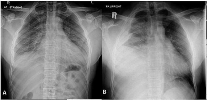
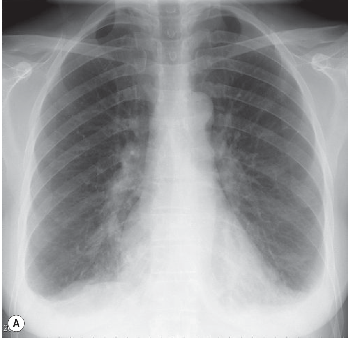
This diagnostic comparison contains two frontal chest X-rays (A and B) illustrating the development of a large pleural effusion. X-ray A, taken on initial admission, shows a relatively normal chest with clear lung fields, sharp costophrenic angles, and a centrally positioned mediastinum. X-ray B, a subsequent study, demonstrates a significant dense opacity in the right lower and middle zones of the hemithorax. This opacity exhibits a classic meniscus sign, curving upward along the lateral chest wall, which is highly characteristic of a large pleural effusion (later confirmed as a hemothorax). The right hemidiaphragm and costophrenic angle are completely obscured by the fluid. There is a subtle mass effect resulting in a slight shift of the mediastinal structures towards the left. The left lung remains clear. These images are used in medical education to demonstrate the radiographic progression of pleural pathology and the visual criteria for identifying fluid accumulation in the pleural space.
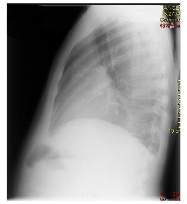
A lateral chest X-ray illustrating a massive re-accumulated pleural effusion in the left hemithorax. The image shows a large, dense area of radiopacity in the lower and middle portions of the chest, which characteristic of a significant fluid collection. This density obscures the left hemidiaphragm, the left costophrenic angle, and the posterior cardiac border. The extensive effusion results in a marked loss of lung volume on the affected side, with visible compression of the lung parenchyma. The right hemidiaphragm and right lung field appear relatively clear by comparison, though the cardiac silhouette is partially silhouette-masked by the adjacent fluid. This diagnostic image demonstrates the hallmark radiological features of massive pleural fluid accumulation, including loss of typical anatomical interfaces and increased thoracic density, serving as a primary example for pulmonary and critical care medicine education regarding pleural space pathology.
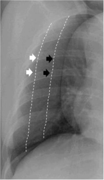
This chest radiograph (X-ray) demonstrates the semiquantification of pleural effusion size within the thoracic cavity. The image depicts a lateral or oblique perspective of the hemithorax, highlighting pleural fluid accumulation and its radiographic density. Two distinct areas are annotated with dotted lines and arrows to differentiate effusion severity. White arrows point to a thinner, less opaque lateral region outlined by a white dotted line, representing a 'small' pleural effusion (occupying less than one-third of the hemithorax). Black arrows point further medially to a broader area of increased radiographic density outlined by a second dotted line, representing a 'moderate' pleural effusion (occupying between one-third and two-thirds of the hemithorax). The visual comparison illustrates how the depth and volume of fluid increase opacity and alter the visible lung field. This pedagogical tool is used in pulmonology and radiology to teach standardized visual estimation of pleural fluid volume based on thoracic occupation ratios.
"pleural effusion" diagnosis management
Pleural Effusion
Epidemiology and Common Causes
- Congestive heart failure (most common transudate)
- Malignancy
- Bacterial pneumonia (parapneumonic effusion)
- Pulmonary embolism
- Cirrhosis (hepatic hydrothorax)
- Tuberculosis (leading cause in endemic areas)
- Nephrotic syndrome, uremia, connective tissue disease, viral infections
Pathophysiology: Transudates vs. Exudates
Transudates
- Ultrafiltrates of plasma; very little protein
- Caused by systemic factors - increased hydrostatic pressure or decreased oncotic pressure
- Examples: CHF, cirrhosis, nephrotic syndrome, hypoalbuminemia, myxedema, peritoneal dialysis, glomerulonephritis
Exudates
-
Protein-rich fluid; reflect intrinsic pleural/pulmonary pathology
-
Caused by local factors - inflammation, altered permeability, or impaired lymphatic drainage
-
Examples: Bacterial pneumonia, TB, malignancy (lung cancer, mesothelioma, lymphoma, metastases), pancreatitis, rheumatoid arthritis, SLE, subphrenic abscess, esophageal rupture, drug reactions, chylothorax
-
ROSEN's Emergency Medicine, Concepts and Clinical Practice
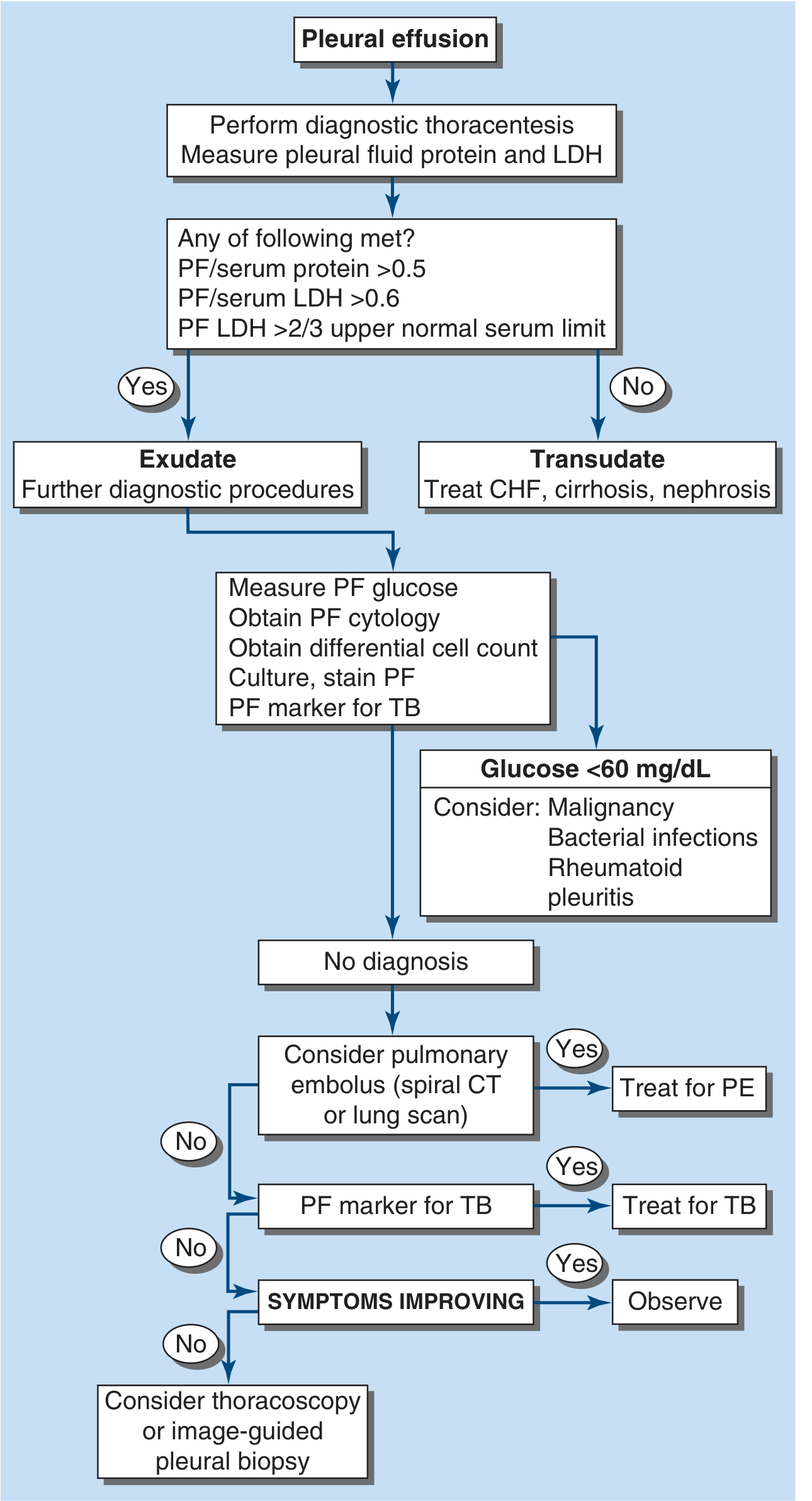
Light's Criteria (Exudate vs. Transudate)
| Criterion | Threshold |
|---|---|
| Pleural fluid protein / serum protein | > 0.5 |
| Pleural fluid LDH / serum LDH | > 0.6 |
| Pleural fluid LDH | > 2/3 upper normal limit for serum LDH |
Important caveat: Light's criteria misidentify ~25% of transudates as exudates ("pseudoexudates"), often due to diuresis. When clinical suspicion favors a transudate but Light's criteria suggest exudate, consider the serum-effusion albumin gradient (>12 g/L favors transudate). - Harrison's, 22E
- pH < 7.3: suggests parapneumonic effusion, malignancy, TB, rheumatoid, systemic acidosis
- pH < 7.0: strongly suggests empyema or esophageal rupture - indication for chest tube
- Glucose < 60 mg/dL: consider malignancy, bacterial infection, rheumatoid pleuritis
- Bloody fluid (hematocrit > 50% peripheral blood): = hemothorax (trauma, neoplasm, pulmonary infarction)
Diagnostic Algorithm
Clinical Features
- Symptoms: Dyspnea (most common), pleuritic chest pain, cough, orthopnea (with large effusions)
- Signs:
- Dullness to percussion over effusion
- Reduced or absent breath sounds
- Decreased tactile fremitus
- Egophony at the fluid-air interface
- Hemodynamic effects: Large effusions (especially right-sided) can cause tamponade-like effects on right ventricular filling; hypoxemia may result from both shunting and low cardiac output - Murray & Nadel's Textbook of Respiratory Medicine
Special Types
| Type | Key Features |
|---|---|
| Parapneumonic | Associated with bacterial pneumonia or lung abscess; exudate |
| Empyema (pyothorax) | Frank pus in pleural space; requires tube drainage |
| Loculated | Adhesions between visceral/parietal pleura; not free-flowing; often seen in TB, empyema, post-surgery |
| Hemothorax | Hematocrit of fluid >50% peripheral blood; trauma most common cause |
| Chylothorax | Milky fluid; triglycerides > 110 mg/dL; lymphatic disruption |
| Hepatic hydrothorax | Complicates 5-10% of cirrhosis; right-sided in 85%; ascitic fluid through diaphragmatic defects; transudative |
| Malignant effusion | Exudate; altered permeability + lymphatic obstruction; send fluid for cytology |
Imaging
Chest X-ray
- Erect PA film: Blunting of costophrenic angle when ≥ 200-300 mL present; meniscus sign (fluid curves up laterally); opacification of lower zones
- Supine: Effusion appears as a diffuse hazy opacity over the lower hemithorax - the costophrenic angles may remain visible
- Subpulmonic effusion: Appears as "high hemidiaphragm" peaking more laterally than usual; >2 cm between stomach bubble and lung (left-sided) is a clue
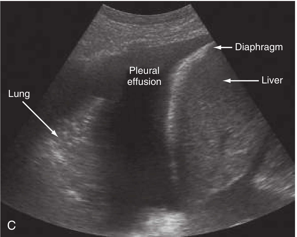
Ultrasound
CT
- Grainger & Allison's Diagnostic Radiology
Management
Diagnostic Thoracentesis
- Indicated for most new, unexplained effusions
- Relative contraindications: coagulopathy, prior pleurodesis, chest wall infection, pleural adhesions
Therapeutic Thoracentesis
- Remove fluid for symptomatic relief
- Massive effusions (>1.5-2 L): may require urgent drainage
- Risk of re-expansion pulmonary edema limits drainage to <1.5 L per session
Chest Tube / Pleural Drainage
- Empyema: Small-bore pigtail catheters (14 Fr) are now first-line; large-bore (28-40 Fr) historically used
- Hemothorax: Large-bore chest tube; surgery if continued bleeding
Specific Treatments
| Cause | Treatment |
|---|---|
| CHF | Diuretics, treat underlying heart failure |
| Cirrhosis (hepatic hydrothorax) | Sodium restriction, diuretics; TIPS (70-80% initial response); indwelling pleural catheter as bridge; VATS pleurodesis in refractory cases |
| Empyema | Antibiotics + drainage; surgery (VATS or open) vs. intrapleural fibrinolytics if complicated |
| Malignant effusion | Therapeutic thoracentesis, pleurodesis (talc or chemical), indwelling pleural catheter |
| TB effusion | Anti-TB therapy |
| Hypothyroidism | Thyroid hormone replacement (effusions typically small and asymptomatic) |
Pleurodesis
- Sclerosing agent (talc, doxycycline) or mechanical abrasion instilled into pleural space to obliterate it
- Used for recurrent malignant effusions or persistent pneumothorax
Key Points Summary
- Most common causes: CHF > malignancy > pneumonia > PE
- Transudate = systemic hemodynamic disturbance; Exudate = local pleural/lung pathology
- Light's criteria (any 1 of 3) distinguish exudate from transudate - ~25% false-positive rate for exudate
- pH < 7.0 in pleural fluid = empyema or esophageal rupture - drain urgently
- Ultrasound is preferred for guiding safe thoracentesis
- Never remove >1.5 L per session (re-expansion pulmonary edema risk)
- Hepatic hydrothorax is right-sided in 85% of cases and is a transudate