
vomitIng reflex
vomiting reflex mechanism neural pathway
The Vomiting Reflex
1. Key CNS Structures
- Located at the caudal end of the fourth ventricle (a circumventricular organ)
- Sits outside the blood-brain barrier, so circulating toxins, drugs, and metabolites can directly activate it
- Contains receptors for dopamine (D2), serotonin (5-HT3), opioids, substance P, acetylcholine, and aquaporin channels
- Axons from the area postrema project to the Nucleus Tractus Solitarius (NTS)
- A critical convergence point receiving input from the pharynx, larynx, GI tract, vestibular system, and area postrema
- The NTS engages distributed neuron groups in the medulla that coordinate the actual motor act of vomiting
- Note: modern neuroscience (Hornby) challenges the old concept of a single "vomiting center" - it is better described as a network of medullary neurons - Adams and Victor's Principles of Neurology, 12th Ed.
- Coordinates the motor mechanisms of vomiting
- Receives afferent input from: the CTZ, vestibular system, pharynx/GI tract, and higher cortical/brainstem structures
- Lippincott Illustrated Reviews: Pharmacology
2. Afferent Inputs (What Triggers the Reflex)
| Source | Mechanism |
|---|---|
| CTZ / Area Postrema | Circulating toxins, drugs (opioids, chemotherapy), metabolic disturbances (uremia, diabetic ketoacidosis) |
| GI tract (vagal/splanchnic afferents) | Mucosal irritation, distension; serotonin released from enterochromaffin cells activates 5-HT3 receptors on vagal fibers |
| Vestibular system | Motion sickness; via H1 and muscarinic receptors |
| Pharynx (gag reflex) | Direct mechanical stimulation of posterior pharynx |
| Higher cortical centers | Anticipatory nausea, fear, pain, unpleasant sights/smells |
| Intracranial pressure | Transmitted pressure to the dorsal medulla |
3. The Motor Sequence of Vomiting
- Abolition of gastric/small intestinal slow waves - normal peristalsis stops
- Reverse (retrograde) peristalsis - begins in the small intestine, moves up into the stomach
- Relaxation of stomach and pylorus
- Forced inspiration against a closed glottis - raises intra-abdominal pressure (Valsalva-like)
- Larynx moves upward and forward + relaxation of the lower esophageal sphincter (LES)
- Glottis closes - protects the airway from aspiration
- Forceful contraction of abdominal muscles - raises intra-abdominal pressure further
- Expulsion of gastric (and sometimes duodenal) contents
Key point: The stomach itself does not actively contract during expulsion. The force comes from abdominal wall and diaphragm contraction. - Adams and Victor's
4. Neurotransmitters and Receptors Involved
| Receptor | Location | Relevance |
|---|---|---|
| D2 (Dopamine) | Area postrema | Target of prochlorperazine, metoclopramide |
| 5-HT3 (Serotonin) | Area postrema + vagal afferents | Target of ondansetron, granisetron, palonosetron |
| H1 (Histamine) | Vestibular pathways | Target of dimenhydrinate, meclizine (motion sickness) |
| M1 (Muscarinic) | Vestibular pathways | Target of scopolamine (motion sickness) |
| NK1 (Substance P/Neurokinin-1) | CNS | Target of aprepitant (delayed CINV) |
5. Antiemetic Drug Classes (Based on Reflex Pathway)
- 5-HT3 antagonists (ondansetron, granisetron): block vagal afferent and CTZ signaling - most effective for chemotherapy/radiation/post-op nausea
- D2 antagonists (prochlorperazine, metoclopramide): block CTZ dopamine receptors
- NK1 antagonists (aprepitant): block substance P in the CNS - used for delayed CINV
- Anticholinergics/antihistamines (scopolamine, meclizine): block vestibular pathway - best for motion sickness, ineffective against CTZ-mediated vomiting
6. Neurological Causes of Vomiting
- Posterior fossa tumors or bleeds
- Raised intracranial pressure (pressure transmitted to dorsal medulla)
- Neuromyelitis optica (aquaporin-4 enrichment in area postrema)
crete a flowchart for this
A detailed medical flowchart of the vomiting reflex with the following structure and content: Title at top: "THE VOMITING REFLEX" Section 1 - AFFERENT TRIGGERS (top row, 5 boxes side by side): - CTZ / Area Postrema: Toxins, drugs, metabolites (outside BBB) - GI Tract (Vagal/Splanchnic): Mucosal irritation, distension, serotonin from enterochromaffin cells via 5-HT3 receptors - Vestibular System: Motion sickness via H1 and muscarinic receptors - Pharynx (Gag Reflex): Mechanical stimulation of posterior pharynx - Higher Cortical Centers: Anticipatory nausea, pain, psychic stimuli All 5 boxes have arrows pointing DOWN to a central box: Section 2 - CENTRAL INTEGRATION (middle, large highlighted box): NUCLEUS TRACTUS SOLITARIUS (NTS) - Convergence point for all afferent signals - Projects to medullary neuron network ↓ VOMITING CENTER (Lateral Reticular Formation of Medulla) - Coordinates motor output Section 3 - KEY RECEPTORS (side panel or box): D2 Dopamine | 5-HT3 Serotonin | H1 Histamine | M1 Muscarinic | NK1 Substance P Section 4 - MOTOR SEQUENCE (bottom, numbered steps in vertical flow): Step 1: Abolition of gastric slow waves → normal peristalsis stops Step 2: Retrograde peristalsis (small intestine → stomach) Step 3: Relaxation of stomach & pylorus Step 4: Forced inspiration against closed glottis → ↑ intra-abdominal pressure Step 5: Larynx moves up & forward + LES relaxes Step 6: Glottis closes → airway protected Step 7: Abdominal muscle contraction → ↑↑ intra-abdominal pressure Step 8: EXPULSION of gastric contents At the bottom, a note box: "RETCHING vs VOMITING: In retching, upper esophageal sphincter stays CLOSED → contents return to stomach. In vomiting, upper esophageal sphincter OPENS → expulsion occurs." Clean medical illustration style, white background, blue and teal color scheme for boxes, orange/red for the central integration section, numbered arrows connecting each step clearly.
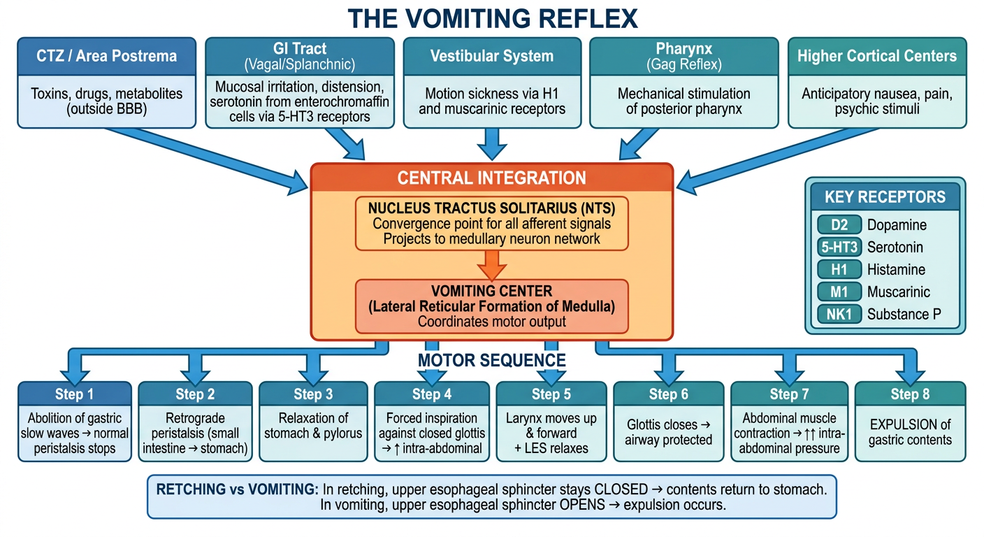
- 5 afferent trigger pathways feeding into the central integration
- NTS + Vomiting Center as the central coordination hub
- Key neurotransmitter receptors (D2, 5-HT3, H1, M1, NK1)
- 8-step sequential motor output from peristalsis reversal to final expulsion
- Retching vs. Vomiting distinction at the bottom
questions that can be asked for urinary bladder by students to the presenters
Questions Students Can Ask Presenters on the Urinary Bladder
Anatomy & Structure
- What are the exact boundaries and relations of the urinary bladder in the male vs. female pelvis? How does its position change as it fills?
- What is the trigone of the bladder - what is its embryological origin, and why does it have a different structure compared to the rest of the bladder wall?
- The trigone is derived from mesonephric ducts while the rest of the bladder is derived from the cloaca - what are the histological differences that result from this dual origin?
- How does the ureter enter the bladder obliquely through the detrusor muscle, and what is the functional significance of this arrangement?
- What is the peritoneal relationship of the urinary bladder, and how does it change when the bladder is full vs. empty?
- What is the blood supply, venous drainage, and lymphatic drainage of the urinary bladder?
Histology
- What are the layers of the urinary bladder wall? How do they compare to other hollow visceral organs?
- What type of epithelium lines the urinary bladder, and what are the unique ultrastructural features (umbrella cells, asymmetric unit membranes) that allow it to accommodate distension?
- Why is there no muscularis mucosae or submucosal layer in the trigone but present in the rest of the bladder wall?
- How are smooth muscle bundles arranged in the detrusor muscle compared to smooth muscle in tubular organs like the gut?
Innervation & Physiology
- What are the three nerve supplies to the urinary bladder - sympathetic, parasympathetic, and somatic - and what are the spinal cord levels for each?
- Sympathetic stimulation relaxes the detrusor and contracts the internal sphincter - through which specific receptors (β3 and α1 adrenergic) is this achieved?
- Parasympathetic stimulation causes bladder contraction through M3 muscarinic receptors and relaxes the internal sphincter via nitric oxide. What drugs target each of these steps?
- What is the role of the pudendal nerve (S2-S4) in voluntary control of micturition? What happens when this nerve is damaged?
- How does the micturition reflex work? What is the role of the pontine micturition center (Barrington's nucleus), and how do higher cortical centers exert voluntary control?
- What is the significance of the "guarding reflex" during bladder filling?
Pathology
- What is a bladder diverticulum? How do congenital and acquired diverticula differ, and what complications can arise from urinary stasis within them?
- Why are women more prone to bacterial cystitis than men? Which organisms are most commonly responsible?
- What is malakoplakia, and what are Michaelis-Gutmann bodies? What is the pathophysiology behind their formation?
- How does chronic schistosomiasis predispose to squamous cell carcinoma of the bladder? What is the sequence of metaplasia to malignancy?
- What is hemorrhagic cystitis, and which drugs/viruses cause it? What is the mechanism by which cyclophosphamide produces bladder toxicity (acrolein)?
- What are the two main molecular pathways of bladder cancer progression, and how do superficial papillary tumors differ from carcinoma-in-situ in terms of genetics and clinical behavior?
Bladder Cancer
- Urothelial (transitional cell) carcinoma accounts for ~90% of bladder cancers - what are the key risk factors including occupational carcinogens (aniline dyes, aromatic amines)?
- What is the TNM staging of bladder cancer, and at what stage does muscle invasion (T2) become clinically critical for management decisions?
- FGFR3 mutations are common in superficial papillary tumors - what targeted therapies have emerged from this finding?
- What is the difference between non-muscle-invasive and muscle-invasive bladder cancer in terms of treatment approach (TURBT + BCG vs. radical cystectomy)?
- Why do superficial bladder tumors frequently recur after transurethral resection, and what surveillance protocols are recommended?
Vesicoureteral Reflux (VUR)
- What is the mechanism by which the oblique course of the ureter through the bladder wall normally prevents vesicoureteral reflux?
- In patients with a shorter intramural ureteral tunnel, reflux occurs - what are the grades of VUR and their clinical consequences, including reflux nephropathy?
- How is primary VUR managed conservatively vs. surgically?
Neurogenic Bladder
- What is neurogenic bladder? How do upper motor neuron (spastic/reflex) bladder and lower motor neuron (flaccid/autonomous) bladder differ in presentation and management?
- What is detrusor-sphincter dyssynergia, and in which neurological conditions does it commonly occur?
- How does diabetes mellitus cause a neurogenic (hypotonic) bladder, and what is its clinical presentation?
Pharmacology & Clinical Applications
- How do anticholinergic drugs (oxybutynin, solifenacin) treat overactive bladder, and what are their side effects?
- What is the mechanism by which alpha-1 blockers (tamsulosin) relieve bladder outlet obstruction in BPH?
- Intravesical BCG is used for non-muscle-invasive bladder cancer - what is its proposed mechanism of action?
- What is interstitial cystitis (bladder pain syndrome)? How does it differ from bacterial cystitis, and what are current treatment options?
Applied / Viva-Style Questions
- A patient presents with painless hematuria - what is your differential diagnosis, and how would you investigate?
- What is the significance of the ureterovesical junction in preventing reflux, and how is it damaged in posterior urethral valves?
- Why does a full bladder protect against radiation injury to adjacent pelvic organs (e.g., during cervical cancer radiotherapy)?

answer the following questions
Answers to All 20 Questions (Oral Cavity / Head & Neck)
1. Explain how jawlines are formed (Growth) - Faith
- Mandibular growth occurs primarily through endochondral ossification at the condylar cartilage (the main growth center) and intramembranous ossification along the periosteal surface
- The condylar cartilage acts as a secondary cartilage - it is the main site of mandibular growth in children, responding to functional stimuli
- Maxillary growth occurs by sutural growth (at sutures connecting to the cranial base) and surface apposition/resorption
- Growth continues until approximately 18-20 years in females and 20-25 years in males
- Hormonal influences (GH, sex hormones) strongly regulate jaw growth - excess GH causes prognathism (acromegaly)
- Genetic factors determine the basic jaw morphology, while functional stimuli (chewing, muscle pull) modify it - The Developing Human, Moore & Persaud
2. Teeth Sensitivity - How is it caused? (Nishuka's question)
- Exposed dentinal tubules (due to enamel loss, gum recession, or cervical abrasion) allow fluid movement within them
- External stimuli (cold, hot, sweet, air blast, touch) cause rapid fluid movement in the dentinal tubules
- This fluid movement stimulates A-delta nerve fibers (odontoblastic processes and free nerve endings at the pulp-dentin junction), triggering a sharp, short-duration pain
- Causes of exposure: enamel erosion (acidic foods), toothbrush abrasion, gingival recession, periodontal disease, tooth bleaching, dental procedures
- Treatment: desensitizing toothpastes (potassium nitrate, strontium chloride), fluoride varnish, dentine bonding agents, laser therapy
3. Relationship between Lymphatic Drainage and Metastasis in the Oral Cavity - Nishuka's question
- The oral cavity drains in a largely predictable, sequential (echelon) fashion through cervical lymph nodes
- Lips/anterior floor of mouth → submental nodes (Level I)
- Cheek, gingiva, hard palate, anterior tongue → submandibular nodes (Level I/II)
- Posterior tongue, floor of mouth, oropharynx → deep cervical nodes (Level II, III)
- Tip of tongue may drain bilaterally (risk of contralateral metastasis)
- Oral squamous cell carcinoma metastasizes first to cervical nodes before systemic spread - this is why neck dissection is performed alongside primary tumor resection
- Sentinel lymph node biopsy can identify first-echelon nodes to detect occult metastasis
- Skip metastasis (bypassing Level I to go directly to Level III/IV) can occur, making complete assessment necessary - Cummings Otolaryngology
4. Veins of the Oral Cavity - Follow the naming pattern like arteries (Nishuka's question)
| Region | Vein | Drains to |
|---|---|---|
| Tongue | Lingual vein | Internal jugular vein |
| Lips/face | Facial vein | Internal jugular / common facial vein |
| Palate | Palatine vein | Pterygoid plexus / facial vein |
| Gingiva/teeth | Dental/alveolar veins | Pterygoid plexus |
| Cheek/pterygoid region | Pterygoid plexus | Maxillary vein → retromandibular vein |
5. Emphasis on the Slide Study - Eroni's point
6. Major Factors Leading to Stones in Salivary Glands - Eroni's question
- Supersaturation of saliva with calcium phosphate and calcium carbonate
- Ductal stasis - reduced salivary flow (dehydration, anticholinergic drugs, fasting)
- Retrograde contamination - oral bacteria and food debris entering the duct
- Organic nidus - inspissated mucus or cellular debris around which mineral salts crystallize
- Altered saliva composition - elevated calcium, phosphate, or protein levels
- Contributing factors: dehydration, gout (urate crystals), hypercalcemia, Sjögren's syndrome
- Cummings Otolaryngology
7. Why is the Submandibular Gland More Prone to Stones than the Parotid Gland? - Eroni's question
- Wharton's duct is long (5 cm) and travels upward against gravity from the gland floor to the floor of the mouth - stasis is promoted
- Wharton's duct has a narrow orifice relative to the duct diameter - further restricting flow
- Submandibular saliva is more viscous - it secretes more mucous (mixed serous + mucous gland) compared to the predominantly serous parotid
- Higher calcium and phosphate concentration in submandibular secretions
- Flow is against gravity for part of its path (the gland lies below the duct exit)
- Compare: Stensen's duct (parotid) is shorter, flows downward/horizontally, and parotid saliva is more watery/serous → less prone to stasis - Cummings Otolaryngology
8. Ganglion, Sica, and Angina - Eroni's question
- Submandibular ganglion - parasympathetic ganglion supplying submandibular and sublingual glands via chorda tympani (CN VII branch) → lingual nerve
- Otic ganglion - parasympathetic ganglion supplying the parotid gland via lesser petrosal nerve (CN IX branch) → auriculotemporal nerve
- Xerostomia (dry mouth) + xerophthalmia (dry eyes) = Sicca syndrome / Sjögren's syndrome
- Caused by autoimmune destruction of salivary and lacrimal glands
- Predisposes to rampant dental caries, difficulty chewing/swallowing, oral infections
- Ludwig's angina = rapidly spreading bilateral cellulitis of the submandibular, sublingual, and submental spaces
- Most often originates from a lower second or third molar dental infection
- Life-threatening due to airway compromise by tongue elevation
- Treatment: immediate airway management + IV antibiotics + surgical drainage
9. Licking Lips Constantly - Eroni's question
- Constant lip-licking is a behavioral/habit that causes angular cheilitis and dry/cracked lips
- Mechanism: saliva contains amylase and other enzymes that macerate and irritate the perioral skin/mucosa when repeatedly applied
- The moisture-evaporation cycle causes dehydration of lip mucosa
- Predisposes to Candidal infection (Candida albicans thrives in the moist corners created)
- Seen in: children (habit), anxious individuals, those with lip-licking habit, nutritional deficiencies (B2, B3, iron, zinc)
- Treatment: stop the habit, emollients/barrier creams, antifungal if Candida present
10. Changes in Periodontal Tissues During Puberty, and What Occurs in Gingivitis? - Eroni's question
- Puberty gingivitis: hormonal changes (estrogen, progesterone) increase vascularity and inflammatory response of the gingiva
- Gingiva becomes swollen, red, bleeding on probing even with minimal plaque accumulation
- Hormones alter the subgingival microbiota (increase in anaerobes like Prevotella intermedia)
- The exaggerated inflammatory response subsides after puberty if plaque is controlled
- Color: red/bluish-red (due to increased vascularity)
- Contour: swollen, rounded margins (blunted papillae)
- Consistency: soft, edematous (loss of stippling)
- Bleeding: bleeds on probing or spontaneously
- Exudate: gingival crevicular fluid increased
- Tooth loss: no bone loss in gingivitis (distinguishes from periodontitis)
11. Wisdom Teeth Come Out Late / Food Taste Different in Old Age - Sheenam's question
- Third molars (wisdom teeth) are the last teeth to mineralize (crown formation: 7-10 years; root completion: 18-25 years)
- Jaw size is largely determined by mid-adolescence; third molars erupt after the jaw has reached near-adult size
- Evolutionary theory: as human jaws became smaller (with cooking/softer foods reducing jaw size over evolution), space became insufficient
- Impaction occurs when insufficient space exists in the arch (most common impaction: mesioangular lower third molar)
- Gustatory decline with aging due to: reduced number of taste buds (from ~10,000 at birth to ~5,000 by 70 years), decreased saliva production (saliva carries tastants to taste buds), reduced olfactory sensitivity (taste is largely smell-dependent), medications, zinc deficiency
- Salty and sweet tastes diminish first; bitter and sour are more preserved
12. Anastomosis of Arteries / Incisional Biopsy - Nishuka's question
- The oral cavity has a rich anastomotic arterial network, primarily from the facial artery and maxillary artery (both branches of the external carotid artery)
- Key anastomoses:
- Facial artery ↔ infraorbital artery (from maxillary artery) - at the face
- Lingual artery ↔ contralateral lingual artery (across the midline of the tongue)
- Greater palatine artery ↔ nasopalatine artery (at the incisive foramen)
- Superior labial artery ↔ inferior labial artery (around lips)
- Clinical significance: these anastomoses mean that ligation of a single vessel may not fully control bleeding; also allows flap survival in reconstructive surgery
- Used for large lesions where complete excision is not practical or would cause deformity
- A representative wedge or section of tissue is taken including the margin between normal and abnormal tissue
- Preferred for: leukoplakia, large ulcers, potentially malignant disorders, lesions where diagnosis must precede definitive treatment
- Contrast with excisional biopsy (complete removal of the lesion - used for small lesions <1 cm)
13. Lister Penal Canal / Explor - Nishuka's question
- Located in the anterior midline of the hard palate, behind the central incisors
- Transmits: nasopalatine nerve (branch of V2, maxillary nerve) and terminal branches of the sphenopalatine artery
- Opens at the incisive foramen on the palatal side and at the floor of the nasal cavity
- Clinically: nasopalatine cyst (the most common non-odontogenic cyst) arises from epithelial remnants within this canal; appears as a heart-shaped radiolucency on periapical X-ray
- Located in the base of the pterygoid process of sphenoid
- Transmits: nerve of the pterygoid canal (Vidian nerve) = greater petrosal nerve (parasympathetic, CN VII) + deep petrosal nerve (sympathetic)
- Connects the foramen lacerum to the pterygopalatine fossa
14. Disease Appears First in the Mouth - Eroni's question
| Disease | Oral Manifestation |
|---|---|
| HIV/AIDS | Oral candidiasis, hairy leukoplakia (EBV), Kaposi's sarcoma, severe periodontitis |
| Leukemia | Gingival hyperplasia, petechiae, spontaneous bleeding |
| Crohn's disease | Cobblestone mucosa, angular cheilitis, aphthous ulcers |
| Addison's disease | Oral melanotic pigmentation |
| Scurvy (Vit C deficiency) | Bleeding swollen gingiva, poor wound healing |
| Measles | Koplik's spots (white spots on buccal mucosa - pathognomonic, appear before the rash) |
| Syphilis | Primary chancre on lip/tongue; "snail track" ulcers in secondary syphilis |
| Pernicious anemia | Smooth/bald tongue (atrophic glossitis) |
| Pemphigus vulgaris | Oral erosions/blisters appear before skin lesions in 60% of cases |
| Diabetes mellitus | Xerostomia, periodontitis, candidiasis, slow-healing ulcers |
15. Cleft Palate Occurs / People Born Without a Tongue - Sheenam & Eroni's question
- Results from failure of fusion of the palatine shelves (lateral palatine processes) with each other and with the nasal septum
- Occurs around week 7-10 of embryonic development
- Isolated cleft palate is etiologically distinct from cleft lip (different developmental process at a different time)
- Types: incomplete (soft palate only), complete (hard + soft palate), submucous cleft
- Associated with: Pierre Robin sequence, velocardiofacial syndrome, Treacher Collins syndrome
- Treatment: palate repair at 9-18 months (before speech development)
- Cleft clip/clam refers to a feeding plate used before surgical repair - The Developing Human
- Extremely rare congenital anomaly (aglossia or microglossia)
- Associated with hypoglossia-hypodactyly syndrome (Hanhart syndrome) and Möbius syndrome
- The tongue develops from lingual swellings of the first arch and the copula/hypobranchial eminence of arches 2, 3, 4
- Failure of these swellings to develop/fuse results in aglossia
- Functional impact: severe impairment of speech, swallowing, and mastication
16. Mumps - How it Occurs, Clinical Correlates / How to Put Jaw Back - Eroni's question
- Caused by mumps virus (Paramyxovirus, RNA virus)
- Spreads via respiratory droplets; incubation: 14-25 days
- Infects primarily the parotid gland (bilateral parotitis in most cases)
- Mechanism: virus infects ductal epithelial cells → inflammatory swelling of parotid
- Clinical features: fever, malaise, tender parotid swelling (lifting the earlobe), pain on chewing/sour foods (which stimulate saliva production)
- Complications: orchitis (post-pubertal males, may cause infertility), oophoritis, aseptic meningitis, encephalitis, pancreatitis, deafness
- Prevention: MMR vaccine (measles, mumps, rubella)
- Dislocation occurs when the condylar head moves anterior to the articular eminence and cannot return
- Reduction technique (Hippocratic method): operator stands in front of patient; thumbs placed on lower molars bilaterally with fingers under the chin; push down and back (inferiorly to disengage the condyle from the eminence, then posteriorly to seat it back in the glenoid fossa)
- Patient's mouth then closes spontaneously
- If recurrent: consider eminectomy or other surgical stabilization
17. Tooth Extraction Pain in Ear - Eroni's question
- The auriculotemporal nerve (branch of V3, mandibular nerve) supplies both the temporomandibular joint, parotid region, and the skin of the temporal region/external ear
- The inferior alveolar nerve (V3) supplies the lower teeth; its sensory fibers travel back through the mandibular foramen to the trigeminal ganglion
- Post-extraction inflammation or irritation of the inferior alveolar nerve can produce referred otalgia (ear pain) because:
- The auriculotemporal nerve shares its ganglion (trigeminal) with the inferior alveolar nerve
- Central sensitization causes referred pain in adjacent dermatomes
- Dry socket (alveolar osteitis) after lower molar extraction is a common cause of severe post-extraction ear pain
- Also: the chorda tympani nerve (CN VII branch) passes through the infratemporal fossa very close to the lower molar roots, explaining ear/taste disturbances after third molar surgery
18. CI → Eroni's question
- Class I (G.V. Black's classification): caries involving pits and fissures of posterior teeth (occlusal surface of molars/premolars), buccal/lingual pits, and palatal pits of upper incisors
- Most common location for caries because pits and fissures trap food and are difficult to clean
- Management: composite resin or amalgam restoration; preventive: pit and fissure sealants
19. Pharyngeal Membrane - Importance / Function - Faith's question
- Formed at the stomodeum (primitive mouth) where the ectoderm of the oral cavity meets the endoderm of the foregut
- Located at the junction of the primitive mouth and the pharynx
- Normally ruptures at approximately day 24-26 of development, establishing continuity between the oral cavity and the GI tract
- Importance:
- Its rupture is essential for normal swallowing and feeding development
- Failure to rupture → rare congenital condition causing oral atresia
- It marks the boundary between ectoderm-derived oral mucosa (anterior) and endoderm-derived pharyngeal/GI mucosa (posterior)
- This boundary is clinically relevant: the parotid gland arises from oral ectoderm, while pharyngeal glands arise from endoderm
- Tumors arising anterior vs posterior to this membrane have different cell origins and behavior
- Homologous structures exist as pharyngeal membranes separating each pharyngeal pouch from its corresponding groove - The Developing Human
20. What is Incisional Biopsy?
- Large lesions (>1 cm) where complete removal would be extensive or disfiguring
- Lesions where the diagnosis must be confirmed before planning definitive treatment
- Suspicious lesions for malignancy (leukoplakia, erythroplakia, non-healing ulcers)
- When multiple areas of a lesion show different appearances (sample the most suspicious area)
- Local anesthesia (inject away from the lesion, not through it)
- Elliptical incision including the junction of normal and abnormal tissue
- Full-thickness tissue sample (includes submucosal tissue - not just epithelium)
- Suture closure of the donor site
- Specimen fixed in 10% formalin and sent for histopathology
- Excisional = complete removal of the entire lesion (used for small lesions, likely benign)
- Incisional = partial removal for diagnosis only (definitive treatment follows after diagnosis)
hepatic encephalopathy
Hepatic Encephalopathy (HE)
Pathophysiology
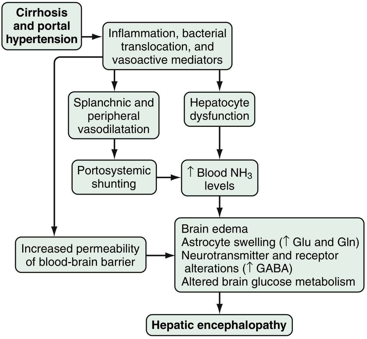
1. Ammonia - The Primary Neurotoxin
- Ammonia is produced primarily in the colon, where gut bacteria metabolize dietary proteins and nitrogen-containing compounds; enterocytes also synthesize ammonia from glutamine
- Normally, ammonia enters the portal circulation and is cleared by hepatocytes (converted to urea via the urea cycle)
- In cirrhosis: two mechanisms cause ammonia accumulation:
- Hepatocyte dysfunction - reduced capacity to metabolize ammonia
- Portosystemic shunting - ammonia bypasses the liver through collateral vessels (varices, TIPS, surgical shunts) and reaches the systemic circulation directly
- Hyperammonemia is found in up to 90% of HE patients (though blood levels correlate imperfectly with severity)
- In portosystemic shunting, skeletal muscle becomes an increasingly important ammonia-detoxifying organ - which is why muscle wasting worsens HE
- Goldman-Cecil Medicine
2. Astrocyte Swelling (Key Cellular Mechanism)
- Ammonia enters the brain (the blood-brain barrier is ~200x more permeable to NH3 gas than NH4+)
- Inside astrocytes, ammonia is converted to glutamine by glutamine synthetase (trapping reaction)
- Rising intracellular glutamine → osmotic astrocyte swelling → cytotoxic brain edema
- Initially, cells compensate by releasing osmolytes (myoinositol); when myoinositol is depleted, even small amounts of ammonia cause swelling
- Astrocyte swelling triggers:
- Generation of reactive oxygen species (ROS)
- Mitochondrial permeability transition (mPT) → collapse of mitochondrial membrane potential → defective oxidative phosphorylation → cessation of ATP synthesis
- Further ROS production (vicious cycle)
- Alzheimer type II astrocytosis - characteristic histological finding in chronic HE: enlarged, pale astrocyte nuclei with prominent nucleoli - Goldman-Cecil Medicine
3. Enhanced GABA-ergic Neurotransmission
- Increased sensitivity of astrocyte peripheral-type benzodiazepine receptors → enhanced neurosteroid production (allopregnanolone, tetrahydrodeoxycorticosterone)
- Neurosteroids are potent positive modulators of GABA-A receptors → enhanced inhibitory neurotransmission → cortical depression
- This explains why benzodiazepines dramatically worsen HE (they act at the same receptor)
- Endogenous benzodiazepine-like ligands have also been found elevated in HE - Sleisenger & Fordtran
4. Other Contributing Mechanisms
| Factor | Effect |
|---|---|
| Manganese | Accumulates in the globus pallidus (seen as T1 hyperintensity on MRI); causes dopaminergic dysfunction, parkinsonian features |
| Systemic inflammation | Bacterial translocation from the gut, cytokines (TNF-α, IL-6) amplify ammonia toxicity - can precipitate HE with even mildly elevated ammonia |
| Increased BBB permeability | Greater extraction of ammonia by cerebellum and basal ganglia |
| False neurotransmitters | Aromatic amino acids (tyrosine, phenylalanine, tryptophan) cross BBB and compete with normal neurotransmitter synthesis |
| Altered serotonin, nitric oxide, opioid peptides | All contribute to neurotransmission disruption |
| Gut microbiome dysbiosis | Cirrhotic patients have altered colonic flora producing more ammoniagenic bacteria |
Classification (ISHEN / World Gastroenterological Association)
| Type | Basis |
|---|---|
| Type A | HE associated with Acute Liver Failure (previously normal liver; symptoms within 8 weeks) |
| Type B | HE due to portosystemic bypass without intrinsic hepatocellular disease |
| Type C | HE in patients with cirrhosis + portosystemic shunting (most common) |
Grading - West Haven Criteria
| Grade | Features |
|---|---|
| 0 (Minimal/Covert HE) | No detectable personality or behavioral changes; detectable only on neuropsychometric tests |
| Grade 1 | Trivial lack of awareness; euphoria or anxiety; shortened attention span; impaired calculation ability |
| Grade 2 | Lethargy or apathy; disorientation (time); obvious personality change; asterixis present (flapping tremor) |
| Grade 3 | Somnolence to semi-stupor; responsive to stimuli; confused; gross disorientation; asterixis may be absent |
| Grade 4 | Coma (unresponsive to verbal or noxious stimuli) |
Asterixis (flapping tremor, "liver flap") - hallmark sign: non-rhythmic, asynchronous flapping of outstretched dorsiflexed hands due to intermittent lapses in sustained posture. Ask patient to "stop traffic" with hands. Reflects metabolic encephalopathy.
Precipitating Factors ("HEPATICS" mnemonic)
| Factor | Mechanism |
|---|---|
| Hemorrhage (GI bleed) | Large nitrogen load from blood in the gut → ammonia production |
| Electrolyte disturbance (hypokalemia, hyponatremia) | Alkalosis increases NH3 (uncharged) → greater BBB penetration |
| Protein excess | Excess dietary nitrogen → more ammonia from gut bacteria |
| Alcohol (binge) | Direct hepatotoxin + sedation |
| Transjugular intrahepatic portosystemic shunt (TIPS) | Blood completely bypasses liver |
| Infection / Inflammation (SBP, pneumonia, UTI) | Systemic inflammation amplifies ammonia neurotoxicity |
| Constipation | Prolonged gut transit → increased bacterial ammonia generation |
| Sedatives/drugs (opioids, benzodiazepines, diuretics) | Direct CNS depression; diuretics cause dehydration + hypokalemia |
Diagnosis
- Clinical: West Haven grading, asterixis, neuropsychometric tests (Stroop test, EncephalApp)
- Blood ammonia: elevated in ~90%; useful for diagnosis/monitoring but does NOT reliably correlate with grade - don't use alone for diagnosis; venous sample (avoid tourniquet, process immediately)
- EEG: slowing of dominant frequency; triphasic waves in moderate HE; useful in subclinical HE
- MRI brain: T1 hyperintensity in globus pallidus (manganese deposition); MR spectroscopy shows elevated glutamine/glutamate (Glx) and decreased myoinositol
- Critical Flicker Frequency (CFF): simple light-based test assessing cortical function; reliable marker of minimal HE
- Exclude other causes: head CT (to rule out bleed, mass), metabolic screen (glucose, electrolytes, renal function, thyroid)
Treatment
1. Identify and Eliminate Precipitating Factors (First Priority)
- Treat GI bleed, infection (SBP, etc.), correct electrolytes, stop offending drugs
2. Reduce Ammonia Production and Absorption
- Non-absorbable disaccharide; metabolized by colonic bacteria to short-chain organic acids
- Lowers colonic pH → NH3 → NH4+ (ionized, non-absorbable) → trapped in colon
- Causes catharsis (accelerates transit, reducing ammonia absorption time)
- Dose: titrated to 2-3 soft stools/day
- Can be given as enema if aspiration risk
- Side effects: bloating, flatulence, diarrhea, electrolyte imbalance
- Oral non-absorbable antibiotic; targets gut ammonia-producing bacteria
- 550 mg twice daily - approved (2010) for prevention of recurrent overt HE
- Superior tolerability vs. lactulose; better neuropsychometric outcomes
- Often used in combination with lactulose in recurrent HE - Sleisenger & Fordtran
3. Nutritional Support
- Protein restriction is NO longer recommended - it is harmful (worsens sarcopenia which reduces ammonia clearance)
- Adequate protein intake: 1.2-1.5 g/kg/day
- Vegetable and dairy proteins are preferable to animal proteins (more favorable calorie-to-nitrogen ratio, more fiber)
- Branched-chain amino acids (BCAA) - leucine, isoleucine, valine: may improve HE by providing nitrogen in a non-ammoniagenic form and by competing with aromatic amino acids at the BBB
- Avoid prolonged overnight fasting; late-night snack is recommended for cirrhotic patients
- Zinc supplementation - zinc is a cofactor for urea cycle enzymes; often deficient in cirrhosis
4. Other Measures
- Flumazenil - reversal of endogenous benzodiazepine-like ligands; short-term benefit only
- Ornithine phenylacetate / L-ornithine L-aspartate - promote ammonia metabolism; under investigation
- Probiotics / fecal microbiota transplant (FMT) - modify gut microbiome to reduce ammoniagenic bacteria; emerging evidence
- Embolization of large portosystemic collaterals - for spontaneous portosystemic shunts in patients without significant portal hypertension
5. Definitive Treatment
- Liver transplantation - generally reverses HE; only definitive cure; HE is itself a criterion for MELD/transplant listing priority
Prognosis
- Overt HE: 1-year survival ~42%; 3-year survival ~23% without transplant
- Minimal HE adversely affects quality of life, driving ability, and significantly increases risk of developing overt HE
- Decompensated cirrhosis with HE: median survival 1.6 years
how does portal hypertension causes edema
How Portal Hypertension Causes Edema (Ascites)
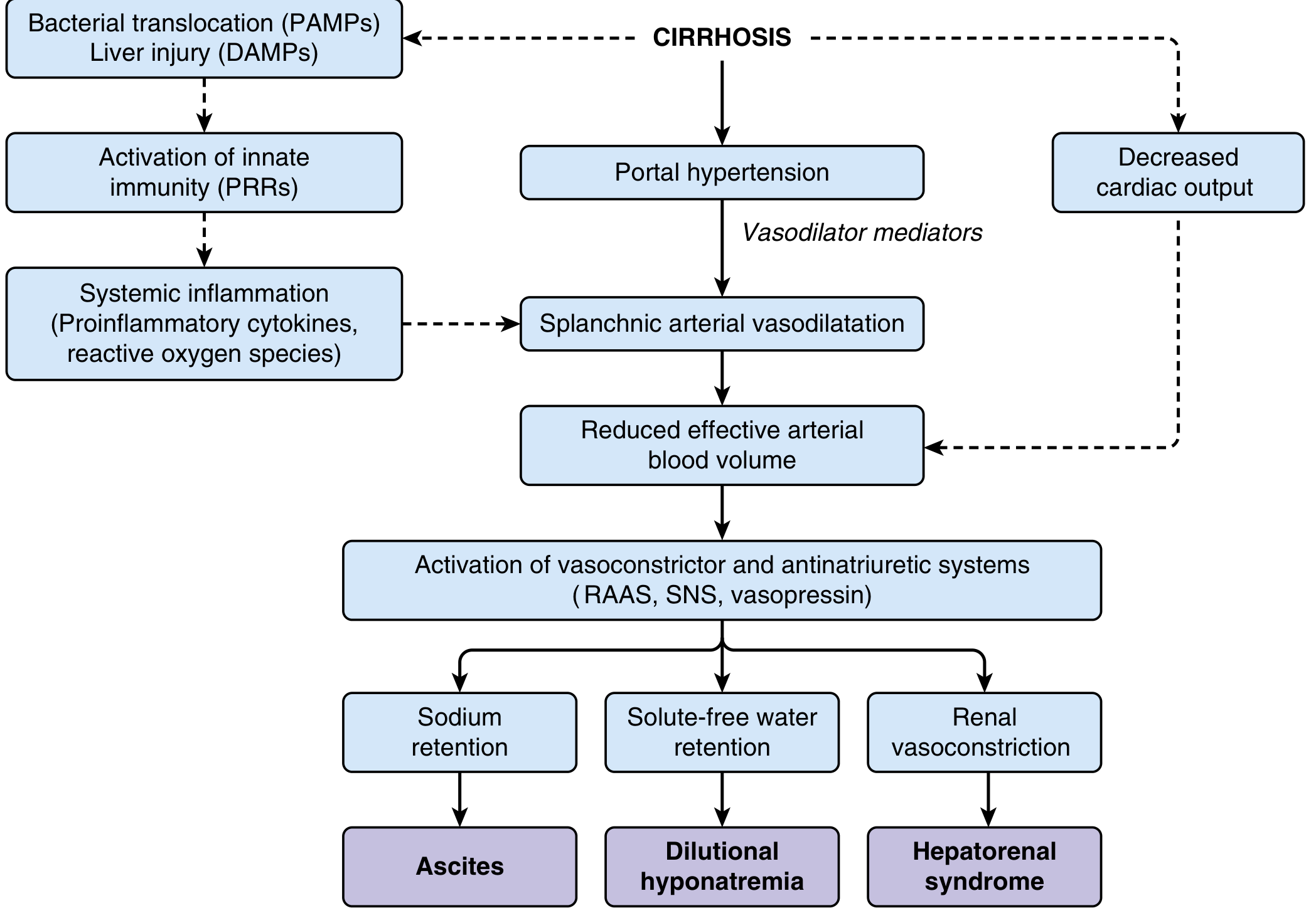
The Pathophysiology Flowchart (from Sleisenger & Fordtran)
Step 1: Increased Intrahepatic Resistance
- Cirrhosis causes architectural distortion of hepatic sinusoids by fibrosis and regenerative nodules
- This creates increased resistance to portal blood flow
- An additional dynamic component arises from contraction of hepatic stellate cells and endothelial dysfunction - in the cirrhotic liver, there is paradoxically reduced intrahepatic NO (vasoconstriction) despite overall systemic NO excess
- Result: portal venous pressure rises - clinically significant portal hypertension = Hepatic Venous Pressure Gradient (HVPG) > 10-12 mmHg
- Ascites formation threshold: HVPG ≥ 12 mmHg - Goldman-Cecil Medicine
Step 2: Splanchnic Arterial Vasodilatation (The "Arterial Vasodilatation Theory")
- Portal hypertension triggers release of vasodilating mediators - primarily nitric oxide (NO), plus carbon monoxide, prostacyclin, TNF-α, endogenous endocannabinoids - into the splanchnic circulation
- Splanchnic arteries dilate → decreased splanchnic vascular resistance → increased splanchnic blood flow → more blood poured into the portal system (worsening portal hypertension in a vicious cycle)
Step 3: Effective Arterial Hypovolaemia ("Underfilling")
- Splanchnic vasodilatation pools blood in the splanchnic vascular bed
- This causes reduced effective arterial blood volume - the body's pressure receptors sense the circulation as "underfilled" even though total blood volume may be normal or increased
- Early cirrhosis: cardiac output increases to compensate → arterial pressure maintained
- Advanced cirrhosis: vasodilatation is so intense that even a high cardiac output cannot compensate → effective arterial hypovolaemia develops
- At very late stages, cardiac output also begins to fall (cirrhotic cardiomyopathy), further reducing effective blood volume - Sleisenger & Fordtran
Step 4: Activation of Sodium-Retaining Neurohumoral Systems
| System | Effect |
|---|---|
| RAAS (Renin-Angiotensin-Aldosterone) | Aldosterone → Na+ and water retention by kidneys; plasma aldosterone elevated in most cirrhotic patients with ascites |
| Sympathetic Nervous System (SNS) | Renal vasoconstriction + tubular Na+ reabsorption |
| ADH / Vasopressin | Free water retention → dilutional hyponatremia |
- These systems "refill" the intravascular space but the fluid leaks out into the peritoneum as fast as it is retained
- Result: continuous formation of ascites (and peripheral edema), dilutional hyponatremia, and - if extreme - renal vasoconstriction → Hepatorenal Syndrome
Step 5: Local Factors Driving Fluid into the Peritoneum
A. Sinusoidal Hypertension (increased hydrostatic pressure)
- Raised portal pressure is transmitted to hepatic sinusoids
- Sinusoidal hydrostatic pressure exceeds plasma oncotic pressure → fluid is forced out of sinusoids → hepatic lymph formation greatly increased
- The thoracic duct's capacity is overwhelmed → lymph weeps off the liver surface into the peritoneal cavity
B. Hypoalbuminaemia (decreased oncotic pressure)
- The diseased liver fails to synthesize albumin
- Low plasma oncotic pressure means fluid is not held within blood vessels
- Normal plasma albumin: 3.5-5 g/dL; in cirrhosis often <2.5 g/dL
- Serum-Ascites Albumin Gradient (SAAG) ≥ 1.1 g/dL confirms portal hypertension as the cause of ascites
C. Increased Hepatic Lymph Flow
- Hepatic lymph production can increase 20-fold above normal
- Exceeds the drainage capacity of hepatic lymphatics → overflow into peritoneum
D. Peritoneal Capillary Leakage
- Splanchnic vasodilatation increases capillary pressure throughout the mesentery and omentum → Starling forces favour fluid transudation into the peritoneal cavity
Summary of the Complete Mechanism
Cirrhosis
   ↓
Fibrosis + nodules → ↑ Intrahepatic resistance
   ↓
Portal hypertension (HVPG > 12 mmHg)
   ↓
Splanchnic vasodilation (NO, prostacyclin, TNF-α)
   ↓
↑ Splanchnic blood flow → ↑ Portal inflow (worsens PH)
   ↓
Effective arterial hypovolaemia (blood "pooled" in splanchnic bed)
   ↓
Activation of RAAS + SNS + ADH (kidneys sense underfilling)
   ↓
Na+ and water retention
   ↓
Combined with:
  • ↑ Sinusoidal hydrostatic pressure (Starling)
  • ↓ Plasma oncotic pressure (hypoalbuminaemia)
  • ↑ Hepatic lymph production (overwhelms drainage)
   ↓
ASCITES + PERIPHERAL OEDEMA
   ↓ (if severe)
Dilutional hyponatraemia + Hepatorenal Syndrome
Why Peripheral Oedema Occurs Too
- Hypoalbuminaemia reduces oncotic pressure system-wide, not just in the portal bed
- Sodium retention (RAAS activation) expands total body fluid
- Inferior vena cava compression by tense ascites raises venous pressure in lower limbs
- With tense ascites, peritoneal fluid can seep through transdiaphragmatic lymphatics into the right pleural space → hepatic hydrothorax
Threshold for Complications
| HVPG (mmHg) | Complication |
|---|---|
| > 5 | Defined as portal hypertension |
| > 10-12 | Ascites, varices develop |
| > 16 | High mortality risk |
| > 20 | Treatment failure and high mortality in variceal bleeding |
in simple summary
How Portal Hypertension Causes Edema - Simple Summary
- High pressure in portal veins pushes fluid out into the abdomen
- Low albumin (liver can't make it) = less "pulling force" to keep fluid in vessels
One-liner to remember:
High portal pressure + low albumin + RAAS sodium retention = fluid leaks out of vessels into tissues and the abdomen.
if two patients present with same degree of hydrostatic pressure whybthey have different degreee of edema
Why Same Hydrostatic Pressure → Different Edema
The Starling Equation (the full picture)
Net filtration = (Pc - Pi) - σ(πc - πi)
| Symbol | Meaning |
|---|---|
| Pc | Capillary hydrostatic pressure (pushes fluid OUT) |
| Pi | Interstitial hydrostatic pressure (pushes fluid IN) |
| πc | Plasma oncotic pressure (pulls fluid IN) |
| πi | Interstitial oncotic pressure (pulls fluid OUT) |
| σ | Reflection coefficient (capillary integrity) |
Reasons Two Patients Differ
1. Different Plasma Albumin (oncotic pressure - πc)
- Patient A: albumin 3.5 g/dL → strong "pull" keeping fluid in vessels → less edema
- Patient B: albumin 1.8 g/dL → weak oncotic pull → fluid pours out easily
- Same pressure, far worse edema in B
- This is why malnourished/nephrotic/cirrhotic patients swell so easily
2. Different Capillary Permeability (σ - reflection coefficient)
- Inflammation, infection, or sepsis makes capillaries "leaky"
- Normally albumin stays inside; in leaky capillaries, albumin escapes into interstitium
- This raises interstitial oncotic pressure (πi) → pulls even more fluid out
- Patient with local infection will have much worse edema than expected from pressure alone
3. Different Lymphatic Drainage Capacity
- Lymphatics normally drain excess interstitial fluid back into circulation
- If lymphatics are blocked (e.g., filariasis, post-surgery, tumour invasion) → fluid accumulates rapidly even with modest pressures
- If lymphatics are healthy and compensating well → moderate pressure rise may cause little visible edema
4. Different Interstitial Compliance
- Some tissues (loose subcutaneous tissue of eyelids, scrotum, ankles) accept large volumes before visible swelling
- Dense tissues (palms, soles) resist fluid accumulation
- Same pressure → visible edema in one person first, not another, depending on tissue composition
5. Different RAAS / Sodium Retention Severity
- Even with the same portal pressure, some patients retain far more sodium than others
- Sodium retention refills the ascites as fast as it forms - so a patient with aggressive RAAS activation has much more fluid accumulation
- Urine sodium excretion varies widely between cirrhotic patients (some excrete 80 mEq/day, others <10 mEq/day)
6. Presence of Hyponatraemia / Water Retention (ADH)
- ADH activation retains free water → dilutes plasma further → lowers oncotic pressure further → worsens edema
7. Renal Function Differences
- Better renal function = more ability to excrete the retained sodium
- Worse renal function (early hepatorenal syndrome) = more sodium/water retention = worse edema
Summary Table
| Factor | Worsens Edema | Protects Against Edema |
|---|---|---|
| Plasma albumin | Low | High |
| Capillary integrity | Leaky (infection, inflammation) | Intact |
| Lymphatic drainage | Blocked | Functioning well |
| RAAS activation | Intense | Mild |
| Renal function | Impaired | Normal |
| Tissue type | Loose (eyelids, ankles, scrotum) | Dense (palms) |
Bottom Line
in simple summary
Why Same Pressure, Different Edema - Simple
- Low albumin → fluid escapes easily → more edema
- Good albumin → fluid stays in → less edema
- Leaky vessels → even small pressure pushes fluid out → more edema
- Good lymphatics → mop it up → less edema
- Blocked lymphatics → fluid accumulates → more edema
One-liner:
Hydrostatic pressure starts the leak. Albumin, capillary integrity, lymphatics, and kidney sodium handling decide how big the flood gets.
is there treatment for brain edema
Treatment of Brain Edema
Types of Brain Edema (Quick Recap)
| Type | Mechanism | Examples |
|---|---|---|
| Vasogenic | Leaky BBB → fluid enters extracellular space | Tumours, abscess, meningitis |
| Cytotoxic | Cell swelling (Na+/K+ pump failure) | Stroke, hypoxia, trauma |
| Interstitial | CSF forced into brain tissue | Hydrocephalus |
| Osmotic | Rapid drop in serum osmolality | Hyponatraemia, dialysis disequilibrium |
Treatment Options
1. Osmotic Therapy - Mannitol (most widely used)
- IV mannitol creates an osmotic gradient → draws water out of brain cells into blood
- Also reduces CSF production by ~50%
- Improves cerebral blood flow rheology
- Has antioxidant effects
- Dose: 0.25-1 g/kg IV (low doses as effective as high, fewer electrolyte problems)
- Caution: prolonged use causes electrolyte imbalance; monitor serum osmolality
- Used in: trauma, stroke (temporizing), any acute raised ICP
2. Hypertonic Saline (3% NaCl)
- Similar osmotic mechanism to mannitol
- May be preferred when patient is hypotensive (mannitol is a diuretic, can drop BP)
- Increasingly used as an alternative or adjunct to mannitol
3. Corticosteroids (Dexamethasone)
- Work only in vasogenic edema - they close the leaky BBB, reducing permeability
- Dexamethasone is the drug of choice
- Highly effective for: brain tumours (especially metastatic), MS relapses, abscess, meningitis
- Do NOT use in: cytotoxic edema from stroke or haemorrhage - shown to be ineffective and harmful (worsen systemic complications, hyperglycaemia worsens infarct)
- MS attacks: methylprednisolone 1 g/day for 3-5 days dramatically reduces BBB enhancement on MRI
4. Hyperventilation
- Lowering CO2 → cerebral vasoconstriction → reduces cerebral blood volume → lowers ICP rapidly
- Short-term/temporizing measure only (minutes to hours)
- Excessive hyperventilation causes ischaemia - not for prolonged use
- Target PaCO2: 30-35 mmHg
5. CSF Drainage
- Intraventricular drain (EVD) mechanically removes CSF → rapidly lowers ICP
- Used in: severe head injury, hydrocephalus, post-operative brain oedema
- Can be difficult to place when ventricles are compressed by severe oedema
6. Acetazolamide / Diuretics
- Reduce CSF production
- Used in idiopathic intracranial hypertension (pseudotumour cerebri)
- Only marginal benefit in other forms of brain oedema
7. Surgical Decompression - Hemicraniectomy
- Removes a large section of skull to allow swollen brain to expand outward
- Used for malignant MCA infarction (massive stroke with life-threatening oedema)
- Pooled trial data (DECIMAL, DESTINY, HAMLET): hemicraniectomy within 48 hours significantly reduces mortality
- Best outcomes in patients under 60; even older patients benefit from survival, though functional recovery is limited - Adams and Victor's Principles of Neurology
8. Positioning and General Measures
- Head of bed at 30° - promotes venous drainage from the brain
- Avoid hypoxia, hypercapnia, hyperthermia, hypoglycaemia - all worsen cerebral oedema
- Maintain normal/slightly elevated sodium (avoid hyponatraemia - worsens cytotoxic oedema)
- Sedation and pain control (agitation and pain raise ICP)
Which Treatment for Which Type?
| Type of Edema | Best Treatment |
|---|---|
| Vasogenic (tumour, infection) | Dexamethasone ✓ |
| Cytotoxic (stroke, hypoxia) | Mannitol / hypertonic saline; hemicraniectomy if massive; steroids are contraindicated |
| Raised ICP (any cause) | Mannitol, hyperventilation (short-term), EVD drain |
| Hydrocephalus | EVD or ventriculoperitoneal (VP) shunt |
| Idiopathic intracranial hypertension | Acetazolamide, weight loss, LP drainage |
| MS relapse | High-dose methylprednisolone |
Key Rule to Remember
Steroids for vasogenic (tumour/infection) - YES Steroids for cytotoxic (stroke/bleed) - NO Mannitol works for both temporarily, but treats neither cause