
Post mortem finding of acute and chronic arsenic poisoning
post mortem findings acute chronic arsenic poisoning forensic pathology
arsenic poisoning post mortem gastric mucosa red velvet appearance
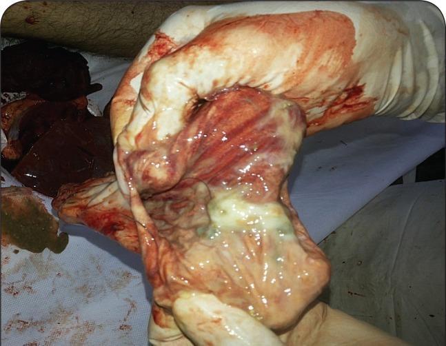
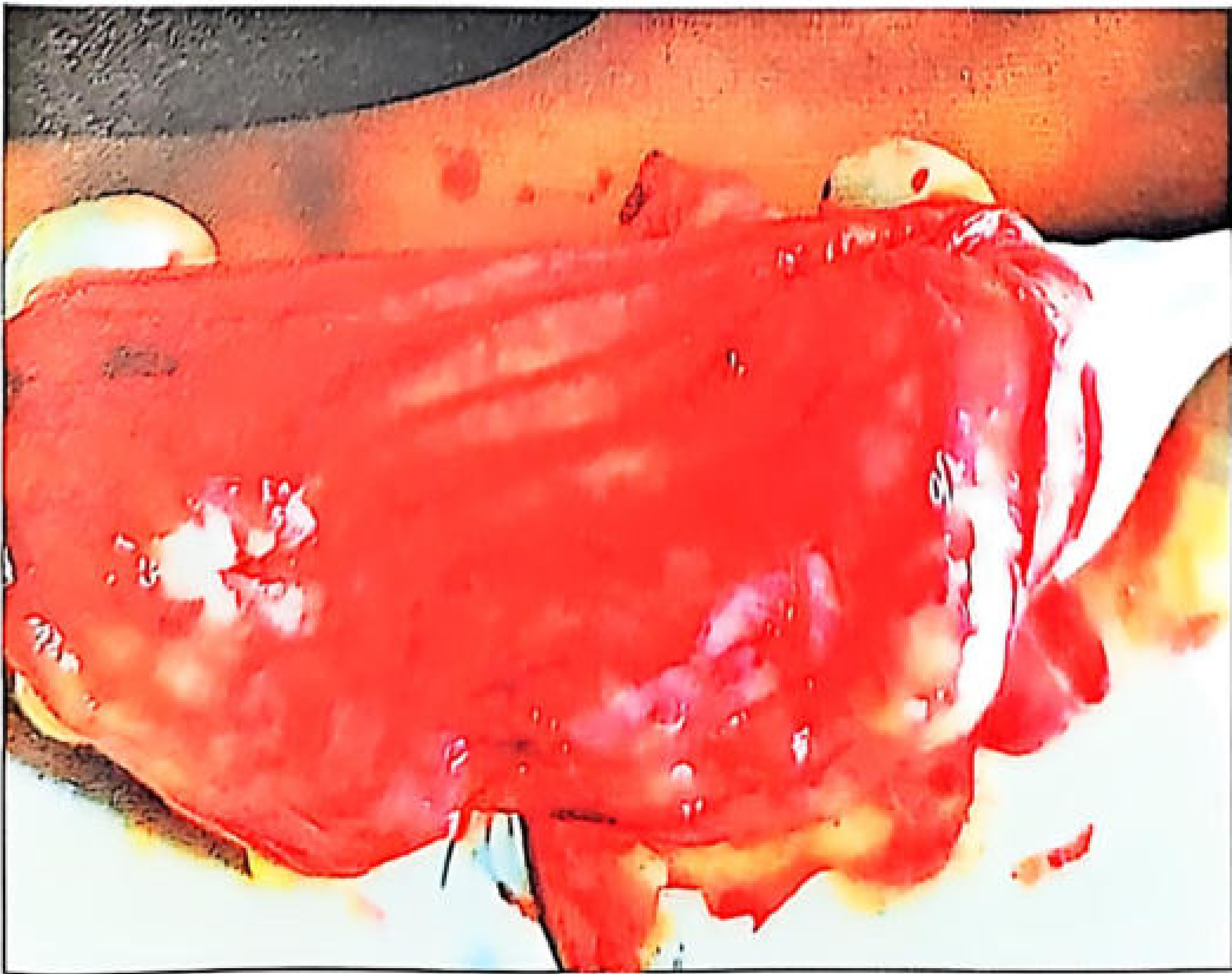
This clinical photograph shows a gross pathological specimen of the human stomach mucosa, likely taken during a post-mortem or surgical examination. The gastric lining exhibits intense, diffuse hyperemia and congestion, characterized by a deep red coloration. There are visible areas of intramural hemorrhage and petechial spots across the mucosal surface. The tissue has a shiny, edematous appearance with prominent rugae. A focal area of greenish-yellow mucoid substance is present, suggesting potential chemical irritation or altered gastric contents. This image serves as an educational example of severe gastrointestinal mucosal injury, often associated with acute gastritis or toxic ingestion, such as aluminium phosphide poisoning. The specimen is being handled by a person wearing white latex gloves in a clinical or forensic setting.
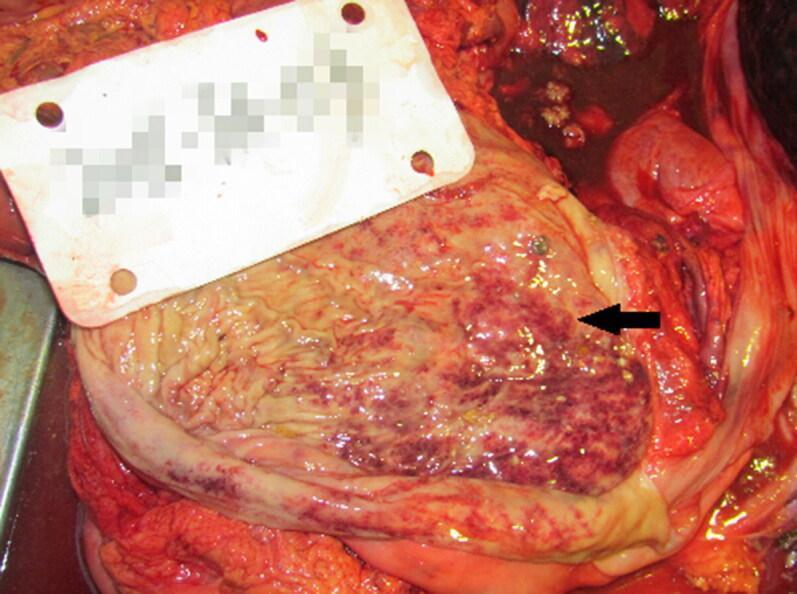
This clinical gross specimen photograph displays the internal surface of a human stomach during an autopsy. The gastric mucosa shows clear signs of pathology characterized by light, focal haemorrhage. The mucosal surface exhibits an uneven, mottled appearance with scattered petechiae and larger areas of ecchymosis, particularly prominent in the region indicated by the black arrow. These haemorrhagic foci appear as dark red to purple discolorations interspersed with the paler, tan-pink tissue of the normal gastric folds (rugae). The irregular distribution of these spots suggests acute mucosal injury or congestion. Such findings in a forensic or clinical pathology context can be associated with various systemic stressors, including acute alcohol poisoning or severe physiological shock. The image serves as a teaching tool for identifying macroscopic signs of gastric mucosal haemorrhage and inflammation during post-mortem examination.
This forensic clinical photograph displays gross autopsy findings from the respiratory system of a deceased individual. Image A shows a longitudinal section of the larynx and trachea, held open with surgical forceps. The mucosa exhibits a characteristic cherry-red coloration, which is a classic post-mortem indicator of carboxyhemoglobinemia associated with carbon monoxide poisoning. Significant deposits of black, granular soot are visible lining the interior of the lower trachea. Image B shows a cross-section of pulmonary tissue, demonstrating intense congestion and a similar cherry-red hue. The primary bronchi and smaller airways contain prominent black carbonaceous material (soot). These findings collectively illustrate the pathological hallmarks of smoke inhalation and carbon monoxide toxicity, typically encountered in fire-related fatalities or enclosed-space combustion. The image serves as a teaching tool for forensic pathology, particularly in identifying indicators of vital reaction (breathing during a fire) versus post-mortem exposure.
This gastroscopic image displays a view of the gastric mucosa, likely within the antrum, showing a relatively healthy appearance following the resolution of prior pathology. The mucosa is characterized by a uniform pinkish-red color with a smooth, glistening surface and normal vascular patterns. There is a distinct absence of the diffuse erythema, edematous thickening, or wall rigidity often associated with scirrhous gastric cancer or ectopic pancreas-related inflammation. The gastric folds appear supple and undistended. In the center, the gastric lumen and the pyloric orifice are visible and patent, with no evidence of stenosis, mass effect, or significant mucosal irregularities. This image serves as a clinical reference for the interval regression of gastric wall thickening in a patient being monitored for recurrent epigastric symptoms.
Post Mortem Findings in Arsenic Poisoning
ACUTE ARSENIC POISONING
External Findings
| Feature | Details |
|---|---|
| General appearance | Body is shrunken due to severe dehydration |
| Rigor mortis | Lasts longer than usual |
| Eyeballs | Sunken/shrunken |
| Skin | Cold, clammy; cyanosis of hands and feet |
| Jaundice | May be present |
Internal Findings
- Mucous membrane of mouth and pharynx is inflamed and ulcerated
- Mouth and pharynx may sometimes be unaffected; oesophagus may show oesophagitis
- Mucosa is swollen, oedematous, congested, and red - either generally or in patches, especially at the pyloric region
- Inner wall tinged with streaks of blood and arsenic particles embedded in mucus
- Mucosa on scraping appears brownish-red or scarlet in colour - "red velvet" appearance
- Congestion most marked along the crest of the rugae and at the greater curvature/cardiac end
- Petechial haemorrhages and submucosal/subperitoneal haemorrhages
- Lines of redness running along the walls; curved lines of submucous haemorrhages
- Sticky mucus covers the mucosa; particles of arsenic may be seen embedded within it
- Small acute ulcerations or large erosions, especially at the pyloric end
- Ulceration, gangrene, and perforation are rarely reported
- In fulminating (very rapid) cases, the stomach and intestines may show no signs of inflammation at all
- Flaccid/flabby; contains large flakes of mucus with very little faecal matter
- Mucosa is pale-violet, oedematous, shows inflammation
- Submucosal haemorrhages along its whole length
- Contains seromucous fluid; intestinal glands are swollen and enlarged
- Caecum and rectum show slight inflammation; mucous membrane is flabby
- Enlarged and congested
- Fatty infiltration and degeneration (cloudy swelling, fatty change)
- Kidneys show glomerulonephritis; acute tubular necrosis common
- In a few days, fatty deposits appear in the heart, liver and kidneys
- Congested and oedematous
- Subpleural ecchymoses
- Contains coagulated blood
- Subendocardial petechial haemorrhages of the left ventricle - this is a characteristic and important finding in arsenic poisoning
- Ecchymosis of endocardium and ventricular muscles
- Brain is congested; ventricles are full of serum
- Oedema of brain with patchy necrosis or haemorrhagic encephalitis in severe cases
- Meninges are congested
CHRONIC ARSENIC POISONING - POST MORTEM FINDINGS
External Findings
- Skin: "Raindrop" pigmentation - finely mottled brown pigmentation on flexures, temples, eyelids, nipples, lower abdomen, neck. Persists for many months
- Palms and soles: Hyperkeratosis with irregular thickening
- Nails: Transverse white lines (1-2 mm width) called Aldrich-Mees lines (Mees' lines) appearing above the base of each fingernail; double lines if more than one exposure episode
- Hair: Patchy or diffuse alopecia; hair analysis shows arsenic deposition
- Oedema: Oedema of face, periorbital region, and ankles
- Jaundice may be present
Internal Findings (Chronic)
| Organ | Findings |
|---|---|
| Stomach | Normal or chronic gastritis; patchy inflammatory redness of rugae; patchy haemorrhagic gastritis with acute and chronic erosions |
| Small intestine | Dilated, reddened, with thickened mucosa |
| Liver | Fatty change, severe necrosis, cirrhosis, hepatomegaly, jaundice (portal hypertension with ascites reported from therapeutic arsenic use) |
| Kidneys | Tubular necrosis, chronic nephritis |
| Lungs | Congestion; haemoptysis-related changes; increased risk of lung cancer |
| Heart | Cardiac failure-related changes; myocardial degeneration |
| Bone marrow | Hypoplasia; normochromic normocytic anaemia; thrombocytopenia; leukopenia; karyorrhexis (bizarre nuclear forms on marrow exam); megaloblastic picture from folate interference |
| Peripheral nerves | Symmetrical sensorimotor polyneuropathy; disintegration of axis cylinder (axonal neuropathy); fragmentation and resorption of myelin - "glove and stocking" distribution |
| Brain | Encephalopathy; cortical changes |
| Skin histology | Bowen's disease (intraepidermal squamous cell carcinoma) as a long-term complication |
| Spleen | In arsine gas exposure: evidence of RBC destruction with deposition of blood pigment throughout |
KEY DISTINGUISHING FEATURES AT AUTOPSY
| Feature | Significance |
|---|---|
| Red velvet stomach mucosa | Classic gross finding in acute poisoning |
| Subendocardial petechial haemorrhages (left ventricle) | Characteristic of arsenic; also seen in phosphorus, barium, mercury poisoning |
| Mees' lines on nails | Hallmark of chronic/subacute exposure |
| Raindrop pigmentation | Chronic arsenic skin finding |
| Arsenic detected in hair/nails/bones | Persists years after death; useful in exhumation cases |
| Delayed putrefaction | Arsenic is a preservative; useful forensic indicator |
| X-ray of GI tract | May show radiopaque arsenic particles |
| Fulminating cases | May show NO GI inflammation despite high blood arsenic levels |
MEDICOLEGAL IMPORTANCE
- Arsenic is the most common homicidal poison used in India; mimics cholera (mass poisoning)
- It is tasteless, odourless, easily available, and small doses are lethal
- Arsenic can be detected in hair, nails, and bones years after death - making it detectable on exhumation
- Hair segmental analysis can time the exposure
- Fatal dose: 180 mg of arsenic trioxide; fatal period: 12-48 hours (can occur in 2-3 hours)
Answer again
Post Mortem Findings in Arsenic Poisoning
ACUTE ARSENIC POISONING
External Findings
| Feature | Details |
|---|---|
| General appearance | Body shrunken due to severe dehydration |
| Rigor mortis | Lasts longer than usual |
| Eyeballs | Sunken/shrunken |
| Skin | Cold, clammy; cyanosis of hands and feet |
| Jaundice | May be present |
Internal Findings
- Mucous membrane of mouth and pharynx is inflamed and ulcerated
- In some cases these may be unaffected; oesophagitis may be present
- Mucosa is swollen, oedematous, congested, red - generally or in patches, especially at the pyloric region
- Inner wall tinged with streaks of blood; arsenic particles embedded in overlying mucus
- Mucosa on scraping appears brownish-red or scarlet - "Red Velvet" appearance
- Congestion is most marked along the crest of the rugae and at the greater curvature/cardiac end
- Petechial haemorrhages and submucosal/subperitoneal haemorrhages
- Lines of redness running along the walls; curved lines of submucous haemorrhages
- Sticky mucus covers mucosa; arsenic particles may be seen embedded within it
- Small acute ulcerations or large erosions, especially at the pyloric end
- Ulceration, gangrene, and perforation are rarely reported
- In fulminating cases, the stomach and intestines may show no signs of inflammation despite high arsenic levels
- Flaccid/flabby; contains large flakes of mucus with very little faecal matter
- Mucosa is pale-violet, oedematous, inflamed
- Submucosal haemorrhages along its whole length
- Contains seromucous fluid; intestinal glands swollen and enlarged
- Caecum and rectum show slight inflammation; mucous membrane flabby
- Enlarged and congested
- Fatty infiltration and degeneration (cloudy swelling, fatty change)
- Kidneys: glomerulonephritis; acute tubular necrosis
- After a few days: fatty deposits appear in heart, liver, and kidneys
- Congested and oedematous
- Subpleural ecchymoses
- Contains coagulated blood
- Subendocardial petechial haemorrhages of the left ventricle - a characteristic and important finding
- Ecchymosis of endocardium and ventricular muscles
- Brain congested; ventricles full of serum
- Oedema of brain with patchy necrosis or haemorrhagic encephalitis in severe cases
- Meninges congested
Toxicological threshold: Arsenic values in liver and blood in excess of 1 mg% are typically present in deaths from acute poisoning.
Putrefaction note: If putrefaction has occurred, yellow streaks appear in the subperitoneal layer of the stomach/intestines (absorbed arsenic converted to arsenic sulphide). Arsenic delays putrefaction and remains detectable in decomposed and charred bodies.
CHRONIC ARSENIC POISONING - POST MORTEM FINDINGS
External Findings
| Feature | Details |
|---|---|
| Skin pigmentation | "Raindrop" type - finely mottled brown pigmentation on flexures, temples, eyelids, nipples, lower abdomen, neck |
| Palms & soles | Hyperkeratosis with irregular thickening |
| Nails | Transverse white lines (Aldrich-Mees lines) 1-2 mm wide above base of each fingernail; double lines if repeated exposures |
| Hair | Patchy or diffuse alopecia; arsenic deposits detectable by segmental hair analysis |
| Oedema | Face, periorbital region, ankles |
| Jaundice | May be present in hepatic involvement |
Internal Findings
| Organ | Findings |
|---|---|
| Stomach | Normal or chronic gastritis; patchy inflammatory redness of rugae; haemorrhagic gastritis with acute and chronic erosions |
| Small intestine | Dilated, reddened, with thickened mucosa |
| Liver | Fatty change, severe necrosis, cirrhosis, hepatomegaly; portal hypertension with ascites |
| Kidneys | Tubular necrosis, chronic nephritis |
| Lungs | Congestion; lung cancer risk (arsenic is a carcinogen) |
| Heart | Myocardial degeneration; cardiac failure changes |
| Bone marrow | Hypoplasia; normochromic normocytic anaemia; thrombocytopenia; leukopenia; karyorrhexis (bizarre nuclear forms); megaloblastic picture from folate interference |
| Peripheral nerves | Symmetrical axonal neuropathy; disintegration of axis cylinder; fragmentation and resorption of myelin; "glove and stocking" distribution |
| Brain | Encephalopathy; cortical changes |
| Skin histology | Bowen's disease (intraepidermal carcinoma) as a long-term complication |
| Spleen | Arsine exposure: destruction of RBCs with blood pigment deposition throughout the organ |
AUTOPSY FINDINGS IN ARSINE GAS POISONING (separate entity)
- Skin is dirty yellow in colour
- Mucous membrane of stomach and intestine is yellow and inflamed
- Liver: small or enlarged, shows fatty degeneration
- Spleen: destruction of RBCs; blood pigment deposits throughout
- Kidneys: enlarged, congested, tubular necrosis
- Lungs: congested and oedematous
KEY DISTINGUISHING FEATURES - SUMMARY TABLE
| Feature | Type | Significance |
|---|---|---|
| Red velvet stomach mucosa | Acute | Classic gross pathology finding |
| Subendocardial petechial haemorrhages (left ventricle) | Acute | Characteristic; also seen in phosphorus, barium, mercury poisoning |
| Mees' lines on nails | Chronic/Subacute | Hallmark of repeated exposure |
| Raindrop pigmentation | Chronic | Pathognomonic skin finding |
| Fulminating cases with no GI changes | Acute | Do not miss - toxicology is essential |
| Arsenic in hair, nails, bones | Both | Persists years after death; key in exhumation cases |
| Delayed putrefaction | Both | Arsenic acts as a preservative |
| X-ray abdomen | Acute | May show radiopaque arsenic particles in GI tract |
MEDICOLEGAL IMPORTANCE
- Most common homicidal poison in India; mimics cholera (mass poisoning scenarios)
- Tasteless, odourless, easily available, small doses sufficient for homicide
- Fatal dose: 180 mg of arsenic trioxide
- Fatal period: 12-48 hours (can occur within 2-3 hours)
- Arsenic is detectable in hair, nails, and bones years after death - including from exhumed and charred bodies
- Segmental hair analysis can time the exposure to specific periods
- Its persistence in tissues makes it one of the most forensically traceable poisons
Compile and give comprehensive answer
post mortem findings acute chronic arsenic poisoning forensic pathology autopsy
arsenic poisoning Mees lines nails chronic
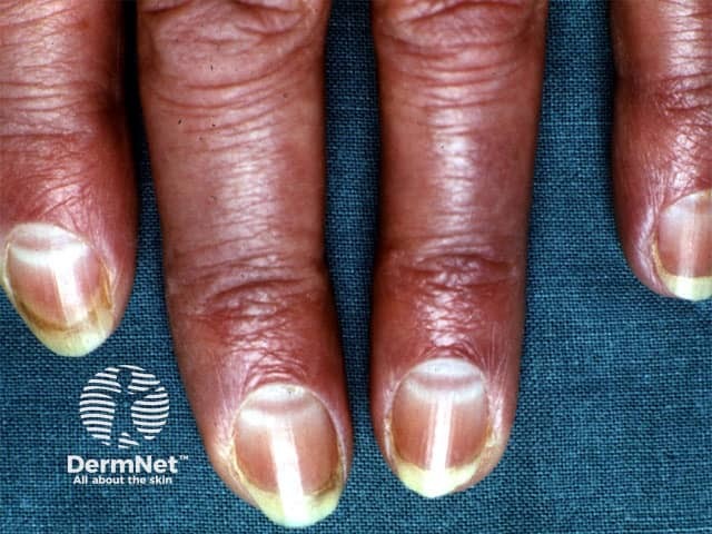
Clinical photography of the nails demonstrating Muehrcke lines. Anterolateral view of several fingernails (index, middle, ring, and little fingers) on both hands. The nail plates show paired, parallel, transverse white bands crossing the lunula region and extending toward the distal edge. These lines are non-palpable and lie within the nail bed/plate interface, giving the appearance of alternating light bands against the surrounding pinkish nail bed. The bands do not accompany surface textural changes and disappear with recovery of serum albumin; they are not true leukonychia. The finding is best explained by hypoalbuminemia leading to edema of the nail bed and reduced capillary perfusion, producing transverse opacities. Associated clinical context includes liver dysfunction, nephrotic syndrome, malnutrition, inflammatory states, or protein loss; systemic edema may be present. Distinguish from Beau lines (transverse ridges within the nail plate from growth arrest) and Mees’ lines (arsenic poisoning) which are embedded in the nail plate rather than the nail bed. This image underscores the dermatologic sign’s diagnostic significance as a noninvasive clue to systemic protein deficiency. Potential clinical use cases include screening for hypoalbuminemia in hospitalized patients, monitoring nutritional status in malnourished individuals, and differential diagnosis of white nail banding in clinical dermatology in practice.
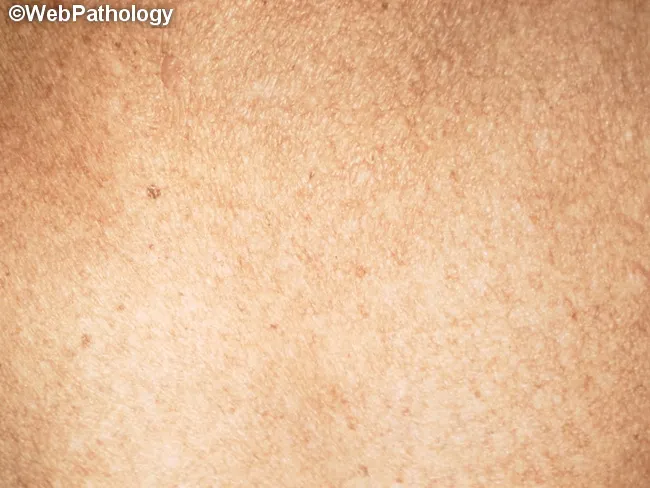
This clinical dermatologic photograph depicts chronic arsenic-related skin pigmentation. Modality: clinical photography; technique: gross macro image with standard lighting. The skin surface shows diffuse, mottled hyperpigmentation characterized by numerous small, irregular brown-to-slate macules and patches arranged in a raindrop pattern across the trunk and proximal extremities. Borders are variable, with some coalescing into larger pigmented fields; texture remains largely normal but may appear slightly rough in exposed areas. The appearance is typical of long-standing arsenic exposure from contaminated groundwater and is often accompanied by palmoplantar keratoses (not shown here). The color contrast between pigmented and adjacent normal skin is pronounced, aiding recognition. Pathophysiology involves arsenic-induced dysregulation of melanocyte activity and epidermal changes, producing “raindrop” hyperpigmentation that may herald cutaneous malignancy risk (basal cell carcinoma, squamous cell carcinoma) with chronic exposure. Clinical significance includes confirming exposure history, guiding public health intervention, and prompting systemic evaluation for arsenic-related toxicity. Differential considerations include postinflammatory hyperpigmentation, tinea versicolor, solar lentigines, and other drug- or metal-induced pigmentary disorders. Clinical correlation with urinary, hair, or nail arsenic levels is recommended, along with surveillance for Mees’ lines and hyperkeratosis. This image is valuable for education, toxicology, environmental health, and dermatology curricula. Useful for clinical training and health.
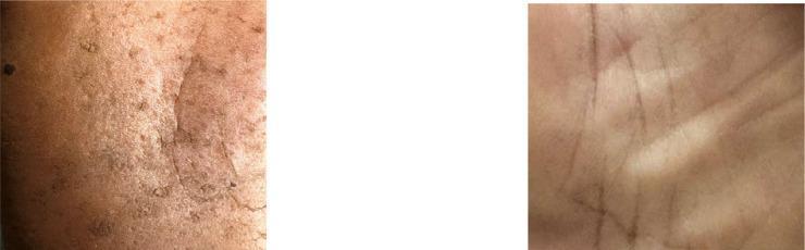
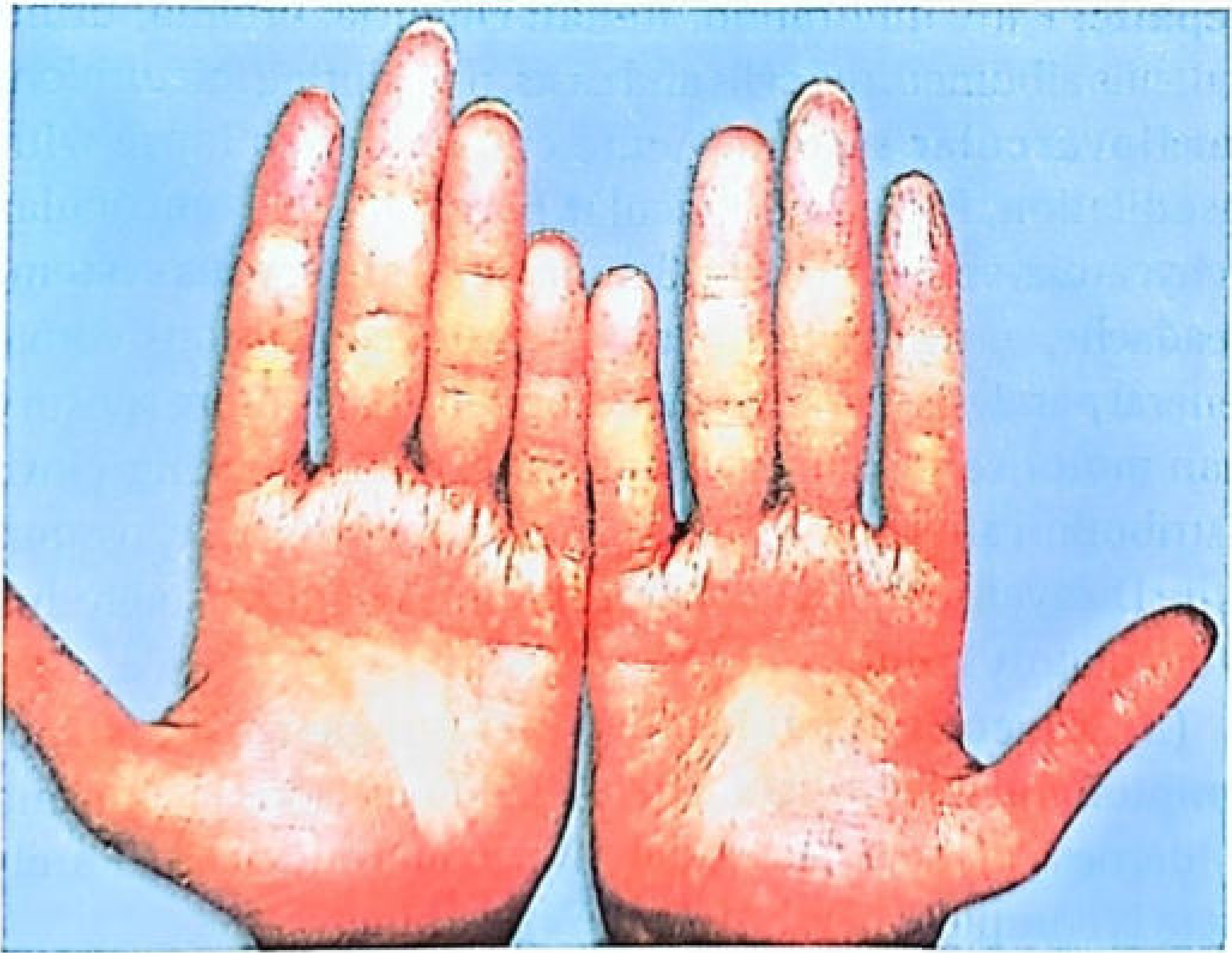
This clinical comparison photograph displays the plantar surfaces (soles) of two different individuals to illustrate skin manifestations of chronic arsenic exposure (arsenicosis). The image on the left shows an affected foot characterized by diffuse hyperpigmentation, mottled melanosis (darkened patches), and palmoplantar hyperkeratosis. Visible features include a rough, thickened skin texture with numerous small, raised, corn-like keratotic papules and irregular desquamation. In contrast, the image on the right depicts a healthy sole with a smooth, uniform skin texture, normal light pigmentation, and clearly defined dermatoglyphic lines without any evidence of lesions, nodules, or abnormal thickening. This comparison serves as an educational tool for identifying the dermatological signs of arsenic poisoning, specifically highlighting the diagnostic transition from healthy tissue to pathological keratotic and pigmentary changes characteristic of environmental or occupational toxin exposure.
Post Mortem Findings in Arsenic Poisoning
A Comprehensive Forensic Account
Key facts: Fatal dose of arsenic trioxide = 180-300 mg | Fatal period = 12-48 hours (can occur in 2-3 hours)
PART I: ACUTE ARSENIC POISONING
A. EXTERNAL POST MORTEM FINDINGS
| Feature | Details |
|---|---|
| General appearance | Body appears shrunken and emaciated due to profound dehydration |
| Rigor mortis | Lasts longer than usual; appears early |
| Putrefaction | Delayed - arsenic has antibacterial action and acts as a tissue preservative |
| Eyeballs | Sunken (shrunken) |
| Skin | Cold, clammy; cyanosis of hands and feet |
| Jaundice | May be present in cases of liver involvement |
| Blood-tinged vomitus | May be found on the body and clothing |
B. INTERNAL POST MORTEM FINDINGS
1. Mouth, Pharynx & Oesophagus
- Usually not affected, but in some cases the mucous membrane is inflamed or ulcerated
- Oesophagitis may be present
2. Stomach - The Most Characteristic Site
- Mucosa is swollen, oedematous, and red - either generally or in patches, especially at the pyloric region
- Mucosa on scraping appears brownish-red or scarlet in colour
- Classic appearance: "Red Velvet" stomach - the mucosa resembles a piece of red velvet
- Congestion is most marked along the crest of the rugae and at the greater curvature/cardiac end
- Petechial haemorrhages scattered over the mucosa
- Lines of redness running along the walls; curved lines of submucous haemorrhages
- Large submucosal and subperitoneal haemorrhages in severe cases
- A mass of sticky mucus covers the mucosa - arsenic particles may be seen embedded in it
- Small acute ulcerations or large erosions, especially at the pyloric end
- Inner wall tinged with streaks of blood and arsenic
- Ulceration, gangrene, and perforation are rarely reported
- Arsenic can penetrate through the stomach wall and appear in the liver, omentum, and endocardium
Fulminating cases (important!): The stomach and intestines may show NO signs of inflammation at all, despite lethal arsenic levels in blood. Always perform toxicology.
3. Small Intestine
- Appears flaccid/flabby
- Contains large flakes of mucus with very little faecal matter
- Mucosa is pale-violet, oedematous, inflamed
- Submucosal haemorrhages along its whole length
4. Large Intestine, Caecum & Rectum
- Contains seromucous fluid; intestinal glands are swollen and enlarged
- Caecum and rectum show slight inflammation
- Mucous membrane is flabby
5. Liver, Spleen & Kidneys
- All three organs are enlarged and congested
- Show fatty infiltration and degeneration (cloudy swelling, fatty change)
- Fatty deposits appear in the heart, liver, and kidneys after a few days
- Kidneys: glomerulonephritis (particularly glomerular type); acute tubular necrosis
6. Lungs
- Congested and oedematous
- Subpleural ecchymoses (characteristic finding)
- Haemorrhages may be found in larynx, trachea, and lung parenchyma in severe cases
7. Heart - A Characteristic Finding
- Contains coagulated blood
- Subendocardial petechial haemorrhages of the left ventricle - this is a hallmark of arsenic poisoning
- Ecchymosis of the endocardium and muscles of the left ventricle
- This finding may be present even when the stomach shows little or no sign of irritation
- Cardiopulmonary toxicity includes congestive cardiomyopathy and non-cardiogenic pulmonary oedema
Note: Subendocardial petechial haemorrhages are also seen in phosphorus, barium, and mercury poisoning, and in heat stroke, acute infectious diseases (influenza), and traumatic asphyxia.
8. Brain & Meninges
- Brain is congested; ventricles are full of serum
- Oedema of the brain with patchy necrosis or haemorrhagic encephalitis in severe cases
- Meninges are congested
9. Putrefaction Changes
- If putrefaction has taken place, yellow streaks are found in the subperitoneal layer of the stomach and intestines
- This is due to absorbed arsenic being converted into arsenic sulphide
C. TOXICOLOGICAL THRESHOLDS (Acute)
| Sample | Significant Level |
|---|---|
| Liver and blood (death) | Arsenic values > 1 mg% typically present |
| Blood | > 1.5 mg/100 mL indicates serious poisoning |
| Urine | Normal < 0.03 mg/L; thousands of mcg in acute poisoning (first 2-3 days) |
| X-ray abdomen | May show radiopaque arsenic particles in GI tract |
D. AUTOPSY FINDINGS IN ARSINE GAS POISONING (separate entity)
| Organ | Finding |
|---|---|
| Skin | Dirty yellow colour (jaundice from haemolysis) |
| Stomach & intestines | Mucous membrane is yellow and inflamed |
| Liver | Small or enlarged; shows fatty degeneration |
| Spleen | Evidence of RBC destruction; deposition of blood pigment throughout |
| Kidneys | Enlarged, congested; tubular necrosis |
| Lungs | Congested and oedematous |
PART II: CHRONIC ARSENIC POISONING
A. EXTERNAL POST MORTEM FINDINGS
Skin
- Finely mottled brown pigmentation - the "raindrop" pattern
- Predominantly on covered parts: flexures, nipples, lower abdomen, temples, eyelids, and neck
- Persists for many months
- Differentiating from Addison's disease: mucous membranes are not pigmented in arsenic (they are in Addison's)
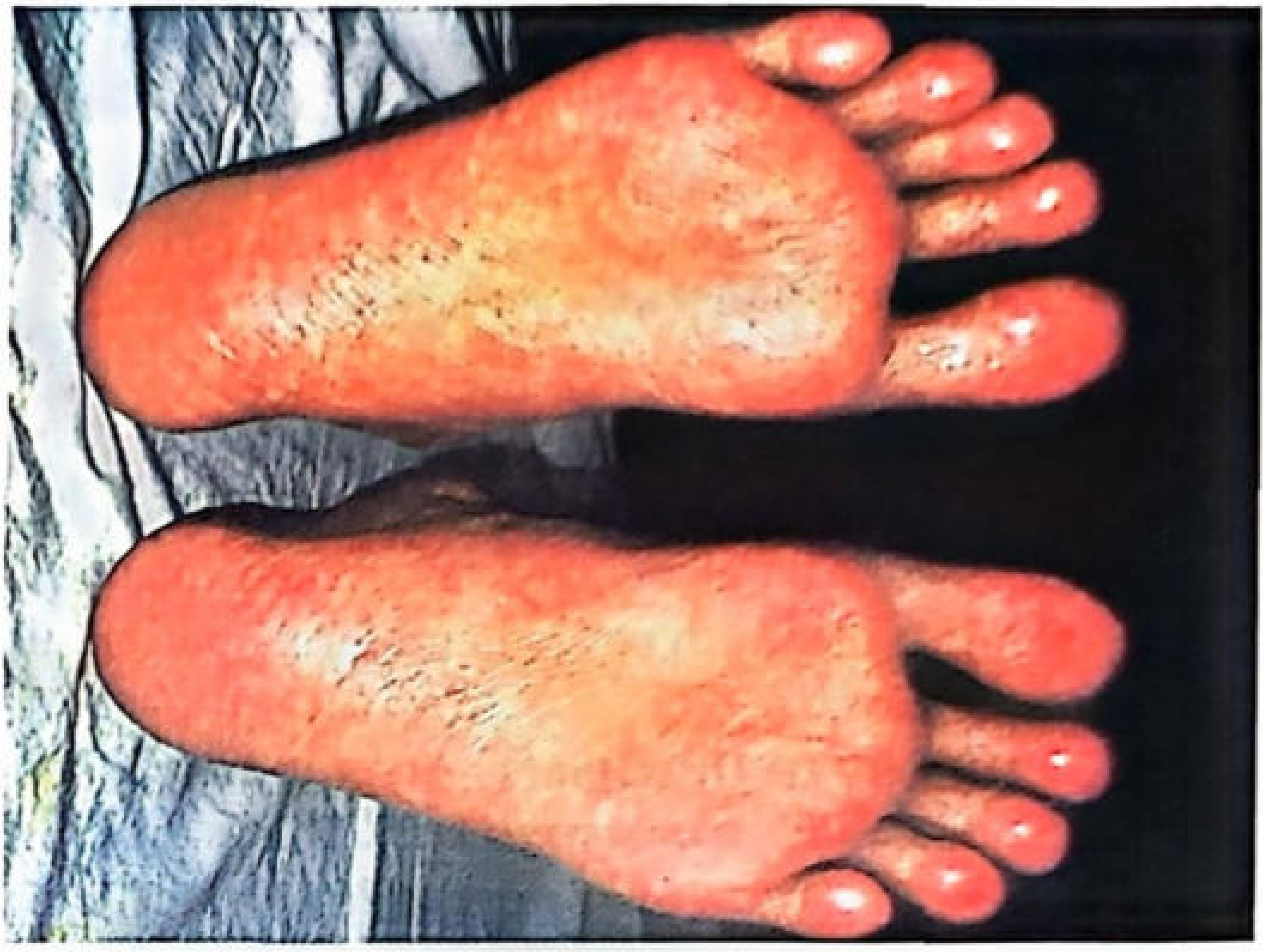
- Palms and soles: irregular thickening - palmoplantar keratoses
- Dry, scaly desquamation over trunk and extremities
- Intraepidermal squamous cell carcinoma as a long-term complication of arsenic exposure
- Indicates systemic neoplastic processes
Nails
- Transverse white striae, 1-2 mm wide, appearing above the base of each fingernail
- Appear approximately 5-6 weeks after acute/repeated exposure
- Represent growth arrest bands in the nail plate (embedded in nail plate, not nail bed)
- Double lines may be seen if more than one exposure episode
- Months after acute poisoning, these bands become visible as they grow distally
- Differentiate from Muehrcke's lines (hypoalbuminaemia) which are in the nail bed and not embedded
Hair
- Patchy or diffuse alopecia
- Arsenic deposits along the hair shaft - segmental hair analysis can date the exposure
Other External Features
- General emaciation and wasting of muscles
- Oedema of face, periorbital region, and ankles
- Jaundice from liver involvement
- Ulceration of the nasal septum (characteristic of occupational exposure)
B. INTERNAL POST MORTEM FINDINGS (Chronic)
| Organ/System | Post Mortem Findings |
|---|---|
| Stomach | Normal or chronic gastritis; patchy inflammatory redness of rugae; patchy haemorrhagic gastritis with acute and chronic erosions; focal ulceration |
| Small intestine | Dilated, reddened, thickened mucosa |
| Liver | Hepatomegaly; fatty degeneration or severe necrosis; non-cirrhotic portal hypertension; cirrhosis; jaundice |
| Kidneys | Tubular necrosis; chronic nephritis; urine may be red or green (haemoglobinuria) |
| Lungs | Congestion; haemoptysis-related changes; lung carcinoma (arsenic is a Group 1 carcinogen for lung cancer) |
| Heart | Myocardial degeneration; cardiac failure changes; peripheral vascular disease |
| Blood/Bone marrow | Bone marrow hypoplasia; normochromic normocytic anaemia; thrombocytopenia; leukopenia; karyorrhexis (bizarre nuclear forms on marrow); megaloblastic picture from folate interference; pancytopenia; basophilic stippling of erythrocytes |
| Peripheral nerves | Symmetrical sensorimotor polyneuropathy; disintegration of axis cylinder (axonal neuropathy); fragmentation and resorption of myelin; "glove and stocking" distribution; wrist drop, foot drop, muscular atrophy |
| Brain | Encephalopathy; cortical changes; severe headache; personality changes |
| Spleen | Congested |
PART III: SIDE-BY-SIDE COMPARISON
| Feature | Acute | Chronic |
|---|---|---|
| Body habitus | Shrunken (dehydration) | Emaciated (wasting) |
| Putrefaction | Delayed | Delayed |
| Stomach | Red velvet; petechiae; submucosal haemorrhages; sticky mucus with arsenic | Chronic gastritis; patchy redness; erosions |
| Intestine | Flaccid; pale-violet; submucosal haemorrhages | Dilated; reddened; thickened mucosa |
| Heart | Subendocardial petechial haemorrhages (left ventricle) | Myocardial degeneration; cardiac failure |
| Liver | Enlarged; fatty degeneration; cloudy swelling | Hepatomegaly; cirrhosis; non-cirrhotic portal hypertension |
| Kidneys | Acute tubular necrosis; glomerulonephritis | Chronic nephritis; tubular necrosis |
| Lungs | Congested; oedematous; subpleural ecchymoses | Congested; carcinoma (long-term) |
| Brain | Congested; haemorrhagic encephalitis | Encephalopathy |
| Skin | Cyanosis; jaundice | Raindrop pigmentation; hyperkeratosis; Bowen's disease; alopecia |
| Nails | No nail changes (appear 5-6 weeks post-exposure) | Aldrich-Mees lines |
| Bone marrow | Pancytopenia (within 1 week) | Hypoplasia; karyorrhexis |
| Nerves | Ascending sensorimotor neuropathy (2-6 weeks delay) | Symmetrical polyneuropathy; glove-stocking |
PART IV: MEDICOLEGAL IMPORTANCE
-
Homicidal use: Most common homicidal poison used in India. Tasteless, odourless, easily available, small doses required. Mass poisoning mimics cholera.
-
Differentiating from cholera at autopsy:
| Feature | Arsenic Poisoning | Cholera |
|---|---|---|
| Pain in throat | Before vomiting | After vomiting |
| Purging | Follows vomiting | Precedes vomiting |
| Stools | Dark, bloody; later rice-watery | Rice-watery only; involuntary jet |
| Tenesmus | Present | Absent |
| Vomitus | Mucus, bile, and blood | Watery, no mucus/bile/blood |
| Conjunctivae | Inflamed | Not inflamed |
| Analysis | Arsenic in excreta | Cholera vibrio |
-
Persistence of arsenic in the body:
- Detected in hair, nails, and bones years after death
- Detected in decomposed and charred bodies
- Segmental hair analysis can time the exposure to specific weeks or months
-
Exhumation cases: Arsenic delays putrefaction and remains detectable long after burial - making it one of the most forensically traceable poisons
-
Advantages as a homicidal poison:
- Tasteless and odourless
- Easily mixed with food, drinks, sweets, milk, pan
- Produces symptoms resembling gastroenteritis or cholera
-
Disadvantages (from criminal perspective):
- Detected even in charred bodies
- Delays putrefaction (draws attention)
- Detected in decomposed bodies
- Found in bones, hair, and nails years after poisoning