
Why Heparin is avoided in pericardial effusion and what is pericardial effusion in esy words
pericardial effusion echocardiogram diagram
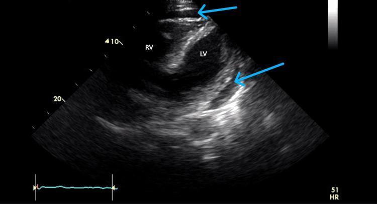
A subcostal four-chamber view echocardiogram demonstrating a moderate circumferential pericardial effusion. The image shows the anatomical arrangement of the heart with the right ventricle (RV) positioned anteriorly, closest to the transducer at the top of the sector, and the left ventricle (LV) located posteriorly. The pericardial effusion is clearly visualized as an anechoic (dark), echo-free space surrounding the myocardium, indicated by blue arrows. This dark space creates a distinct separation between the echogenic (bright) pericardium and the heart tissue. This imaging modality is essential for evaluating pericardial fluid volume and assessing for signs of cardiac tamponade, such as diastolic collapse of the right heart chambers. The subcostal view is particularly useful in clinical practice for visualizing the effusion through the acoustic window provided by the liver.
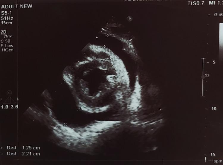
A 2D transthoracic echocardiogram in the parasternal short-axis view demonstrating a significant pericardial effusion. The diagnostic image shows a large, circumferential anechoic (echo-free) space between the parietal pericardium and the visceral pericardium (epicardium), indicating fluid accumulation around the myocardium. Electronic calipers and distance markers are visible, quantifying the thickness of the effusion at two points: 1.25 cm and 2.21 cm. The myocardium appears as a homogenous echogenic structure, and there is no visible evidence of internal echoes or fibrin strands within the fluid space. The pericardium presents as a bright, hyperechoic boundary. This clinical imaging finding is representative of a large pericardial effusion, which carries a risk of progression to cardiac tamponade. Key educational concepts include ultrasound diagnosis of pericardial disease and the quantification of pericardial fluid volume.
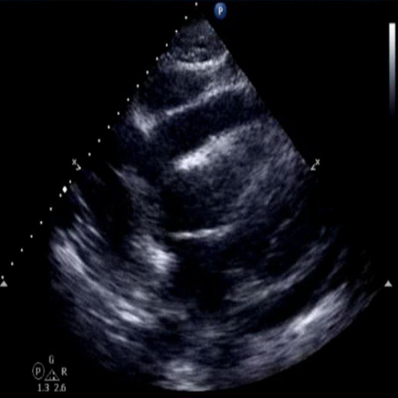
Diagnostic Image: This subcostal view echocardiogram demonstrates a moderate to large circumferential pericardial effusion. The effusion is identified as a prominent anechoic (dark), echo-free space located between the hyper-echogenic pericardium and the epicardial surface of the heart. The fluid collection surrounds the cardiac chambers, most notably the left ventricle, creating a clear separation between the heart and the surrounding pleural and diaphragmatic interfaces. The cardiac silhouette appears centralized within the fluid collection, and the significant depth of the anechoic space suggests a substantial volume of fluid accumulation. This finding is clinically significant for evaluating pericardial tamponade physiology in patients presenting with shortness of breath, low voltage on EKG, or pulsus paradoxus. Key educational concepts include ultrasound identification of fluid in the pericardial sac, subcostal windows in emergency medicine or cardiology, and the distinction between pericardial and pleural fluid collections.
Pericardial Effusion - Explained Simply & Why Heparin is Avoided
What is Pericardial Effusion? (In Easy Words)
- The sac gets stretched and fills up
- The fluid starts pressing on the heart from outside
- The heart has less room to expand and fill with blood
- If enough fluid collects, it can squeeze the heart so tightly it cannot pump properly - this is called cardiac tamponade, which is a medical emergency
What Causes It?
| Cause | Frequency |
|---|---|
| Idiopathic or viral (most common) | 37% |
| Cancer (malignant) | 19% |
| Iatrogenic / trauma | 13% |
| Tuberculosis or bacterial | 6% |
| Acute heart attack (MI) | 6% |
| Autoimmune (lupus, etc.) | 4% |
| Kidney failure (uremia) | 4% |
| Radiation-induced | 2% |
Why is Heparin Avoided in Pericardial Effusion?
The Mechanism
- Heparin prevents blood from clotting in the pericardial space
- Any small bleeds in the pericardium keep going instead of stopping naturally
- Blood/hemorrhagic fluid accumulates in the pericardial sac
- This dramatically increases the volume of fluid pressing on the heart
- This can rapidly escalate to cardiac tamponade - where the heart is so compressed it cannot function
Direct Textbook Quotes
"Heparin should be avoided in the setting of pericarditis with or without pericardial effusion because it may lead to pericardial hemorrhage."
- The Washington Manual of Medical Therapeutics
"Because of the propensity to hemorrhage in pericardial fluid, hemodialysis should be performed without heparin."
- Harrison's Principles of Internal Medicine, 22nd Edition (2025)
"Anticoagulation with heparin or warfarin should be discontinued..."
- Goldman-Cecil Medicine
A Specific & Important Clinical Context: Uremic Pericarditis
What to Use Instead?
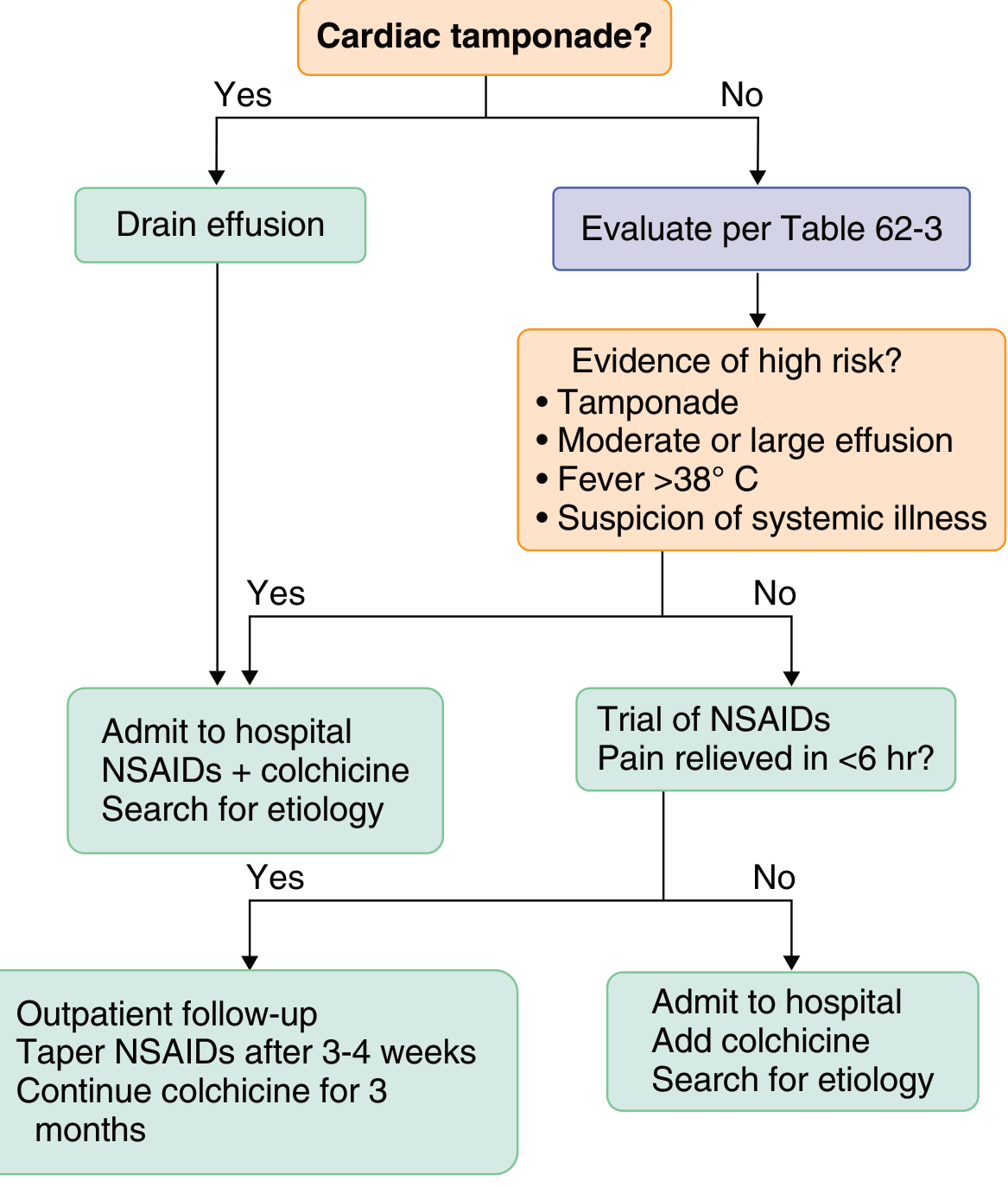
- The underlying cause of the effusion is treated (dialysis for uremia, NSAIDs/colchicine for pericarditis, etc.)
- If anticoagulation is absolutely needed for another reason (like a clot), an IVC filter (Greenfield filter) may be placed to prevent pulmonary embolism, as noted in Harrison's
- Pericardiocentesis (draining the fluid with a needle) is done if the effusion is large or causing tamponade
Echocardiogram View of Pericardial Effusion