
Ewing's sarcoma is a metaphysical disease or diaphysical
Ewing's sarcoma diaphysis bone X-ray
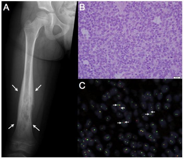
This composite educational image illustrates the multimodal diagnostic features of Ewing sarcoma, a malignant primary bone tumor. Panel A displays an anteroposterior X-ray of a pediatric femur showing an expansile, permeative, and destructive lesion within the diaphysis, marked by white arrows. Panel B provides a histopathological view (H&E stain, 400x magnification) revealing a dense proliferation of monomorphic 'small round blue cells' characterized by high nuclear-to-cytoplasmic ratios and scanty cytoplasm, arranged in sheets. Panel C demonstrates a break-apart Fluorescence in situ Hybridization (FISH) assay used for molecular confirmation. The presence of separated red (5'-EWSR1) and green (3'-EWSR1) fluorescent signals (white arrows) indicates a chromosomal rearrangement of the EWSR1 locus on chromosome 22, while fused orange signals represent intact alleles. This progression from gross radiology to cellular morphology and molecular genetics highlights the integrated diagnostic approach required for bone sarcomas in children and young adults.
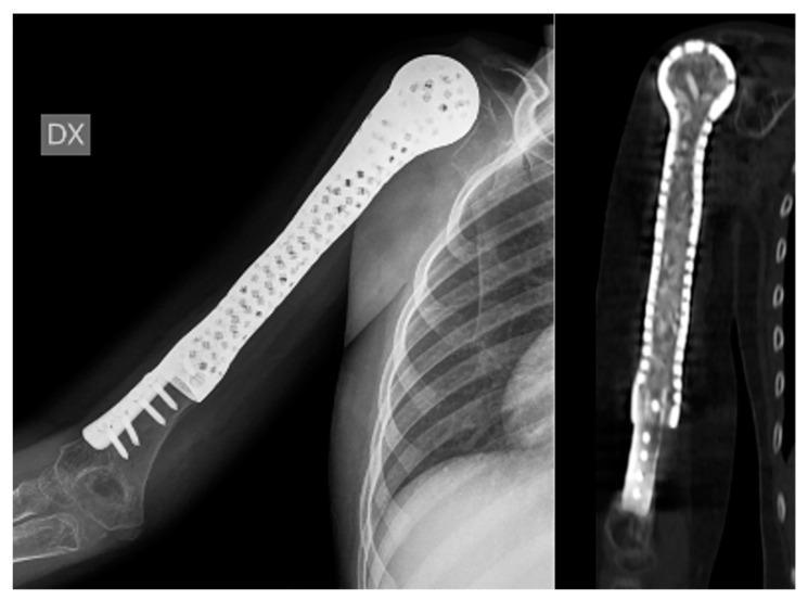
This composite diagnostic image features an anteroposterior X-ray (left) and a coronal CT reconstruction (right) of a pediatric humerus following limb-salvage surgery for Ewing’s sarcoma. The image demonstrates a 3D-printed, custom-made hollow prosthesis replacing the proximal and mid-diaphysis of the humerus while preserving the distal native bone and elbow joint. The prosthesis exhibits a distinctive porous structure with multiple radiolucent perforations throughout its radiopaque shell, designed to facilitate revascularization of internal bone grafts from surrounding soft tissues. The distal end of the implant features a wide collar that circumferentially overlaps the remaining humeral bone, secured by multiple transverse screws. The CT scan confirms the hollow nature of the internal chamber, which is packed with morselized allograft cortico-cancellous bone chips, visible as heterogenous, semi-dense material within the high-density prosthetic shell. This setup highlights orthopedic oncological techniques for pediatric reconstruction, focusing on biological integration and joint preservation.
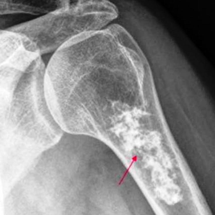
Anteroposterior X-ray of the proximal humerus showing an aggressive, intramedullary osteolytic lesion. The lesion is primarily located within the metaphysis and extends into the proximal diaphysis. It exhibits a mottled, permeative appearance with poorly defined, irregular borders, indicating a narrow zone of transition. There is evidence of cortical thinning and a 'moth-eaten' pattern of bone destruction, characteristic of malignant processes such as Ewing sarcoma. A red arrow highlights the area of most significant radiolucency and architectural disruption within the medullary canal. The image demonstrates key radiological signs used in orthopedic oncology to differentiate aggressive primary bone tumors from benign lesions.
Ewing's Sarcoma - Location
Primary Location: Diaphysis (shaft of long bones)
- Robbins Pathology: "Ewing sarcoma usually arises in the diaphysis of long tubular bones, especially the femur and the flat bones of the pelvis."
- Schwartz's Principles of Surgery: "The typical presentation is a tumor in the diaphysis of long bones, especially the femur."
- Bailey & Love's Surgery: "It tends to arise in the diaphysis..."
- S. Das Manual on Clinical Surgery: "Ewing's tumour grows from the diaphysis."
- Grainger & Allison's Diagnostic Radiology: "Ewing sarcoma tends to be an intramedullary lesion in the diaphysis."
Important Nuance
"Classically, Ewing sarcoma appears radiographically as a destructive lesion in the diaphysis of a long bone... In reality, Ewing sarcoma more often originates in the metaphysis of a long bone but frequently extends..."
Classic X-ray Finding
Quick Comparison Table for Bone Tumors by Location
| Tumor | Typical Location |
|---|---|
| Ewing's Sarcoma | Diaphysis (classic) |
| Osteosarcoma | Metaphysis |
| Giant Cell Tumor | Epiphysis (extends to metaphysis) |
| Chondroblastoma | Epiphysis |
| Simple bone cyst | Metaphysis |
| Enchondroma | Metaphysis/diaphysis (small bones) |