
Last conversation
Endoscopic dissectomy
endoscopic discectomy lumbar spine
Endoscopic Discectomy
Overview
- Campbell's Operative Orthopaedics 15th Ed 2026
Approaches
1. Transforaminal Endoscopic Lumbar Discectomy (TELD)
- Central, posterolateral, foraminal, or extraforaminal herniations at L1-L4, and sometimes at L5-S1 if anatomy permits
- Preferred when facet joint preservation is important (zygapophyseal joint not violated)
- Patient prone or lateral decubitus (lateral allows intraoperative straight-leg-raise testing)
- Performed under monitored anesthesia care (MAC) with intermittent propofol - patient remains awake enough to communicate
- An 18-20 gauge needle is advanced obliquely into Kambin's triangle at the foramen
- A diagnostic transforaminal epidural injection is placed first - profound relief predicts good surgical outcome
- Guidewire placed, then sequential dilators advanced under fluoroscopy
- A 1-cm skin incision is made
- Foramen is opened either by percutaneous reamers (fluoroscopic guidance) or a diamond burr under direct endoscopic vision
- Disc material is removed once the Wagner arch (inferior pedicle + superior articular process) and herniation are identified
- Decompression endpoint: nerve root pulsates to heartbeat without deformity; patient reports resolution of radicular pain
- Wound closed with single subcuticular stitch + skin glue
- Shower same day; discharge once ambulatory and able to void
- Driving restricted until postoperative day 2 or off narcotics (often only NSAIDs needed)
- Trunk stabilization therapy starts at 2 weeks
2. Interlaminar Endoscopic Lumbar Discectomy (IELD)
- Primarily for posterolateral herniations at L5-S1
- Can be used at higher levels if the interlaminar window is wide enough
- Facet-sparing technique (used when lateral recess decompression is NOT needed)
- Patient prone; can be done under general anesthesia or MAC
- Laminae, medial pedicular line, and midline are marked fluoroscopically
- 18-gauge needle placed at the trailing edge of the L5 lamina
- 8 mL of 1% lidocaine with epinephrine infiltrated to ease soft tissue dissection
- 1-cm incision made; guidewire, then dilators placed
- Approach angle: more medial-to-lateral for lateral herniations, more lateral-to-medial for central herniations
- Postoperative care is identical to the transforaminal approach
Combined Approach
- Maintaining direct light-based visualization of anatomy and pathology
- Minimizing iatrogenic instability (zygapophyseal joint preserved)
- Avoiding the need for general anesthesia
Advantages Over Open/Microdiscectomy
| Feature | Endoscopic | Traditional |
|---|---|---|
| Incision size | ~1 cm | 3-5 cm |
| Tissue disruption | Minimal (dilator-based) | Muscle stripping |
| Anesthesia | MAC often sufficient | Usually general |
| Discharge | Same day | 1-2 days |
| Return to activity | 2 weeks | 4-6 weeks |
| Inflammatory response | Lower CRP/IL-6 | Higher |
| Iatrogenic instability | Minimal | Higher risk if facets violated |
Complications
-
Dural tear (CSF leak): One advantage of endoscopic/tubular discectomy is the small dead space - minor dural tears can often be treated with fibrin glue alone without root herniation
- Management: fibrin glue, 3-day course of acetazolamide (Diamox) to reduce CSF production; bed rest only if positional headaches present; blood patch for persistent spinal headache
- Pseudomeningocele can rarely cause persistent pain from nerve root compression
-
Epidural bleeding: Controlled by adjusting fluid pressure and flow through the endoscope pump (not to exceed diastolic blood pressure)
-
Recurrence: Percutaneous endoscopic lumbar discectomy has been used successfully for recurrent disc herniations
-
Operative failure: Conversion to open surgery may be required if anatomy is unfavorable or adequate decompression cannot be achieved endoscopically
Additional Considerations
- Large herniations / lateral recess or foraminal stenosis: May require additional open exposure - hemilaminectomy, total laminectomy, or facetectomy. If >50% of combined bilateral facets removed, consider fusion.
- Far lateral disc herniation: A long tubular retractor at the correct trajectory is useful; approached via removal of the intertransverse ligament lateral to the spinal canal.
- Intradural disc herniation (rare): May require transdural approach; higher risk of CSF leak and intradural scarring.
- Fat grafting: Reserved for dural repairs and small laminar defects - does not improve clinical outcomes but reduces dural scarring.
Recent Evidence
- A 2025 network meta-analysis comparing treatments for lumbar disc herniation found endoscopic approaches competitive with microdiscectomy (PMID 40611244)
- A 2025 meta-analysis of percutaneous transforaminal endoscopic discectomy confirmed favorable outcomes in lumbar disc herniation (PMID 40553167)
- A 2024 systematic review of complications in endoscopic spine surgery in European Spine Journal provides a comprehensive complication profile (PMID 37587257)
- A 2023 meta-analysis on anesthesia type (epidural vs. local) for percutaneous endoscopic lumbar discectomy showed both are viable options (PMID 37348062)
Endoscopic images while endoscopic dissectomy
"cdn.orris.care" in /textbooks/9780443117633
1. Intraoperative Endoscopic View - Working Channel Field of View

The circular field represents the endoscopic portal view. The surgeon sees structures directly illuminated through the working channel - this is the hallmark view of endoscopic spine surgery.
2. Epidural Space Entry - Ligamentum Flavum Dissection
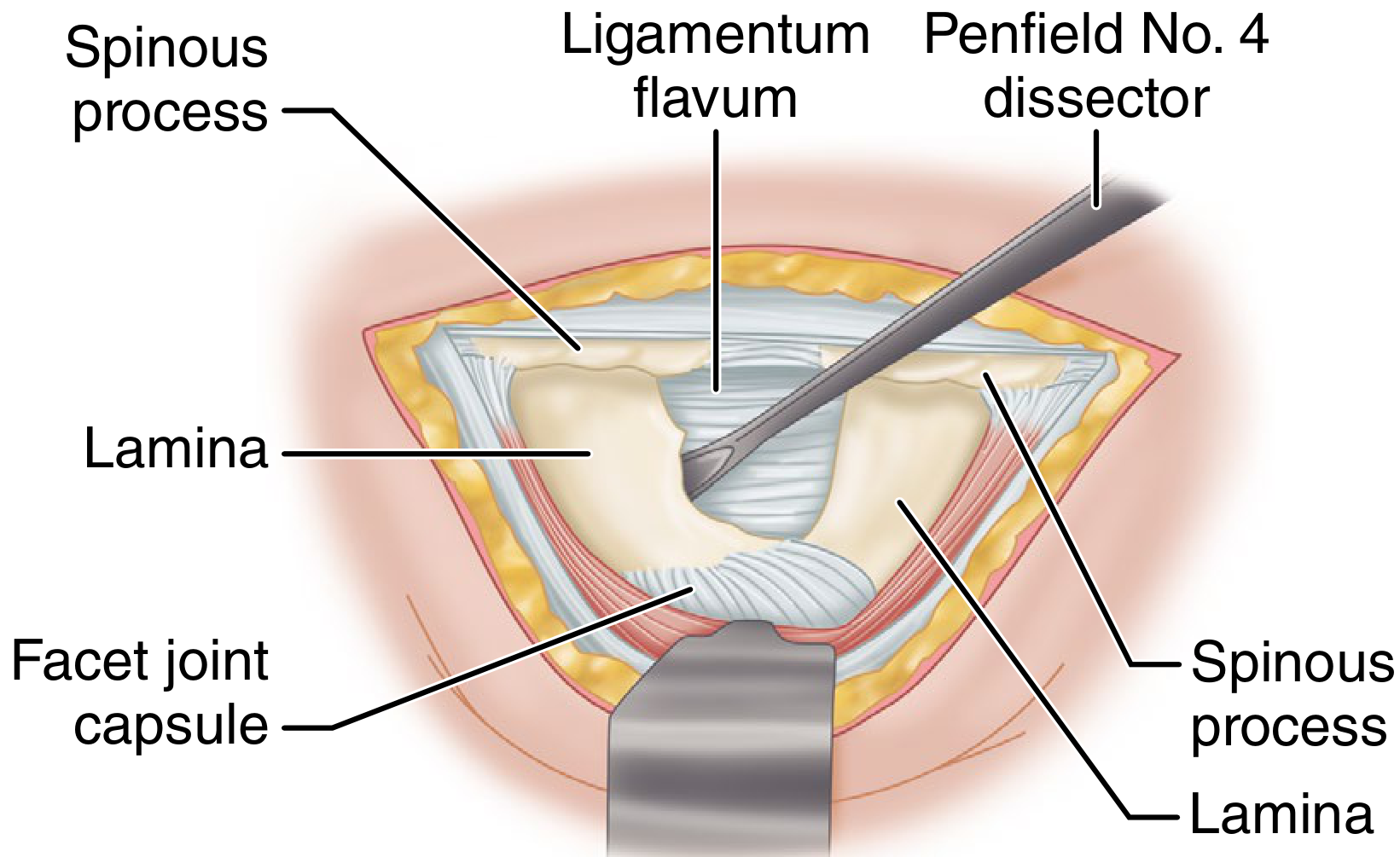
Fig. 46.12A - Entry to the epidural space is the critical initial step. The ligamentum flavum is carefully peeled off the leading edge of the caudal lamina with a Penfield dissector.
3. Ligamentum Flavum Resection with Kerrison Rongeur
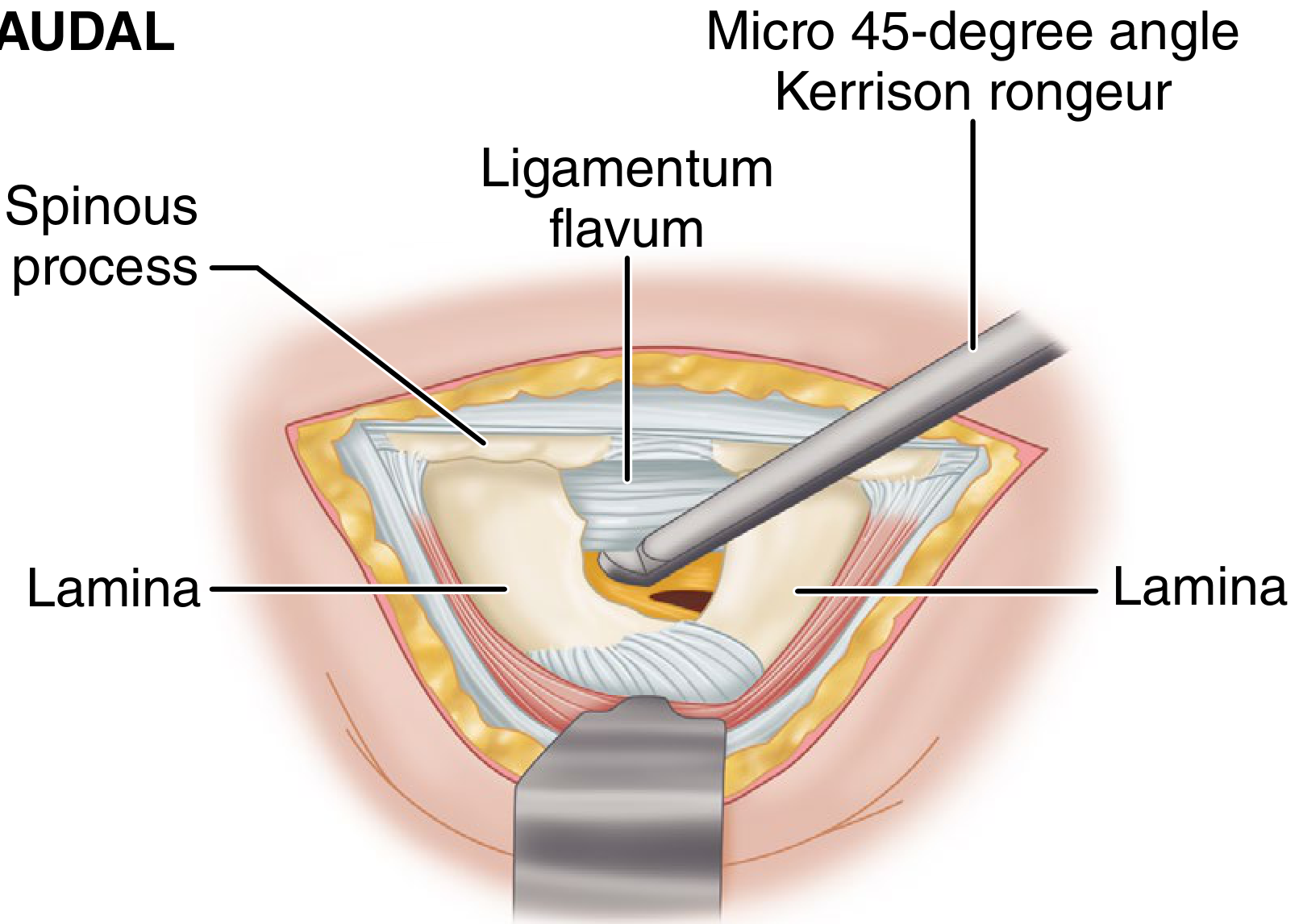
Fig. 46.12B - The Kerrison rongeur bites through the ligamentum flavum to widen the entry window. In endoscopic surgery, an endoscopic punch is used instead to fenestrate the ligamentum flavum through the working cannula.
4. Nerve Root Retraction and Disc Fragment Removal
- Left panel: Penfield dissector entering the herniated nucleus pulposus (HNP), with the nerve root, suction retractor, lamina, and facet labeled
- Right panel: Disc forceps removing disc material with the nerve root retracted, showing the disc, vertebra, and adjacent nerve structures
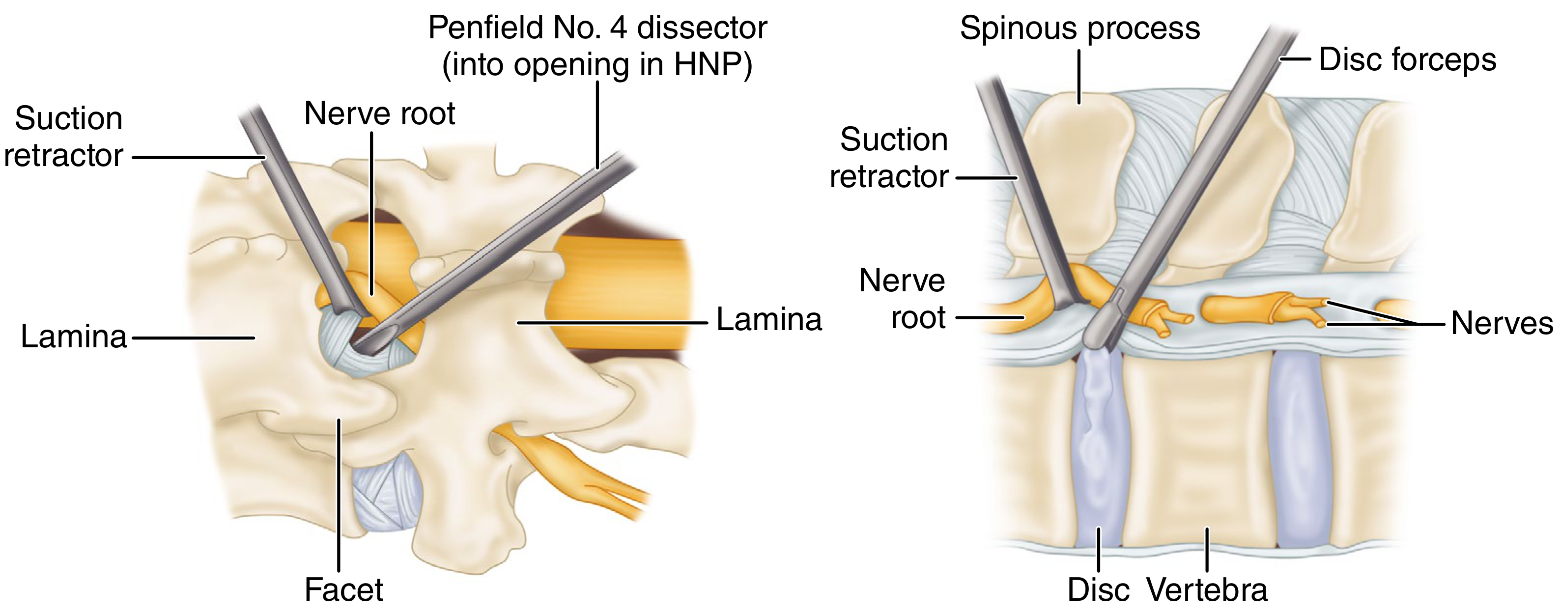
Fig. 46.13 - Dilation of the annular defect to facilitate disc fragment removal. HNP = Herniated Nucleus Pulposus. In the endoscopic technique, these same maneuvers are performed through the working channel under fluid-coupled direct visualization, with dedicated endoscopic instruments (graspers, probes) replacing open instruments.
Key Endoscopic Landmarks to Identify Intraoperatively
| Structure | Significance |
|---|---|
| Wagner arch (inferior pedicle + superior articular process) | Bony landmark confirming correct foraminal position |
| Herniated disc fragment | Target for removal |
| Traversing nerve root | Protected; must be visible and free after decompression |
| Dura | Should pulsate with heartbeat when decompression is complete |
| Epidural fat | Normal filler tissue; cleared to expose nerve root |
| Annular defect | Opening through which nucleus pulposus has herniated |
Endoscopic view along with normal diagram
Medical educational diagram showing two panels side by side. LEFT PANEL labeled "Normal Lumbar Disc Anatomy (Transverse Cross-Section)" showing a clean anatomical cross-section of a lumbar vertebra with labeled structures: vertebral body, intervertebral disc (nucleus pulposus and annulus fibrosus), spinal canal, dural sac, nerve roots (L4, L5), ligamentum flavum, lamina, facet joint (superior articular process), pedicle, epidural fat, Kambin's triangle (highlighted in green with label). RIGHT PANEL labeled "Endoscopic View Through Working Cannula (Transforaminal Approach)" showing a circular scope field-of-view as seen during endoscopic discectomy, with labeled structures visible: Wagner arch (inferior pedicle + superior articular process forming a bony arch at top), herniated disc fragment (white/yellow bulging mass), traversing nerve root (yellowish tubular structure being gently retracted), epidural fat (white fluffy tissue), annular defect (torn annulus), endoscopic instruments (grasper tip entering from side). Clean clinical illustration style, white background, clear annotation lines, professional medical textbook quality.
Endoscopic Discectomy: Endoscopic View + Normal Anatomy
Generated Diagram: Normal Anatomy vs. Endoscopic Field of View
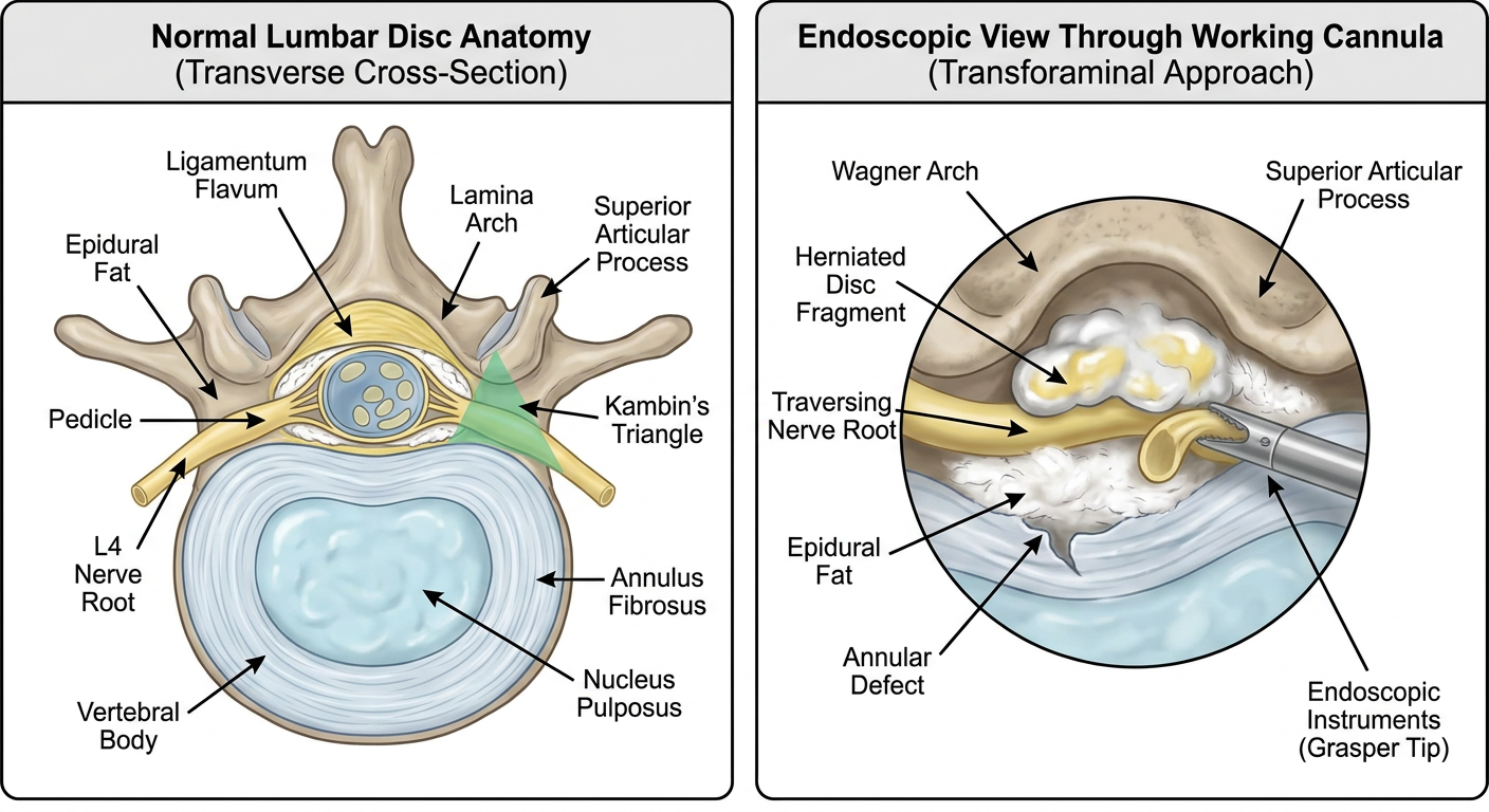
From Campbell's Operative Orthopaedics (15th Ed 2026)
What the Surgeon Sees Through the Endoscope - Key Landmarks
| Step | Structure Identified | Significance |
|---|---|---|
| 1 | Epidural fat | First soft tissue seen after entering the canal |
| 2 | Wagner arch (inferior pedicle + SAP) | Bony roof confirming correct foraminal position - transforaminal approach |
| 3 | Herniated disc fragment | White/yellow firm material under the nerve root |
| 4 | Traversing nerve root | Orange/yellowish tubular structure - must be protected |
| 5 | Annular defect | Torn posterior annulus through which disc herniated |
| 6 | Dura mater | Bluish-white pulsating sac - do not violate |
| 7 | Decompressed nerve root | At endpoint - pulsates freely to heartbeat, no deformity |
Decompression endpoint (Technique 46.5): The nerve root pulsates to heartbeat without deformity AND the awake patient reports resolution of their typical radicular pain.