
Eczema
Reading File
Now let me search the medical library and PubMed in parallel for comprehensive eczema coverage.
Finding Sources
Searching PubMed
"atopic dermatitis"[MeSH Terms] AND treatment
Now let me read the most authoritative dermatology sources in parallel - Fitzpatrick's and Andrews'.
Reading File
Reading File
Searching PubMed
"atopic dermatitis" AND "dupilumab OR biologics"
Good. Now let me read more of Andrews' on pathogenesis/clinical features/treatment, plus Fitzpatrick's clinical section.
Reading File
Reading File
Good. Let me now read the clinical features and treatment sections from Andrews' (the most comprehensive source here).
Reading File
Reading File
Reading File
Reading File
Excellent - I have very rich content. I now have enough to compose a thorough, well-organized answer. Let me also fetch the clinical images to include.
Loading Image
Loading Image
I now have comprehensive information to write a thorough, well-organized response. Here is the full overview:
Atopic Dermatitis (Eczema)
Atopic dermatitis (AD) is a chronic, inflammatory skin disease characterized by intense pruritus and a relapsing-remitting course. It is the most common inflammatory skin condition worldwide, and is the first manifestation of the "atopic march" - a progression through which skin sensitization may precede asthma, allergic rhinoconjunctivitis, and food allergies.
Epidemiology
AD prevalence is approximately 30% in the most developed nations and exceeds 10% in many countries, giving a worldwide cumulative prevalence of around 20%. Rates plateaued in developed nations in the 1990s but continue to rise in developing countries. Key risk factors include:
- High latitude and low annual sun exposure
- Western diet
- First-born child, cesarean section delivery
- Prenatal/early postnatal antibiotic exposure
- Urban, indoor ("clean") environments - the hygiene hypothesis
About 50% of cases appear in the first year of life (but rarely before 2 months), the vast majority within the first 5 years. Only 6-14% of patients are first diagnosed after age 18.
- Andrews' Diseases of the Skin, p. 83
Pathogenesis
AD results from a convergence of barrier defects and immune dysregulation:
1. Skin Barrier Defect
The filaggrin gene (FLG) on chromosome 1q21 encodes a protein essential for normal stratum corneum function. Null mutations in FLG:
- Reduce "natural moisturizing factor" (NMF), causing xerosis
- Increase transepidermal water loss (TEWL)
- Impair ceramide-rich lipid bilayers that retain water
Between 42-79% of persons with one or more FLG null mutations will develop AD. However, 40% of carriers never develop AD, confirming multifactorial causation. Ichthyosis vulgaris is caused by the same FLG mutations and is frequently associated with AD.
2. Th2 Immune Dysregulation
AD patients demonstrate a prominent Th2 phenotype:
- Elevated IgE and eosinophilia
- Key cytokines: IL-4 and IL-13 drive inflammatory cell recruitment and perpetuate the inflammatory loop
- IL-31 (produced by Th2 and Th22 cells) binds directly to nerve fibers, causing itch, and also downregulates filaggrin expression
- Thymic stromal lymphopoietin (TSLP), produced by keratinocytes, drives Th2 CD4+ T cell development
- The JAK-STAT pathway is critical in overactivation of the Th2 response and itch signaling
Th17 cells also contribute - IL-17 and IL-22 are elevated in AD lesions. Chemokines CCR4/CCR10 and their ligands TARC/CCL17 recruit T cells to atopic skin, and serum TARC levels correlate with disease severity.
- Andrews' Diseases of the Skin, p. 83-84; Fitzpatrick's Dermatology, p. 212
Clinical Manifestations
AD is classically divided into three age-based stages, with pruritus as the universal hallmark - often described as "the itch that rashes":
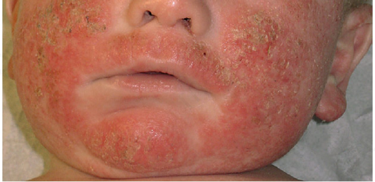
Infantile AD (2 months - 2 years)
- Begins on the cheeks, scalp, and forehead
- Weeping, crusted, erythematous plaques
- Often spares the diaper area (covered/protected skin)
Childhood AD (2-10 years)
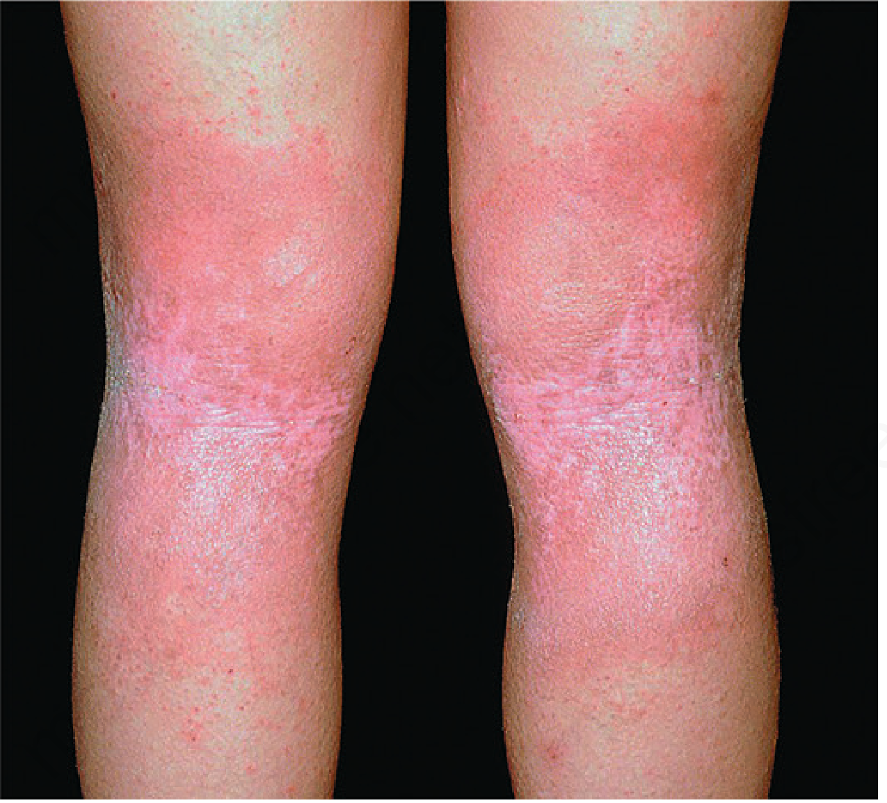
- Lesions shift to flexural surfaces: antecubital and popliteal fossae, flexor wrists, ankles, eyelids, neck
- Less exudative; more lichenified (thickened, indurated plaques)
- Isolated, excoriated 2-4 mm papules scattered on uncovered skin
- The "itch-scratch cycle" becomes established: scratching induces lichenification and secondary infection, which perpetuates further itch
Adolescent/Adult AD
- Involves antecubital and popliteal fossae, face, neck, upper chest, dorsal hands/feet
- Lesions tend to be dry, scaly, lichenified, or prurigo-like papules
- Severe, widespread involvement can cause growth retardation in children and significant psychological comorbidity
- Andrews' Diseases of the Skin, p. 85-86
Diagnostic Criteria (Hannifin & Rajka)
Major criteria - must have 3 of 4:
- Pruritus
- Typical morphology and distribution (flexural lichenification in adults; facial/extensor in infants)
- Chronic or chronically relapsing dermatitis
- Personal or family history of atopic disease (asthma, allergic rhinitis, AD)
Minor criteria - must also have 3+:
- Xerosis, ichthyosis, hyperlinear palms, keratosis pilaris
- IgE reactivity (positive RAST/prick test)
- Early age of onset
- Tendency toward skin infections (especially Staphylococcus and HSV)
- Nipple eczema, cheilitis, recurrent conjunctivitis
- Dennie-Morgan infraorbital fold
- Anterior subcapsular cataracts, keratoconus
- Orbital darkening, facial pallor/erythema
- Pityriasis alba
- Itch when sweating; intolerance to wool
- White dermatographism or delayed blanch
- Food hypersensitivity
- Andrews' Diseases of the Skin, p. 85 (Box 5.1)
Complications
| Complication | Notes |
|---|---|
| Eczema herpeticum | Disseminated HSV superinfection - a dermatologic emergency |
| Bacterial superinfection | Staphylococcus aureus colonization universally increased; may trigger flares |
| Molluscum contagiosum | Widespread spread in AD skin |
| Growth retardation | Severe widespread AD in children |
| Cataracts/keratoconus | Eye complications from chronic AD |
| Sleep disruption | Nocturnal itch severely impacts quality of life |
| Psychological | Anxiety, depression, behavioral issues especially in children |
Atopic March
Because AD often precedes other atopic conditions, it has been proposed as the first step in the atopic march: skin sensitization through defective barrier → airways sensitization (asthma, allergic rhinitis) → digestive tract sensitization (eosinophilic esophagitis). Whether AD is causally upstream remains unproven but is plausible, motivating early and effective treatment to potentially prevent the march. The underlying genetic predisposition (including FLG mutations) is shared across all atopic conditions.
Treatment
Step 1 - Skin care fundamentals
- Moisturizers/emollients: Cornerstone of management. Applied at least twice daily, immediately after bathing. Thick emollients (petrolatum, ceramide-containing creams) are preferred
- Bathing: Lukewarm water, gentle fragrance-free cleansers; pat dry
- Trigger avoidance: Wool, lipid solvents, sweating, emotional stress, relevant aeroallergens (dust mites, pet dander, tobacco smoke), and confirmed food allergens
Step 2 - Topical anti-inflammatory agents
- Topical corticosteroids (TCS): First-line anti-inflammatory. Low-potency (e.g., hydrocortisone 1%) for face/skin folds; moderate-to-high potency (triamcinolone, betamethasone) for body. Risks of prolonged use include skin atrophy, striae, HPA suppression
- Topical calcineurin inhibitors (TCIs): Tacrolimus (0.03%, 0.1%) and pimecrolimus. Steroid-sparing agents; preferred for face, eyelids, skin folds. No skin atrophy
- Topical PDE4 inhibitor: Crisaborole - approved for mild-to-moderate AD in patients ≥2 years
- Topical JAK inhibitor: Ruxolitinib cream - approved for mild-to-moderate AD in patients ≥12 years
Step 3 - Phototherapy
- Narrowband UVB (NB-UVB) is preferred; also broadband UVB, UVA1, PUVA
- Useful for moderate-to-severe AD not controlled with topicals
Step 4 - Systemic agents
- Dupilumab (anti-IL-4Rα, blocks IL-4 and IL-13 signaling): The first approved biologic for AD; approved for moderate-to-severe AD in patients as young as 6 months. Most well-supported by evidence
- Other biologics: Tralokinumab (anti-IL-13), lebrikizumab (anti-IL-13), nemolizumab (anti-IL-31Rα, specifically targets itch)
- JAK inhibitors: Upadacitinib, abrocitinib, baricitinib - oral agents approved for moderate-to-severe AD in adults/adolescents. Carry a class boxed warning (serious infections, malignancy, MACE, thrombosis)
- Traditional immunosuppressants: Cyclosporine (most rapid onset), methotrexate, azathioprine, mycophenolate mofetil - used when biologics unavailable or as bridging therapy
- Systemic corticosteroids: Short courses only for acute flares; chronic use not appropriate
- Andrews' Diseases of the Skin, p. 83-90; Fitzpatrick's Dermatology
Prevention
- Early emollient use (starting before 3 weeks of life) reduces AD development in high-risk infants by ~50% - strong evidence supporting the skin barrier hypothesis
- Probiotics during/after pregnancy show approximately 14% reduction in AD incidence
- Early peanut introduction in high-risk infants (those with severe AD or egg allergy): consider testing at 4-6 months, then controlled introduction, to reduce peanut allergy risk by ~86%
- House dust mite avoidance does NOT reduce AD incidence; switching to soy formula is not protective
- Andrews' Diseases of the Skin, p. 84
Recent Evidence
A 2024 living systematic review and network meta-analysis in JAMA Dermatology (PMID 39018058) found dupilumab and JAK inhibitors (upadacitinib, abrocitinib) to be the most effective systemic treatments for moderate-to-severe AD, with JAK inhibitors showing faster onset and higher short-term EASI response rates, but dupilumab maintaining a more favorable long-term safety profile. An earlier 2023 systematic review in JACI (PMID 37678577) confirmed dupilumab as the systemic agent with the best benefit-to-risk balance overall.
Sources: Andrews' Diseases of the Skin, 13th ed.; Fitzpatrick's Dermatology, 9th ed.; JAMA Dermatology 2024 (PMID 39018058); J Allergy Clin Immunol 2023 (PMID 37678577)
This is a shared conversation. Sign in to Orris to start your own chat.