
Create a **20-slide postgraduate-level seminar presentation** on: # MICROBIOLOGY OF DENTAL CARIES The presentation should be **scientifically accurate, visually rich, concise, and suitable for a dental seminar**. ## Overall design * Use a **professional dental/medical academic theme**. * Keep **text minimal**: maximum 4–6 concise bullet points per slide. * Prioritize **high-quality scientific images, SEM/microscopy images, clinical dental photographs, diagrams, flowcharts, illustrations and infographics**. * Aim for approximately **60–70% visual content and 30–40% text**. * Do not fill slides with paragraphs. * Use clear labels for microorganisms and biological processes. * Use italic formatting for bacterial species names. * Use consistent terminology throughout. * Include **figure captions/source citations** wherever external images are used. * Prefer images from **textbooks, peer-reviewed articles, CDC, NIH/PMC, Wikimedia Commons or other reliable academic sources**. * Avoid decorative stock images that do not contribute scientifically. ## Slide structure ### Slide 1 — Introduction to Dental Caries Cover: * Definition of dental caries * Multifactorial and biofilm-mediated nature * Role of microorganisms, diet, tooth and time * Acid production and demineralization **Images:** healthy vs carious tooth, dental plaque/biofilm, *Streptococcus mutans* SEM, demineralization/remineralization diagram. ### Slide 2 — Dental Plaque & Oral Biofilm Cover: * Acquired pellicle * Initial bacterial adhesion * Colonization and coaggregation * Biofilm maturation * Extracellular polymeric substance (EPS) **Images:** biofilm formation sequence, SEM/3D biofilm, bacteria embedded in EPS, plaque on tooth surface. ### Slide 3 — Oral Microbiota Cover: * Diversity of oral microorganisms * Major bacterial groups * Different oral niches * Microbial balance and homeostasis **Images:** oral microbiome illustration, mixed microbial SEM, different bacterial morphologies. ### Slide 4 — Microbial Theories of Dental Caries Explain: * Specific plaque hypothesis * Non-specific plaque hypothesis * Ecological plaque hypothesis * Current understanding **Images:** three-theory comparison infographic, specific vs mixed microbial community, ecological shift diagram. ### Slide 5 — Cariogenic Microorganisms Introduce major organisms: * *Streptococcus mutans* * *Streptococcus sobrinus* * *Lactobacillus* spp. * *Actinomyces* spp. * Other acidogenic/aciduric organisms **Images:** individual SEM/microscopy images of major organisms with labels. ### Slide 6 — Streptococcus mutans Cover: * Morphology and characteristics * Colonization of tooth surfaces * Acidogenicity * Aciduricity * Role in caries initiation **Images:** high-quality SEM/TEM of *S. mutans*, bacterial morphology, tooth colonization illustration. ### Slide 7 — Virulence Factors of S. mutans Cover: * Adhesion * Glucosyltransferases * Extracellular polysaccharide production * Biofilm formation * Acidogenicity and aciduricity * Intracellular polysaccharide storage **Images:** virulence-factor infographic, glucan/biofilm formation diagram, molecular illustrations. ### Slide 8 — Streptococcus sobrinus & Other Streptococci Cover: * *S. sobrinus* * Mutans streptococci * Salivary and plaque colonization * Contribution to cariogenic biofilm **Images:** comparative microscopy of *S. mutans* vs *S. sobrinus*, biofilm images. ### Slide 9 — Lactobacillus & Actinomyces Cover: * Characteristics of *Lactobacillus* spp. * Acidogenic/aciduric nature * Role in caries progression * Role of *Actinomyces* spp., particularly in root caries **Images:** microscopy of Lactobacillus and Actinomyces, comparison chart, root-caries illustration. ### Slide 10 — Biofilm Formation & Bacterial Adhesion Explain: * Acquired pellicle * Adhesins * Initial colonizers * Coaggregation * EPS production * Mature cariogenic biofilm **Images:** stepwise biofilm formation diagram, SEM/confocal images, bacterial adhesion illustration. ### Slide 11 — Carbohydrate Fermentation & Acid Production Explain: * Fermentable carbohydrates * Glycolysis * Organic acid production * Mainly lactic acid * Plaque acidification **Images:** sugar → bacterial metabolism → acid pathway, glycolysis/fermentation diagram, molecular illustration. ### Slide 12 — Stephan Curve & Critical pH Cover: * Plaque pH after sugar exposure * Rapid pH fall * Critical pH * Salivary buffering * Recovery phase **Images:** large, clear Stephan curve, pH scale, enamel surface illustration. ### Slide 13 — Demineralization & Remineralization Explain: * Acid-mediated mineral loss * Hydroxyapatite dissolution * Salivary calcium/phosphate * Fluoride-mediated remineralization * Balance between mineral loss and gain **Images:** enamel cross-section, hydroxyapatite diagram, demineralization/remineralization infographic. ### Slide 14 — Ecological Plaque Hypothesis Make this a major conceptual slide. Explain: * Frequent sugar exposure * Low plaque pH * Selection of acidogenic and aciduric organisms * Microbial dysbiosis * Transition from health to disease **Images:** healthy biofilm → environmental change → dysbiosis → cariogenic biofilm, ecological balance illustration. ### Slide 15 — Pathogenesis of Dental Caries Show the entire process: **Tooth surface → pellicle → bacterial adhesion → biofilm → sugar → acid → ↓pH → demineralization → lesion → cavitation** **Images:** large step-by-step illustrated pathway with minimal text. ### Slide 16 — Microbiology of Different Caries Types Compare: * Enamel caries * Dentin caries * Root caries * Early childhood caries **Images:** clinical photographs and cross-sectional diagrams for each type. ### Slide 17 — Role of Diet, Saliva & Fluoride Explain: * Frequency of fermentable carbohydrate intake * Salivary flow and buffering * Antimicrobial components of saliva * Fluoride and remineralization * Environmental factors affecting the biofilm **Images:** diet/sugar exposure, salivary gland/saliva illustration, fluoride-enamel interaction. ### Slide 18 — Microbiological Diagnosis & Detection Include: * Plaque sampling * Microbiological/culture methods * Molecular methods * PCR/16S rRNA sequencing * Microbiome-based approaches **Images:** plaque collection, bacterial culture, PCR workflow, sequencing/microbiome illustration. ### Slide 19 — Microbiological Prevention & Control Cover: * Mechanical plaque control * Fluoride * Dietary modification * Biofilm disruption * Antimicrobial approaches * Maintaining a healthy oral microbial ecology **Images:** toothbrush/plaque control, fluoride action, healthy biofilm, prevention infographic. ### Slide 20 — Conclusion & Key Take-Home Messages Give 5 concise take-home points: * Dental caries is a biofilm-mediated disease. * Caries involves a complex microbial community. * Acidogenicity and aciduricity are key microbial properties. * Frequent carbohydrate exposure promotes ecological dysbiosis. * Prevention aims to restore and maintain microbial and mineral balance. Include a **small integrated final diagram** connecting: **Biofilm → Sugar → Acid → Low pH → Dysbiosis → Demineralization → Caries** Also include a **References** section with the major textbooks and peer-reviewed articles used. ## Important scientific requirements * Do NOT present *Streptococcus mutans* as the sole causative organism. * Emphasize the **ecological plaque hypothesis and polymicrobial nature of dental caries**. * Distinguish between organisms involved in **caries initiation and progression**. * Clearly explain the relationship between **biofilm, carbohydrate metabolism, acid production, pH and mineral loss**. * Use current scientific terminology and avoid outdated claims. * Add **in-slide citations** in a small readable font. * Add a final reference list using **Vancouver style**. * Do not fabricate references, DOI numbers, study findings or image sources. ## Visual priority For every slide, prioritize: **Scientific images > diagrams/flowcharts > concise text** Use **multiple relevant images per slide whenever appropriate**, especially: * SEM/TEM images * Clinical dental photographs * Microbial morphology * Biofilm images * Molecular/biochemical illustrations * Pathogenesis diagrams * Comparative diagrams * Flowcharts The final presentation should look like a **high-quality postgraduate dental microbiology seminar**, not a generic AI-generated presentation.
Research the microbiology of dental caries for a postgraduate dental seminar presentation, using the medical textbook library (query_library_db per the medical-library skill protocol, read_file for full paragraph context) and pubmed_search/pubmed_detail for recent (last 2 years) systematic reviews/meta-analyses on the ecological plaque hypothesis and cariogenic microbiome. Produce a structured research dossier covering these 20 topics (matching seminar slides), each with 4-6 concise, scientifically accurate bullet-point facts suitable for slide text, PLUS a textbook citation (Book Title, p. XXX or section) for each topic where available, PLUS any relevant PMIDs from recent literature that support or update the concept: 1. Introduction to Dental Caries (definition, multifactorial biofilm-mediated disease, host-diet-microbiota-time, acid production/demineralization) 2. Dental Plaque & Oral Biofilm (acquired pellicle, initial adhesion, coaggregation, biofilm maturation, EPS) 3. Oral Microbiota (diversity, major bacterial genera, oral niches, homeostasis) 4. Microbial Theories of Dental Caries (specific plaque hypothesis, non-specific plaque hypothesis, ecological plaque hypothesis, current consensus) 5. Cariogenic Microorganisms overview (S. mutans, S. sobrinus, Lactobacillus spp., Actinomyces spp., other acidogenic/aciduric organisms - Bifidobacterium, Veillonella, Scardovia wiggsiae etc.) 6. Streptococcus mutans (morphology, colonization, acidogenicity, aciduricity, role in initiation) 7. Virulence factors of S. mutans (adhesins, glucosyltransferases GtfB/C/D, EPS/glucan production, biofilm formation, acidogenicity/aciduricity, intracellular polysaccharide storage) 8. Streptococcus sobrinus & other mutans streptococci 9. Lactobacillus & Actinomyces (role in caries progression and root caries specifically) 10. Biofilm formation & bacterial adhesion (stepwise mechanism: pellicle, adhesins, early colonizers e.g. Streptococcus sanguinis/gordonii, coaggregation, EPS, mature biofilm) 11. Carbohydrate fermentation & acid production (fermentable carbohydrates, glycolysis, lactic acid, plaque acidification) 12. Stephan curve & critical pH (classic Stephan 1944 curve, critical pH ~5.5 for enamel, salivary buffering, recovery phase) 13. Demineralization & Remineralization (hydroxyapatite dissolution, salivary calcium/phosphate, fluoride mediated remineralization -fluorapatite) 14. Ecological Plaque Hypothesis in depth (frequent sugar exposure, low pH selection pressure, dysbiosis, health-disease transition) - this is a key conceptual slide, get strong textbook/journal support 15. Pathogenesis of dental caries stepwise pathway (tooth surface -> pellicle -> adhesion -> biofilm -> sugar -> acid -> pH drop -> demineralization -> lesion -> cavitation) 16. Microbiology of different caries types (enamel caries, dentin caries, root caries, early childhood caries - microbial differences) 17. Role of diet, saliva & fluoride (carbohydrate frequency, salivary flow/buffering, antimicrobial saliva components e.g. lysozyme/lactoferrin/sIgA, fluoride remineralization) 18. Microbiological diagnosis & detection (plaque sampling, culture methods e.g. mitis salivarius agar, molecular methods, PCR, 16S rRNA sequencing, microbiome approaches) 19. Microbiological prevention & control (mechanical plaque control, fluoride, dietary modification, biofilm disruption, antimicrobials e.g. chlorhexidine, probiotics, maintaining healthy oral microbial ecology) 20. Conclusion key take-home messages + Vancouver-style reference list (5-8 major references: textbooks used from the library plus 2-3 real peer-reviewed articles found via pubmed with correct PMID/citation - do NOT fabricate any reference) IMPORTANT: Do not fabricate page numbers, DOIs, or study findings. If a specific textbook page isn't found, cite the book title and section heading only. If pubmed search returns nothing relevant for a topic, state that clearly rather than inventing a citation. Return your final answer as a clearly organized dossier (topic 1-20, each with bullets + citation), followed by a final Vancouver-style reference list of all sources actually used (textbooks + PubMed articles with real PMIDs).
Streptococcus mutans scanning electron micrograph
dental plaque biofilm formation on tooth surface
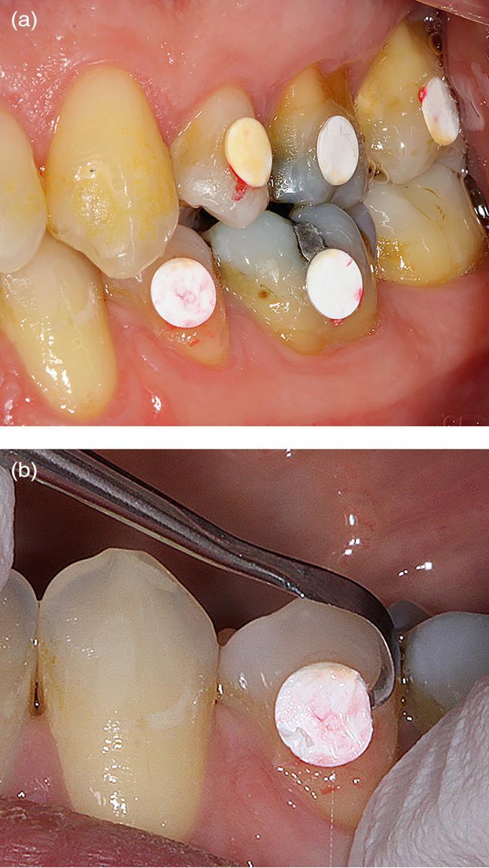
Two-panel clinical intraoral photograph demonstrating an experimental dental study on plaque accumulation. Panel (a) shows the buccal surfaces of maxillary and mandibular teeth (premolars and molars) with several white, circular expanded polytetrafluoroethylene (e-PTFE) membrane discs bonded to the enamel. The teeth exhibit natural yellowish staining, dark amalgam restorations, and mild gingival inflammation near the cervical margins. Panel (b) provides a close-up view of the mechanical retrieval process. A curved metal dental instrument, such as a scaler or probe, is positioned to carefully detach a single membrane disc from the buccal surface of a mandibular tooth. The retrieved membrane shows a slight reddish-pink discoloration, representing 4 hours of early biofilm formation and potentially accumulated oral fluids. The image illustrates a methodology for in vivo biofilm sampling and microbiological analysis in periodontology and dental research.
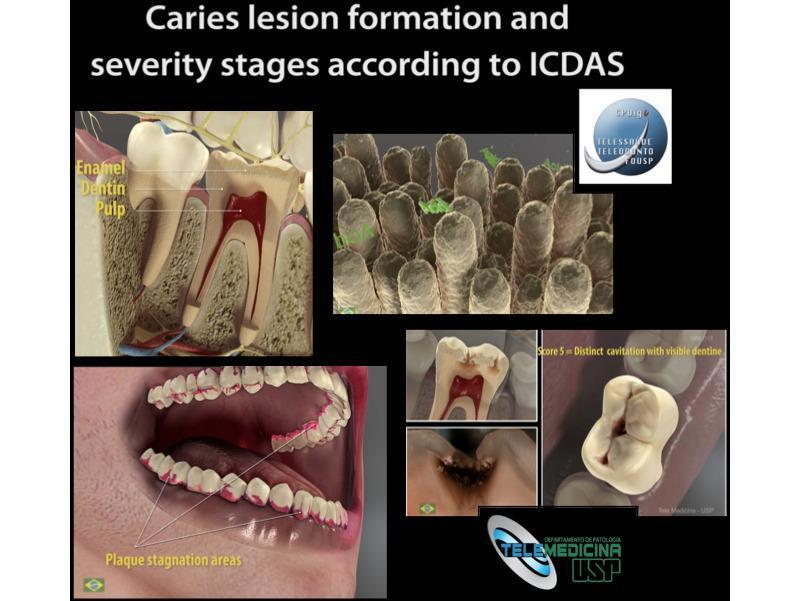
Educational infographic and 3D animation stills from the Virtual Man Project regarding caries lesion formation and severity stages according to the International Caries Detection and Assessment System (ICDAS). The visual contains four main sections: (1) A sagittal cross-section of a human tooth labeling primary anatomical layers including the enamel (outer white layer), dentin (middle beige-brown layer), and pulp chamber (central red vascular tissue). (2) A microscopic view representing dental biofilm or enamel rods. (3) A clinical 3D model of a human mouth with the mandible and maxilla, highlighting 'plaque stagnation areas' in red along the cervical margins (gumline) and interproximal spaces. (4) A panel demonstrating clinical progression of caries, including a view of 'Score 5: Distinct cavitation with visible dentine' on an occlusal surface. This resource is designed for dental education, illustrating the pathophysiology of demineralization, biofilm accumulation, and the standardized classification of dental decay severity.
dental caries carious tooth cavity clinical photograph
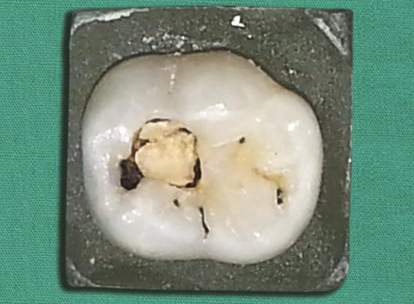
This clinical photograph displays a human primary molar tooth isolated in a dark mounting block for dental study. The tooth exhibits a significant carious lesion on the occlusal surface. The primary pathology is a large, central cavitation containing yellowish-brown carious dentin, which appears soft and necrotic. Dark staining and demineralization are visible along the margins of the cavity, indicating progressive dental decay. Additionally, secondary features include smaller dark pits and fissures with incipient caries or staining. The tooth crown displays multi-cusp morphology characteristic of a posterior tooth. This image serves as a visual reference for dental caries classification, demonstrating advanced enamel and dentin destruction in primary dentition, likely used for teaching diagnostic criteria in pediatric dentistry or restorative treatment planning.
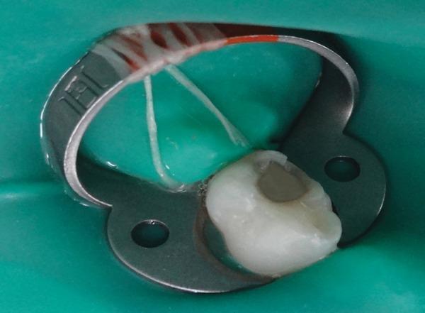
This clinical photograph demonstrates an intraoperative view of a molar undergoing dental treatment. The tooth is isolated using a green rubber dam system to maintain a dry, sterile field and prevent contamination during the procedure. A stainless steel rubber dam clamp (endodontic clamp) is secured around the cervical region of the tooth to stabilize the dam. The occlusal surface of the molar shows an extensive, prepared cavity involving the dentin, characteristic of dental caries removal. The internal walls of the cavity appear irregular and off-white, indicating the excavation of softened carious tissue. This procedural image illustrates a standard operative dentistry protocol for caries management, ensuring aseptic conditions before microbiological sampling or restoration with materials such as glass ionomer cement. The target audience includes dental students and practitioners focusing on restorative dentistry and endodontic isolation techniques.
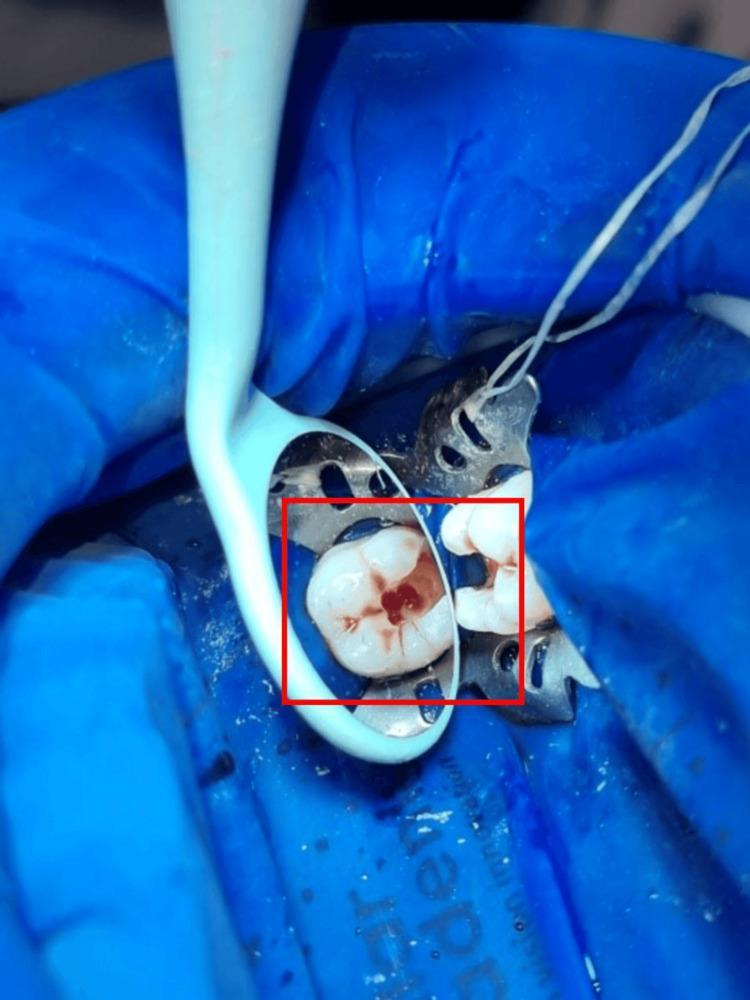
This clinical photograph displays a procedural step in restorative dentistry or endodontics, focusing on a lower left first molar following carious dentin removal and access cavity preparation. The tooth is isolated using a blue rubber dam held by a metallic dental dam clamp. The occlusal surface of the molar shows a prepared cavity with distinct reddish-brown discoloration within the dentin structure, typical of chronic carious lesions or pulpal involvement, while the surrounding enamel remains intact and whitish. A dental mirror is positioned to provide an indirect view of the distal and occlusal aspects of the preparation. Adjacent to the tooth, a metallic matrix band is visible, indicating preparation for a proximal restoration. This image serves as an educational tool for demonstrating pulp chamber de-roofing, caries excavation, and the necessity of proper field isolation using a rubber dam for aseptic dental procedures.
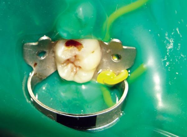
This intraoral clinical photograph shows a preoperative view of a posterior tooth, likely a molar, undergoing dental caries management. The operative field is isolated using a green rubber dam and a metal rubber dam clamp for moisture control and patient safety. The tooth demonstrates a significant carious lesion on the occlusal surface, characterized by a large cavity with dark brown and reddish-brown discolored dentin, indicative of active dental decay. The surrounding enamel appears irregular and thinned. A metal Tofflemire-style matrix band and retainer are positioned around the tooth, stabilized with a yellow interproximal wedge, to establish proper proximal contours during the restorative process. This image serves as an educational example of clinical cariology, isolation techniques, and the preparatory stages for operative dentistry using conventional or chemomechanical caries removal methods.
tooth enamel demineralization remineralization hydroxyapatite diagram
Lactobacillus bacteria microscopy
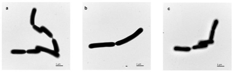
This diagnostic image consists of three Transmission Electron Microscopy (TEM) frames (a, b, and c) showing the morphology of Lactobacillus casei FJSWX3-L3 bacteria at a 1 m scale. The bacteria are depicted as rod-shaped (bacilli) cells arranged in short chains and clusters. Frame (a) shows cells cultured with arabinose, and frame (b) shows cells cultured with EDTA; both conditions demonstrate relatively smooth, well-defined cell boundaries with minimal visible surface accumulation. In contrast, frame (c) displays cells cultured in standard MRS medium, where the bacterial surfaces are surrounded by a prominent, irregular layer of flocculent material, identified as exopolysaccharides or surface substances. This comparison illustrates how different fermentation conditions and chelating agents like EDTA can modulate the production of the bacterial capsule or surface layer, which has significant clinical and biotechnological implications for bacterial stress resistance and lyophilization (freeze-drying) survival rates.
This diagnostic image is a Scanning Electron Microscopy (SEM) micrograph at 1000x magnification illustrating the microstructure of corn starch granules, relevant to biomedical nutrition and prebiotic research. The image displays the diverse morphology of starch granules, characterized by a mixture of round, polygonal, and dome-shaped structures. The surface textures vary from smooth to rough, with visible evidence of irregular and broken granule forms. Such visuals are used in medical laboratory science to study modified corn flour (MCF) and its impact on nutritional components like amylose, amylopectin, and resistant starch (RS). Understanding these structural changes is clinically significant for evaluating the efficacy of prebiotics in stimulating the viability of probiotic bacteria such as Lactobacillus plantarum in the human digestive system. The image serves as a comparison point for various modification techniques including acid hydrolysis, annealing, autoclaving-cooling cycles, and enzymatic debranching.
This scientific illustration and diagnostic image combination explores the influence of gut microbiota on human physiology. The left panel shows a fluorescent microscopy image of the gut mucosa overlaid with a biochemical pathway. Key beneficial bacteria—including Lactobacillus, Bifidobacterium, Akkermansia muciniphila, and Faecalibacterium prausnitzii—are shown producing Short-Chain Fatty Acids (SCFAs). These SCFAs modulate gene transcription via histone deacetylase inhibition and activate G-protein coupled receptors GPR41/43 on enteroendocrine L-cells, triggering the release of GLP-1 and GLP-2. Additionally, genera like Barnesiella and Clostridium are linked to reduced antibiotic resistance. The right panel features a systemic diagram of a human figure, illustrating the 'Gut–Brain–Skin axis.' It maps gut-derived influences to host metabolism (energy balance), behavior and psychopathology (mood and cognition), skin barrier integrity, and the modulation of the immune response. The diagram highlights how probiotic interventions can reinforce the gut barrier and decrease systemic antibiotic resistance, representing current concepts in microbiome-based therapeutics and metabolic health.
Actinomyces root caries
oral microbiome bacterial diversity diagram
early childhood caries clinical photograph
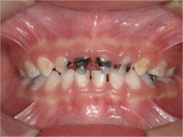
This clinical photograph displays a frontal intraoral view of the primary dentition in a pediatric patient, illustrating severe Early Childhood Caries (ECC). The maxillary anterior teeth are most severely affected, showing extensive cavitation, structural loss, and dark brown to black necrotic decay, particularly involving the central and lateral incisors. The crowns of several teeth appear eroded and irregular in shape. Yellowish and white-spot demineralization is visible on adjacent teeth, indicating varying stages of the caries process. The mandibular teeth appear less involved but show some early lesions. The surrounding gingival tissue is generally pink with relatively healthy margins and no overt signs of acute inflammation or abscess formation visible in this frame. This image serves as a classic educational example of the progression of severe early childhood caries (formerly known as baby bottle tooth decay) and its characteristic distribution patterns in the primary dentition.
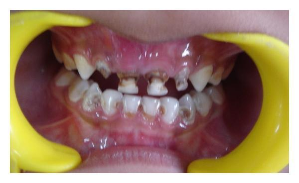
An intraoral clinical photograph of a pediatric patient (4 years old) demonstrating severe, multi-surface dental caries characteristic of Early Childhood Caries (ECC). The maxillary anterior deciduous teeth, including the central and lateral incisors (teeth 51, 52, 61, 62), show advanced destruction of the crown structure with significant cavitation and deep dark brown to black necrotic dentin. The mandibular arch also exhibits widespread caries, primarily affecting the labial and occlusal surfaces of the deciduous incisors, canines, and molars, with visible yellowish demineralization and focal brown lesions at the cervical margins. The maxillary gingiva displays signs of moderate inflammation and erythema. This image serves as a clinical example of nursing bottle syndrome or aggressive childhood caries requiring comprehensive restorative and endodontic intervention.
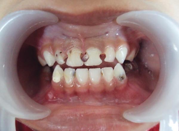
This clinical photograph shows a preoperative intraoral view of a pediatric patient (5-year-old male) using plastic cheek retractors to expose the primary dentition. The image demonstrates severe Early Childhood Caries (ECC). The upper central and lateral incisors (teeth 51, 52, 61, 62) exhibit extensive structural destruction, with significant loss of enamel and dentin on the mesial and distal proximal surfaces, appearing as deep, darkened carious lesions. Dark brown to black cavitation is visible, particularly between the central incisors. The lower anterior dentition displays relatively better alignment, though localized areas of discoloration and incipient carious lesions are present on the mandibular incisors and canines. The gingival tissues appear pink and generally healthy without significant recession, although localized inflammatory changes are visible near the severely decayed maxillary teeth. This image illustrates a typical clinical presentation of bottle-induced or early childhood caries requiring comprehensive restorative or endodontic intervention in a pediatric dentistry context.
dental biofilm extracellular polymeric substance confocal microscopy
acquired enamel pellicle bacterial adhesion coaggregation
"ecological plaque hypothesis" dental caries
cariogenic microbiome dysbiosis
"Streptococcus mutans" virulence factors glucosyltransferase review
root caries microbiology older adults review
probiotics dental caries prevention
Scardovia wiggsiae Bifidobacterium severe early childhood caries
oral microbiome 16S rRNA sequencing caries detection
"Stephan" in /textbooks
"critical pH" in /textbooks
"aciduric" in /textbooks
"acquired pellicle" in /textbooks
PMID: 40933845
PMID: 40499711
PMID: 39154338
PMID: 41409473
PMID: 38451156
PMID: 37275173
PMID: 35389204
Marsh ecological plaque hypothesis dental biofilm dysbiosis
fluoride remineralization enamel caries
PMID: 39362658
PMID: 15153690
give up to now what u prepeared
can u give now