


25-Year-old young patient presents to the OPD with complaints of fever, coughing, and, uh, lower respiratory tract infection symptoms. Uh, he has a history of smoking, uh... Uh, these are the reports. This is the CBC of the patient. And the X-ray findings. Current clinical suspicion is of, uh, pneumonia. Differentially diagnose, uh, clinically stage. Use the treatment algorithm and management algorithm. And plan treatment. Including medication.
"community-acquired pneumonia"[MeSH Terms] AND "drug therapy"[MeSH Terms]
pneumonia chest X-ray consolidation infiltrate
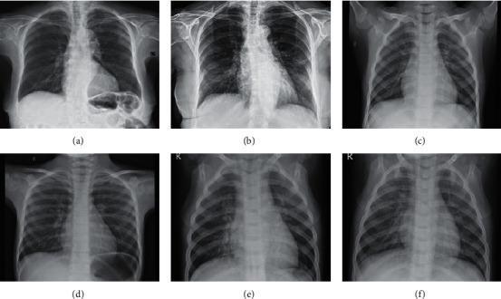
A comparative diagnostic display containing six posteroanterior (PA) chest X-ray radiographs labeled (a) through (f), categorized by pathological findings. Images (a) and (b) represent COVID-19 pneumonia, demonstrating bilateral ground-glass opacities and increased pulmonary density with ill-defined margins, characteristic of viral interstitial infiltrate. Images (c) and (d) represent normal controls, showing clear lung fields, well-defined vascular markings, and sharp costophrenic angles without evidence of consolidation or effusion. Images (e) and (f) depict other types of non-COVID pneumonia, showing varying degrees of focal consolidation and bronchovascular thickening. This comparison serves as a clinical imaging reference for differentiating viral pneumonia patterns from normal thoracic anatomy and other inflammatory lung conditions. The anatomical structures visible include the lungs, heart, rib cage, clavicles, and diaphragm, providing a comprehensive view of the thoracic cavity for radiological training and diagnostic classification.
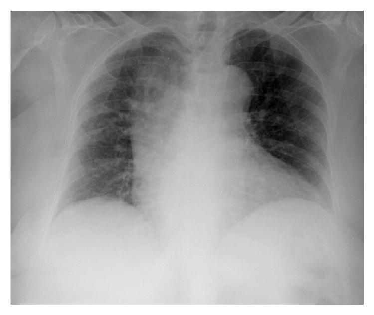
This diagnostic image is a posterior-anterior (PA) view chest X-ray demonstrating characteristic findings of pulmonary infection. The primary pathology is a focal area of increased opacity located in the right upper lobe, consistent with an alveolar infiltrate or lobar consolidation. The opacity is dense and somewhat irregular in shape, partially obscuring the superior right mediastinal border. Additionally, there is a milder, less defined area of increased opacity in the left lower lung field, suggestive of a basal infiltrate. The heart shadow is visible, though its right border is obscured by the adjacent consolidation (positive silhouette sign). Bony structures, including the ribs, clavicles, and vertebral column, are intact. The diaphragmatic domes are visualized, although the right hemidiaphragm is less sharply defined due to overlying pulmonary density. This image is an educational example of bacterial pneumonia presentation, specifically illustrating lobar consolidation patterns in the context of Moraxella catarrhalis infection.
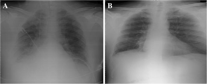
This comparison chart features two chest X-rays (CXR) illustrating the clinical progression and resolution of pulmonary pathology. Image A is a supine anteroposterior (AP) chest X-ray obtained in an intensive care unit (ICU) setting. It demonstrates a prominent right-sided basal pulmonary infiltrate, characterized by increased opacity and haziness in the lower lung field, partially obscuring the diaphragm and pulmonary vasculature. Medical devices, including a central venous catheter or monitoring line, are visible traversing the left hemithorax, reflecting the patient's acute clinical status. Image B is a posteroanterior (PA) chest X-ray taken prior to discharge. It shows a complete resolution of the previously seen basal infiltrate. The lung fields appear radiolucent and clear, with well-defined costophrenic angles and visible pulmonary markings. This side-by-side comparison is intended for medical education to demonstrate radiographic signs of pulmonary consolidation (such as those associated with pulmonary embolism or pneumonia) and subsequent improvement following therapeutic intervention.
Clinical Assessment: 25-Year-Old Male, Fever + Cough + LRTI
STEP 1 - DATA SYNTHESIS
CBC Findings (08/08/2026)
| Parameter | Result | Flag | Normal Range | Interpretation |
|---|---|---|---|---|
| WBC | 5.8 × 10³/µL | Normal | 4.0-12.0 | Within normal limits |
| NEU% | 62.0% | Normal | 50-80% | Normal |
| LYM% | 21.1% | Low | 25-50% | Lymphopenia |
| MON% | 14.8% | H | 2-10% | Monocytosis |
| EOS% | 2.0% | Normal | 0-5% | Normal |
| RBC | 5.71 × 10⁶/µL | Normal | 4.0-6.2 | Normal |
| HGB | 14.1 g/dL | Normal | 11-17 | Normal |
| MCH | 24.7 pg | L | 26-34 | Microcytic |
| MCV | ~72 fL | Low | 80-100 | Microcytosis |
| PLT | 268 × 10³/µL | Normal | 150-400 | Normal |
Key CBC Interpretation
- WBC is 5.8 - notably NOT elevated, which is a critical finding. Classical bacterial pneumonia typically causes leukocytosis (>15,000). A normal WBC with lymphopenia + monocytosis in a symptomatic patient strongly points toward atypical/viral etiology or an early bacterial pneumonia not yet producing a left shift.
- Lymphopenia - seen in viral infections (influenza, COVID-19, other respiratory viruses), intracellular pathogens (Mycoplasma, Chlamydophila), and early stages of serious bacterial infections.
- Monocytosis (14.8%) - reactive; seen in subacute infections, Mycoplasma, viral pneumonias, and TB. A very important clue here.
- Microcytosis with low MCH - likely pre-existing iron deficiency anemia or thalassemia trait (unrelated to acute illness but warrants follow-up).
- Erythroblasts on morphology - may indicate stress erythropoiesis.
Chest X-Ray Findings (10/08/2026)
- No focal consolidation or lobar opacification - absence of the classical dense white wedge-shaped opacity of typical bacterial/lobar pneumonia
- No pleural effusion - costophrenic angles are sharp
- No pneumothorax
- Increased bronchovascular markings bilaterally - consistent with interstitial/bronchopneumonia pattern
- Normal cardiac silhouette - no cardiomegaly
- Normal diaphragmatic domes - no hyperinflation (argues against COPD/obstructive pattern despite smoking history)
STEP 2 - DIFFERENTIAL DIAGNOSIS
| Rank | Diagnosis | Supporting Features | Against |
|---|---|---|---|
| 1. Atypical (Mycoplasma) Pneumonia | Age 25, insidious onset, no lobar consolidation, lymphopenia, monocytosis, normal WBC | - | |
| 2. Viral Pneumonia (Influenza A/B, COVID-19, RSV) | Normal/low WBC, lymphopenia, bilateral interstitial pattern, smoker (increased susceptibility) | No specific seasonal mention | |
| 3. Chlamydophila pneumoniae CAP | Young adult, mild presentation, normal WBC, atypical pattern | Requires serology/PCR to confirm | |
| 4. Early Typical Bacterial CAP (S. pneumoniae) | Fever, productive cough, LRTI | No lobar consolidation, WBC not elevated, no neutrophilia | |
| 5. Pulmonary Tuberculosis | Young male, monocytosis, subacute course, India/endemic setting | No upper lobe infiltrate visible, no cavitation | |
| 6. Acute Bronchitis (± secondary pneumonia) | Smoker with LRTI, normal CXR, viral etiology | Clinical overlap with LRTI symptoms | |
| 7. Legionella pneumophila | LRTI + low WBC possible | Usually more severe systemic symptoms, hyponatremia, older age |
STEP 3 - CLINICAL SEVERITY STAGING
CURB-65 Score (Harrison's Principles of Internal Medicine 22e, p.1069)
| Criterion | Patient | Score |
|---|---|---|
| C - Confusion (new onset) | Not documented - assume absent | 0 |
| U - Urea > 7 mmol/L | Not available | 0 (assume normal) |
| R - Respiratory Rate ≥ 30/min | Not documented | 0 (assume <30) |
| B - BP systolic ≤90 or diastolic ≤60 | Not documented | 0 (assume normal) |
| 65 - Age ≥ 65 years | 25 years old | 0 |
PSI Class
Classification: Non-Severe CAP - Outpatient Eligible
STEP 4 - INVESTIGATIONS TO ORDER
Immediate (Outpatient)
- SpO2 pulse oximetry - if <92%, reassess for admission
- Sputum Gram stain + culture (if productive cough) - though yield is low
- Cold agglutinin test - positive in Mycoplasma pneumoniae (~50% sensitivity)
- Mycoplasma IgM serology - confirms atypical etiology
- COVID-19 PCR / Influenza rapid antigen - for viral exclusion
- CRP / ESR - baseline inflammatory markers
- Serum urea and electrolytes (for CURB-65 completion, and to rule out SIADH - which pneumonia can cause)
- AFB sputum smear x3 - given monocytosis pattern, endemic region, and age - to rule out TB
Consider if No Improvement in 48-72h
- Chest HRCT (more sensitive for subtle interstitial disease)
- Legionella + pneumococcal urinary antigen tests
- Respiratory multiplex PCR panel
STEP 5 - TREATMENT ALGORITHM & MANAGEMENT PLAN
CAP Severity-Based Treatment Algorithm
CAP confirmed (clinical + radiological)
         |
    CURB-65 Score
    /            \
  0-1              2           ≥3
 (Mild)       (Moderate)    (Severe)
Outpatient     Admit ward    ICU/HDU
Treatment Plan (Outpatient, Non-Severe CAP - Atypical Suspected)
First-Line Antibiotic Regimen
- Azithromycin 500 mg PO on Day 1, then 250 mg PO OD on Days 2-5 (Z-Pack)
- OR Clarithromycin 500 mg PO BD × 7 days
- Levofloxacin 750 mg PO OD × 5 days
- OR Moxifloxacin 400 mg PO OD × 5 days
- Amoxicillin-clavulanate 625 mg PO TDS + Azithromycin 500 mg PO OD × 5-7 days
Azithromycin 500 mg Day 1, then 250 mg Days 2-5 (5-day course) is the recommended first-line choice.
Supportive Medications
| Drug | Dose | Indication |
|---|---|---|
| Paracetamol (Acetaminophen) | 500-1000 mg PO TDS/QID | Fever, myalgia |
| Ibuprofen (if no GI contraindication) | 400 mg PO TDS with food | Anti-inflammatory, fever |
| Mucolytic - Ambroxol/Bromhexine | 30 mg PO TDS | Productive cough |
| Salbutamol MDI (if wheeze/bronchospasm) | 2 puffs TDS PRN | Bronchospasm, especially given smoker status |
| Oral rehydration / adequate fluid intake | 2-3 L/day | Hydration, mucus clearance |
Smoking Cessation
Return Precautions / "Red Flag" Instructions (When to Return or Admit)
- Oxygen saturation falls below 92% (obtain home oximeter or return for check in 24h)
- Respiratory rate increases to >30/min
- Confusion or altered consciousness
- Inability to take oral medications
- No improvement after 48-72 hours of antibiotics
- Worsening fever >72h despite antibiotics (consider hospital admission and IV therapy)
Follow-Up Plan
| Timeline | Action |
|---|---|
| 48-72 hours | Clinical review - check for defervescence and symptom improvement |
| 5-7 days | Review serology results (Mycoplasma IgM, COVID/flu) |
| 4-6 weeks | Repeat CXR to confirm radiological clearance (important in smokers to rule out underlying malignancy or structural disease) |
| 3 months | AFB result review; spirometry if LRTI recurrence (to assess for early COPD in smoker) |
SUMMARY TABLE
| Domain | Finding |
|---|---|
| Working Diagnosis | Community-Acquired Pneumonia - Atypical (Mycoplasma likely) |
| CBC Key Finding | Normal WBC, Lymphopenia, Monocytosis - atypical/intracellular pathogen pattern |
| CXR | Bilateral increased bronchovascular markings, no lobar consolidation - interstitial/atypical pattern |
| CURB-65 | 0 - Low severity, outpatient eligible |
| PSI Class | Class I-II |
| Antibiotic | Azithromycin 500 mg Day 1, 250 mg Days 2-5 |
| Supportive | Paracetamol, mucolytic, hydration, salbutamol PRN |
| Special Concern | Rule out TB (monocytosis, endemic area); Smoking cessation counseling |
| Follow-up CXR | 4-6 weeks post-treatment (mandatory in smokers) |
- Harrison's Principles of Internal Medicine 22e, pp. 1068-1072 (CAP diagnosis, CURB-65, severity staging, antibiotic therapy)
- Textbook of Family Medicine 9e, pp. 321-322 (CAP clinical presentation, treatment, PSI/CURB-65)
- Kurotschka et al. (2024) - Network Meta-Analysis of oral antibiotics for CAP [PMID: 38360961]
- Ghadimi et al. (2025) - Empiric antibiotic therapy for moderate-to-severe CAP [PMID: 40602496]
Disclaimer: This is a clinical decision-support analysis intended for use by a qualified healthcare professional. All treatment decisions must be made by the treating physician based on full clinical examination, local antibiotic resistance patterns, and patient-specific factors not captured in this report.