


Find answer of questions for anatomy ospe
ulnar claw hand deformity
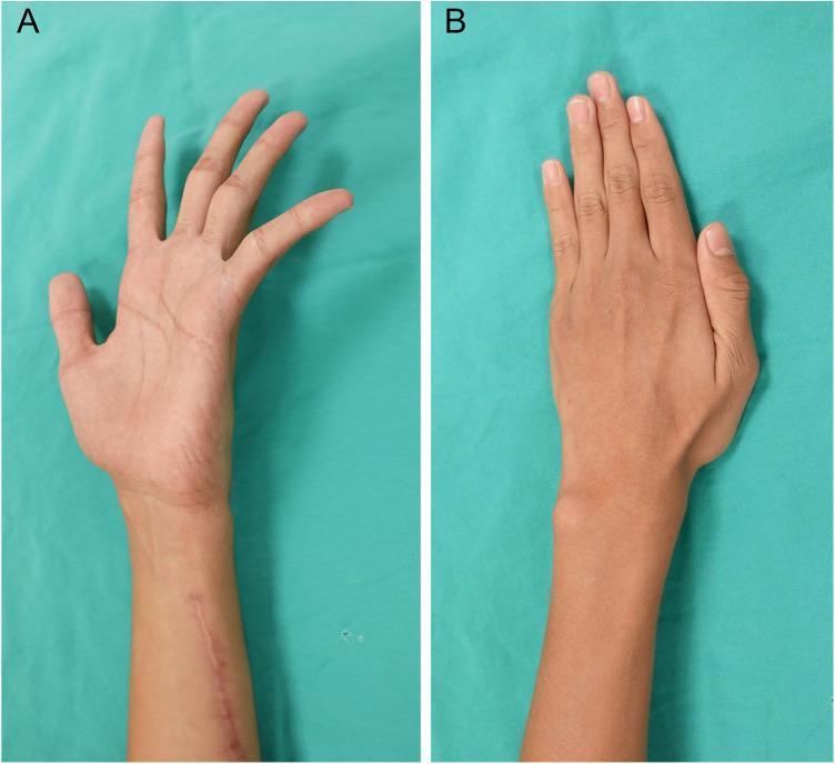
This clinical photograph shows a side-by-side comparison of a left hand and forearm following surgical treatment for ulnar neuropathy. Image A displays the hand in a volar view, highlighting a persistent ulnar claw hand deformity where the 4th and 5th fingers exhibit hyperextension at the metacarpophalangeal joints and flexion at the interphalangeal joints. A linear surgical scar is visible on the distal volar forearm. Image B displays the dorsal view of the same hand six months postoperatively, demonstrating a complete resolution of the claw deformity. The 4th and 5th fingers now maintain a neutral, extended posture aligned with the other digits. There is no visible atrophy of the intrinsic hand muscles or hypothenar eminence, indicating successful reinnervation or functional recovery. This comparison illustrates the clinical outcomes of ulnar nerve repair or transfer in a pediatric patient following traumatic injury and subsequent nerve palsy.
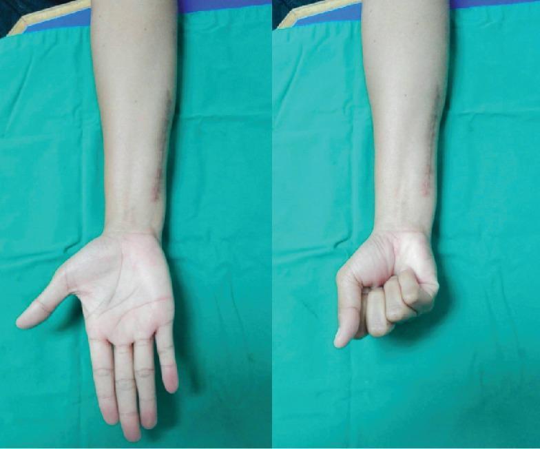
Clinical photograph of a right forearm and hand presented in two side-by-side views demonstrating post-operative recovery of motor function. The images show the volar (palmar) aspect of the extremity. On the ulnar side of the distal forearm, a well-healed, linear surgical scar is visible, corresponding to a previous ulnar shortening osteotomy site. The left panel illustrates the hand in an extended position with fingers abducted, demonstrating successful active extension and preservation of the palmar architecture. The right panel shows the hand flexed into a complete, tight fist, indicating full recovery of the flexor digitorum superficialis and profundus tendon functions. There is no evidence of residual edema, erythema, or visible deformity such as the 'claw hand' associated with ulnar nerve palsy. This clinical visual serves as an educational example of functional restoration following revision surgery and hardware replacement (titanium for stainless steel) in the context of metal hypersensitivity.
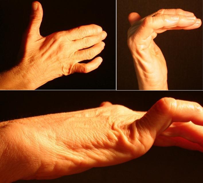
This composite of clinical photographs demonstrates a 'claw hand' (main en griffe) deformity, a characteristic sign of ulnar nerve neuropathy. The images show a right hand from dorsal, palmar, and lateral perspectives. Key visual features include hyperextension at the metacarpophalangeal (MCP) joints and flexion at the proximal and distal interphalangeal (IP) joints, most pronounced in the fourth and fifth digits. There is noticeable atrophy of the hypothenar eminence and hollowing of the interosseous spaces on the dorsal surface, indicating wasting of the intrinsic hand muscles (interossei and ulnar-innervated lumbricals). The thenar eminence appears relatively preserved, though subtle flattening of the adductor pollicis region may be present. This presentation is consistent with distal ulnar nerve compression, such as within Guyon’s canal, leading to motor deficit and muscle denervation. These clinical signs are essential for diagnosing peripheral nerve entrapment syndromes and distinguishing them from proximal lesions or central nervous system pathologies.
OSPE Answers: Ulnar Claw Hand
Q1. Identify the above clinical condition
Q2. Name the Nerves involved in the above clinical condition
- Supplies the medial two lumbricals (3rd and 4th), all interossei, hypothenar muscles, and adductor pollicis
- When injured, loss of these intrinsic muscles produces the claw deformity
If BOTH ulnar + median nerves are injured, all four fingers claw = "Full/Complete Claw Hand."
Q3. Name the Muscles involved in this clinical condition
| Muscle | Role lost |
|---|---|
| 3rd and 4th Lumbricals | Flex MCP joints, extend IP joints of ring and little fingers |
| All Interossei (dorsal + palmar) | Flex MCP, extend IP joints; abduction/adduction of fingers |
| Hypothenar muscles (Abductor digiti minimi, Flexor digiti minimi, Opponens digiti minimi) | Move little finger |
| Adductor Pollicis | Adducts thumb; its loss causes thumb hyperextension |
Q4. Write the characteristic features (deformities) of this clinical condition
- Hyperextension at Metacarpophalangeal (MCP) joints - due to unopposed action of Extensor digitorum (extrinsic extensor)
- Flexion at Proximal and Distal Interphalangeal (PIP & DIP) joints - due to unopposed action of Flexor digitorum superficialis and profundus
- Deformity is most pronounced in ring and little fingers (4th and 5th digits) - because 3rd and 4th lumbricals are ulnar-innervated
- Hollowing of interosseous spaces in the dorsum of the hand (interossei atrophy within 2-3 months)
- Hypothenar wasting (flattening of the hypothenar eminence)
- Thumb hyperextension - due to loss of adductor pollicis, with dominance of extensor pollicis longus
- Paradox of ulnar claw: Deformity is LESS severe with high (proximal) lesions because FDP to ring and little fingers is also paralysed (so IP flexion is reduced). More severe with distal (wrist) lesions = "ulnar paradox"
- Sensory loss over ulnar half of ring finger, entire little finger, and ulnar side of palm
Q5. Name any two causes responsible for this clinical condition
- Fracture/dislocation at the medial epicondyle of the humerus (most common) - the ulnar nerve is exposed in the ulnar groove at the elbow
- Cubital tunnel syndrome - chronic compression between the two heads of Flexor carpi ulnaris
- Lacerations at the wrist (Guyon's canal injury)
- Ulnar tunnel syndrome (chronic pressure, e.g., from tools/handlebar cyclist's palsy)
- Leprosy (common in South Asia - bilateral claw hand)
- Pressure from resting arm on hard surface
Q6. Demonstrate the action of:
- Action: Adduct the fingers toward the middle finger (3rd digit)
- They adduct index, ring, and little fingers
- Test: "Card test" - patient holds a card between adjacent straight fingers; examiner tries to pull the card out
- Action: Abduct the fingers away from the middle finger
- 1st dorsal interosseous abducts the index; 2nd dorsal interosseous abducts middle finger to radial side; 3rd to ulnar side; 4th abducts ring finger
- Test: Ask patient to spread fingers apart against resistance; feel for the first interosseous space contraction
- Action: Flex the MCP joints AND simultaneously extend the IP joints (PIP + DIP) - the "L-shaped" movement
- 1st and 2nd lumbricals = Median nerve; 3rd and 4th lumbricals = Ulnar nerve
- Test: Hold proximal phalanx extended and ask patient to extend the middle and distal phalanges (Bunnell's intrinsic test)
Sources: THIEME Atlas of General Anatomy & Musculoskeletal System, p. 5005; S Das Manual on Clinical Surgery 13th Ed, p. 145; Gray's Anatomy for Students

Also this
- A man with unilateral facial drooping/asymmetry - classic Bell's Palsy (Facial nerve palsy)
- A diagram of thoracocentesis/pleural tap - needle inserted along upper border of lower rib
STATION A: Facial Nerve Palsy
Q1. Identify the Clinical Condition and mention the nerve involved
Q2. Name the branches of the nerve involved after emerging from the stylomastoid foramen
- Posterior auricular nerve - to occipitalis, posterior auricular, transverse & oblique auricular muscles
- Digastric branch - to posterior belly of digastric
- Stylohyoid branch - to stylohyoid muscle
| Branch | Supply |
|---|---|
| Temporal | Frontalis, orbicularis oculi (upper), corrugator supercilii |
| Zygomatic | Orbicularis oculi (lower), zygomaticus |
| Buccal | Buccinator, orbicularis oris, muscles of upper lip |
| Marginal mandibular | Depressor anguli oris, muscles of lower lip |
| Cervical | Platysma |
Q3. Write the functional components of the nerve involved
| Component | Type | Function |
|---|---|---|
| SVE (Special Visceral Efferent) | Motor | Muscles of facial expression, stapedius, stylohyoid, posterior digastric |
| GVE (General Visceral Efferent) | Parasympathetic | Lacrimal gland (via greater petrosal nerve), submandibular & sublingual glands (via chorda tympani) |
| SVA (Special Visceral Afferent) | Taste | Anterior 2/3 of tongue (via chorda tympani → lingual nerve) |
| GVA (General Visceral Afferent) | Sensation | Soft palate, nasal cavity |
| GSA (General Somatic Afferent) | Cutaneous sensation | Skin of concha of ear (small area) |
Q4. Name 2 muscles which are paralysed in this condition
- Orbicularis oculi - cannot close the eye (lagophthalmos)
- Orbicularis oris - cannot purse lips / food collects in cheek
- Frontalis - cannot wrinkle forehead
- Buccinator - food pockets between cheek and teeth
- Zygomaticus - cannot smile on affected side
- Platysma - cannot wrinkle neck skin
Q5. Name the branch of this nerve which carries taste sensation
- Carries taste (SVA) from the anterior 2/3 of the tongue
- Travels with the lingual nerve (branch of V3)
- Taste fibers synapse at the nucleus tractus solitarius
- Also carries preganglionic parasympathetic fibers to submandibular and sublingual glands
Q6. Demonstrate the action of Frontalis and Buccinator muscle
- Action: Elevates the eyebrows and wrinkles the forehead horizontally
- Demonstrate: Ask patient to "raise your eyebrows" or "look surprised"
- In Bell's palsy: forehead cannot be wrinkled on the affected side (key feature distinguishing LMN from UMN palsy - in UMN palsy, forehead is SPARED because it has bilateral cortical representation)
- Action: Compresses the cheeks against the teeth; assists in chewing by keeping food between teeth; important in blowing/whistling
- Demonstrate: Ask patient to "puff out your cheeks and blow" or "whistle"
- In Bell's palsy: cheek puffs on the affected side cannot be maintained; food collects in the vestibule (between cheek and teeth)
How the nerve is related to Middle Ear Cavity (written note in image): The facial nerve passes through the facial canal in the petrous part of the temporal bone. Its horizontal (tympanic) segment runs along the medial wall of the middle ear cavity (just above the oval window). The chorda tympani crosses the middle ear cavity between the malleus and incus. This is why middle ear infections (otitis media) or surgery can injure CN VII.
STATION B: Thoracocentesis (Pleural Tap)
Q1 (=Q8 in image). Why is the needle inserted along the UPPER BORDER of the LOWER RIB?
- Intercostal artery - to prevent hemorrhage/hemothorax
- Intercostal vein - to prevent bleeding
- Intercostal nerve - to prevent pain and nerve injury
Q2. Mention the exact site of the procedure (Thoracocentesis)
- Patient position: Seated, leaning forward (fluid collects at the base posteriorly)
- Landmark: In line with the inferior tip of the scapula (which corresponds to the 7th intercostal space)
- Never below the 9th intercostal space posteriorly - to avoid injury to abdominal organs (diaphragm, spleen, liver)
Sources: Gray's Anatomy for Students; Localization in Clinical Neurology 8th ed., p. 779; Tietz Textbook of Laboratory Medicine 7th ed., p. 1628This is a continuation of the thoracocentesis station with 3 more questions. Let me get the detailed anatomical answers from the textbooks.
STATION B (continued): Thoracocentesis
Q1. Identify the Procedure given in the picture
Q6. Locate the 8th Intercostal Space in the Mid-Axillary Line
- Start at the angle of Louis (sternal angle) - this is at the level of the 2nd costal cartilage (2nd rib joins the sternum here)
- Count the ribs downward from the 2nd rib: 3rd, 4th, 5th, 6th, 7th, 8th
- The 8th intercostal space lies between the 8th and 9th ribs
- Follow this space laterally to the mid-axillary line - a vertical line drawn midway between the anterior and posterior axillary folds
- The inferior tip of the scapula corresponds to approximately the 7th rib / 7th intercostal space (patient's arm at side)
- So the 8th intercostal space is one space below the scapular tip in the mid-axillary line
- This is a commonly used site for pleural tap, being within the "safe triangle" (bounded by the anterior border of latissimus dorsi, lateral border of pectoralis major, and a horizontal line at the level of the nipple)
Q4. Enumerate the structures pierced by the needle in this procedure (Thoracocentesis)
| Order | Structure |
|---|---|
| 1 | Skin |
| 2 | Superficial fascia (with subcutaneous fat) |
| 3 | Deep fascia |
| 4 | Serratus anterior / Latissimus dorsi (depending on exact site - in mid-axillary line) |
| 5 | External intercostal muscle |
| 6 | Internal intercostal muscle |
| 7 | Innermost intercostal muscle |
| 8 | Endothoracic fascia (loose connective tissue layer between muscles and pleura) |
| 9 | Parietal pleura |
| 10 | Pleural cavity (fluid aspirated here) |
Note: The visceral pleura and lung are NOT pierced - the needle stops once it enters the pleural cavity and fluid is aspirated.
Sources: Gray's Anatomy for Students - Intercostal Spaces, p. 186-187; Tietz Textbook of Laboratory Medicine 7th ed. - Thoracentesis, p. 1628; Fishman's Pulmonary Diseases and DisordersThe image shows a foot/leg with the foot plantarflexed and inverted - the classic Foot Drop (Talipes Equinovarus posture) due to common peroneal nerve injury. Let me get full textbook details.
OSPE Station 20-22: Foot Drop
Q1. Identify the clinical condition shown in the picture
Q2. Which nerve is involved in this condition?
- The common peroneal nerve is a branch of the sciatic nerve (L4, L5, S1, S2)
- It winds around the neck of the fibula (fibular head) superficially, just under the skin
- This exposed position makes it the most commonly injured nerve in the lower limb
| Branch | Supplies |
|---|---|
| Deep peroneal nerve | Anterior compartment muscles (tibialis anterior, extensor digitorum longus, extensor hallucis longus, peroneus tertius) + sensation between 1st and 2nd toes |
| Superficial peroneal nerve | Lateral compartment muscles (peroneus longus, peroneus brevis = ankle evertors) + sensation over anterolateral leg and dorsum of foot |
Q3. Which movement is lost in this condition?
| Lost Movement | Muscle Paralysed | Compartment |
|---|---|---|
| Dorsiflexion of ankle (most prominent loss) | Tibialis anterior | Anterior |
| Extension of toes | Extensor digitorum longus & brevis, Extensor hallucis longus | Anterior |
| Eversion of foot | Peroneus longus & brevis | Lateral |
| Extension of great toe | Extensor hallucis longus | Anterior |
- Plantarflexion (gastrocnemius, soleus)
- Inversion (tibialis posterior)
- Toe flexion
- Foot drops into plantarflexion (unopposed action of calf muscles)
- Foot also inverts (unopposed tibialis posterior) = talipes equinovarus posture
- Patient cannot clear the foot from the ground while walking
- Fracture/dislocation of neck of fibula
- Prolonged squatting or leg crossing (compression at fibular head)
- Tight plaster cast
- Pressure during surgery (lithotomy position)
- Leprosy
Sources: S Das Manual on Clinical Surgery 13th Ed, p. 145-146; Harrison's Principles of Internal Medicine 22nd Ed - Peroneal Neuropathy; Adams and Victor's Principles of Neurology 12th EdThese are 3 more questions (Q4, Q5, Q6) continuing the Foot Drop / Common Peroneal Nerve station. Let me get the textbook details for muscles, fibula relation, and ankle joint movements.
OSPE Stations 23-25: Foot Drop (continued)
Q4. Name the muscles that are paralysed in this condition (Any 5)
| Muscle | Action lost |
|---|---|
| Tibialis anterior | Dorsiflexion + inversion of foot |
| Extensor digitorum longus | Extension of lateral 4 toes + dorsiflexion |
| Extensor hallucis longus | Extension of great toe + dorsiflexion |
| Peroneus tertius | Dorsiflexion + eversion |
| Extensor digitorum brevis | Extension of toes |
| Muscle | Action lost |
|---|---|
| Peroneus longus | Eversion of foot + plantarflexion |
| Peroneus brevis | Eversion of foot |
Any 5 to name: Tibialis anterior, Extensor digitorum longus, Extensor hallucis longus, Peroneus longus, Peroneus brevis
Q5. The injured nerve is related to which part of the fibula?
- After arising from the sciatic nerve in the popliteal fossa, the common peroneal nerve passes laterally and posteriorly around the neck of the fibula
- It lies directly on the periosteum of the fibular neck, with only skin and subcutaneous tissue covering it - making it extremely superficial and vulnerable
- It passes under the fibular tunnel (origin of peroneus longus) to divide into its two terminal branches
- A fracture of the neck of fibula is the most common cause of common peroneal nerve injury
- Also vulnerable to pressure injuries here (plaster cast, prolonged squatting, leg crossing)
- Any direct blow or stretch to the fibular neck region can injure this nerve
Q6. Demonstrate the movements of the ankle joint / Q2. Which nerve is involved?
Movements of the Ankle Joint:
| Movement | Range | Muscles | Nerve |
|---|---|---|---|
| Dorsiflexion (pulling foot up) | ~25° | Tibialis anterior, Extensor digitorum longus, Extensor hallucis longus | Deep peroneal nerve |
| Plantarflexion (pointing foot down) | ~35° | Gastrocnemius, Soleus, Tibialis posterior, Flexor digitorum longus, Flexor hallucis longus | Tibial nerve |
Note: Inversion/Eversion occur at the subtalar (subtaloid) joint, NOT at the ankle joint itself. Abduction/Adduction occur at the midtarsal joints.
- Dorsiflexion: Ask patient to pull the foot upward (toward the shin) - this is LOST in foot drop
- Plantarflexion: Ask patient to point the foot downward (standing on tiptoes) - this is PRESERVED
Which nerve is involved (Q2 repeated):
- Dorsiflexion is lost because the deep peroneal nerve (branch of common peroneal) is injured
- Plantarflexion is intact because the tibial nerve is unaffected
- This selective loss of dorsiflexion with preserved plantarflexion = hallmark of common peroneal nerve injury
Sources: S Das Manual on Clinical Surgery 13th Ed, p. 145-146, 261; Harrison's Principles of Internal Medicine 22nd Ed - Peroneal NeuropathyThe diagram clearly labels the condition itself - Hemiplegia. Let me get full textbook details on the affected part and anatomy.
OSPE Station: Hemiplegia
Q1. Identify the Clinical Condition in the given picture
- Arm bent at elbow, hand spastic or floppy, of little use (UMN pattern - flexors spastic in upper limb)
- Walks on tiptoe or outside of foot on affected side (UMN pattern - extensors spastic in lower limb)
- One side completely affected, the other side completely/almost normal
- This is the hemiplegic posture - arm held in flexion, leg held in extension (Wernicke-Mann posture)
Q2. The affected part is flagged in the given specimen. Name the affected part.
What is the Internal Capsule?
- Medially: Caudate nucleus and thalamus
- Laterally: Lentiform nucleus (putamen + globus pallidus)
Parts of the Internal Capsule:
| Part | Contents | Clinical relevance |
|---|---|---|
| Anterior limb | Frontopontine fibers, anterior thalamic radiations | |
| Genu | Corticobulbar fibers (to cranial nerve motor nuclei) | Facial, tongue, pharyngeal weakness |
| Posterior limb | Corticospinal fibers (to spinal cord), thalamocortical sensory fibers | Contralateral hemiplegia |
| Retrolenticular | Optic radiations | Visual field defects |
| Sublenticular | Auditory radiations |
Why does internal capsule lesion cause Hemiplegia?
- In the posterior limb, all the corticospinal fibers from the entire motor cortex are packed tightly together in a small area
- A small lesion (even a tiny lacunar infarct) here can knock out ALL corticospinal fibers at once
- This produces complete contralateral hemiplegia affecting face, arm, and leg together
- Because the UMN is damaged, it results in spasticity, hyperreflexia, and extensor plantar response (Babinski sign)
Features of UMN (Capsular) Hemiplegia:
| Feature | Explanation |
|---|---|
| Contralateral weakness | Corticospinal fibers decussate at medullary pyramids |
| Spastic paralysis | Loss of inhibitory UMN control → hypertonicity |
| Arm: flexor spasticity (bent elbow, wrist flexed) | Flexors stronger than extensors in upper limb |
| Leg: extensor spasticity (extended, walking on tiptoe) | Extensors stronger than flexors in lower limb |
| Hyperreflexia | Exaggerated deep tendon reflexes |
| Extensor plantar (Babinski +ve) | UMN sign |
| No muscle wasting (initially) | LMN intact |
| Face involved (lower face) | Corticobulbar fibers in genu; upper face spared (bilateral cortical supply) |
Common causes of capsular hemiplegia:
- Stroke - occlusion of lenticulostriate arteries (branches of middle cerebral artery) - most common
- Hypertensive hemorrhage - most common site of intracerebral bleed is the internal capsule/basal ganglia
- Tumors pressing on the capsule
- Demyelination
Sources: Adams and Victor's Principles of Neurology 12th Ed, p. 72; Localization in Clinical Neurology 8th Ed; Harrison's Principles of Internal Medicine 22nd Ed - Stroke SyndromesThis is Q3 continuing the Hemiplegia station - asking for the relations of the Internal Capsule. Let me get the precise anatomical relations from textbooks.
Q3. Give the Relations of the Affected Part (Internal Capsule)
Overall Relations (All Parts)
| Side | Related Structure |
|---|---|
| Medial | Head of caudate nucleus (anteriorly) + Thalamus (posteriorly) |
| Lateral | Lentiform nucleus (Putamen + Globus Pallidus) throughout |
Part-by-Part Relations
1. Anterior Limb
| Relation | Structure |
|---|---|
| Medially | Head of caudate nucleus |
| Laterally | Lentiform nucleus (putamen) |
2. Genu ("Knee")
- Occurs at the level of the foramen of Monro (interventricular foramen)
- Medially: junction of caudate and thalamus
- Laterally: lentiform nucleus
3. Posterior Limb (most important - site of hemiplegia)
| Relation | Structure |
|---|---|
| Medially | Thalamus |
| Laterally | Lentiform nucleus (globus pallidus) |
4. Retrolenticular part
- Behind the lentiform nucleus
- Contains optic radiations (to visual cortex)
5. Sublenticular part
- Below the lentiform nucleus
- Contains auditory radiations and temporopontine fibers
Summary Diagram (as a mnemonic):
         MEDIAL side
    Caudate ← ANTERIOR LIMB → Lentiform
                 GENU
    Thalamus ← POSTERIOR LIMB → Lentiform
         LATERAL side
Blood Supply of the Internal Capsule
| Artery | Part supplied |
|---|---|
| Lenticulostriate arteries (branches of Middle Cerebral Artery - MCA) | Posterior limb (most of internal capsule) |
| Anterior choroidal artery (from internal carotid) | Posterior limb (posterior part) |
| Recurrent artery of Heubner (from Anterior Cerebral Artery) | Anterior limb |
Clinical pearl: The lenticulostriate arteries are called "arteries of cerebral hemorrhage" because hypertension causes them to rupture, leading to capsular hemiplegia - the most common cause of stroke-related hemiplegia.
Contents of the Internal Capsule (fibers passing through)
| Part | Fibers |
|---|---|
| Anterior limb | Frontopontine tract, anterior thalamic radiation |
| Genu | Corticobulbar (corticonuclear) tract - to cranial nerve motor nuclei |
| Posterior limb | Corticospinal tract, thalamocortical (sensory) radiations, corticorubral, corticopontine |
| Retrolenticular | Optic radiations (geniculocalcarine), parietopontine fibers |
| Sublenticular | Auditory radiations, temporopontine fibers |
Sources: Neuroanatomy through Clinical Cases 3rd Ed, p. 773; Adams and Victor's Principles of Neurology 12th Ed; Goldman-Cecil Medicine - CerebrovascularThis is Q4 continuing the Hemiplegia/Internal Capsule station - asking for the motor fibres passing through the internal capsule. This can be answered directly and precisely from what was already retrieved.
Q4. Name the Motor Fibres Passing Through the Flagged Affected Area (Internal Capsule)
1. Corticospinal Tract (Pyramidal Tract)
- Location: Posterior limb of internal capsule
- Origin: Primary motor cortex (area 4), premotor cortex (area 6), somatosensory cortex
- Destination: Anterior horn cells of spinal cord (lower motor neurons)
- Function: Voluntary movement of limbs and trunk
- Somatotopic arrangement in posterior limb (anterior to posterior): Face → Arm → Trunk → Leg
- Damage → Contralateral hemiplegia of arm and leg
2. Corticobulbar Tract (Corticonuclear Tract)
- Location: Genu of internal capsule
- Origin: Motor cortex (face area, lower area 4)
- Destination: Motor nuclei of cranial nerves (V, VII, IX, X, XI, XII) in the brainstem
- Function: Voluntary movement of face, tongue, pharynx, larynx
- Damage → Contralateral lower facial weakness (upper face spared - bilateral cortical supply), dysarthria, dysphagia
3. Corticopontine Tract (Frontopontine + Temporopontine)
- Location: Anterior limb (frontopontine) and posterior limb (temporopontine, parietopontine)
- Origin: Frontal lobe (frontopontine) / Temporal & parietal lobes (temporo/parietopontine)
- Destination: Pontine nuclei → cerebellar cortex (via middle cerebellar peduncle)
- Function: Connects cerebral cortex to cerebellum for coordination of voluntary movement
4. Corticorubral Tract
- Location: Posterior limb
- Origin: Motor cortex
- Destination: Red nucleus (midbrain) → rubrospinal tract
- Function: Modulation of motor activity (especially flexor tone in upper limb)
Summary Table
| Motor Fibre | Location in Internal Capsule | Destination | Function |
|---|---|---|---|
| Corticospinal | Posterior limb | Spinal cord (LMN) | Limb voluntary movement |
| Corticobulbar | Genu | Cranial nerve nuclei | Face/tongue/throat movement |
| Frontopontine | Anterior limb | Pons → Cerebellum | Motor coordination |
| Temporopontine/Parietopontine | Posterior limb | Pons → Cerebellum | Motor coordination |
| Corticorubral | Posterior limb | Red nucleus | Flexor modulation |
Key exam point: The two most important motor tracts to name are:
- Corticospinal tract (posterior limb) - for hemiplegia of limbs
- Corticobulbar tract (genu) - for facial weakness and dysarthria
Sources: Neuroanatomy through Clinical Cases 3rd Ed, p. 773; Adams and Victor's Principles of Neurology 12th Ed; Gray's Anatomy for StudentsQ4 was already answered in the previous response. Here is the answer to Q5 directly from the information already retrieved:
Q5. Mention Any Two Arteries Supplying the Affected Part (Internal Capsule)
Any Two of the Following:
1. Lenticulostriate Arteries (Most Important)
- Origin: Lateral branches of the Middle Cerebral Artery (MCA) - M1 segment
- Part supplied: Posterior limb of internal capsule (the most critical motor part), putamen, caudate nucleus, corona radiata
- Clinical significance: These are called "arteries of cerebral haemorrhage" (Charcot's arteries) - rupture in hypertension is the most common cause of capsular hemiplegia
- They are end arteries with no collateral supply, making their territory especially vulnerable
2. Anterior Choroidal Artery
- Origin: Internal Carotid Artery (ICA), just above the origin of posterior communicating artery
- Part supplied: Posterior limb of internal capsule (posterior portion), optic tract, lateral geniculate body, hippocampus
- Occlusion produces: contralateral hemiplegia + hemianesthesia + homonymous hemianopia (classic triad)
3. Recurrent Artery of Heubner (bonus - for completeness)
- Origin: Anterior Cerebral Artery (ACA)
- Part supplied: Anterior limb of internal capsule, head of caudate, anterior putamen
Summary Table
| Artery | Origin | Part of Internal Capsule |
|---|---|---|
| Lenticulostriate arteries | Middle Cerebral Artery | Posterior limb (main supply) |
| Anterior choroidal artery | Internal Carotid Artery | Posterior limb (posterior part) |
| Recurrent artery of Heubner | Anterior Cerebral Artery | Anterior limb |
For exam - the two most important arteries to name are:
- Lenticulostriate arteries (from MCA)
- Anterior choroidal artery (from ICA)
Sources: Goldman-Cecil Medicine - Anterior Cerebral Arteries; Color Atlas of Human Anatomy Vol 3 - Cerebrovascular Systems; Harrison's Principles of Internal Medicine 22nd Ed - Stroke SyndromesThis is a classic anatomy question. I can answer this comprehensively from the textbook content already retrieved, supplemented by core neuroanatomy knowledge.
Q6. Trace the Corticospinal Fibres Pathway
ORIGIN (Upper Motor Neuron - UMN)
- Primary motor cortex - Precentral gyrus, Brodmann area 4 (~30%)
- Premotor cortex - Brodmann area 6 (~30%)
- Somatosensory cortex - Postcentral gyrus, areas 1, 2, 3 (~40%)
PATHWAY - Step by Step
CEREBRAL CORTEX (Areas 4, 6, 1, 2, 3)
         ↓
CORONA RADIATA
(fibres converge into a fan-shaped structure)
         ↓
POSTERIOR LIMB OF INTERNAL CAPSULE
(fibres arranged: face → arm → trunk → leg,
anterior to posterior)
         ↓
CEREBRAL PEDUNCLE (CRUS CEREBRI) - midbrain
(occupies middle 3/5 of the crus)
         ↓
BASIS PONTIS (PONS)
(fibres scattered by transverse pontine fibres
and pontine nuclei into bundles)
         ↓
MEDULLARY PYRAMIDS (MEDULLA OBLONGATA)
(fibres reunite into compact pyramids
on ventral surface of medulla)
         ↓
DECUSSATION OF PYRAMIDS
(at junction of medulla and spinal cord)
85-90% of fibres CROSS to opposite side
         ↓
    ┌────────────────────┐
    ↓                    ↓
LATERAL              ANTERIOR
CORTICOSPINAL        CORTICOSPINAL
TRACT                TRACT
(crossed, 85-90%)    (uncrossed, 10-15%)
Runs in             Runs in
lateral funiculus   anterior funiculus
         ↓                    ↓
Decussates at
respective spinal
cord levels
         ↓
ANTERIOR HORN CELLS of SPINAL CORD
(Lower Motor Neuron - LMN)
         ↓
MUSCLE (via peripheral nerve)
Detailed Description
1. Cortex → Internal Capsule
- Fibres from motor cortex descend through the corona radiata (white matter of hemisphere)
- Converge into the posterior limb of internal capsule
- Somatotopic order (anterior to posterior in posterior limb): Face, Arm, Trunk, Leg
2. Internal Capsule → Brainstem
- Exit internal capsule into the cerebral peduncle (crus cerebri) of the midbrain
- Occupy the middle 3/5 of the crus cerebri (medial 1/5 = frontopontine, lateral 1/5 = temporopontine)
- Pass through the basis pontis - scattered by pontocerebellar fibres but reunite
- Form the pyramids on the ventral surface of the medulla oblongata
3. Pyramidal Decussation (Key event)
- At the lower end of the medulla (cervicomedullary junction), fibres cross the midline
- ~85-90% cross = form the Lateral Corticospinal Tract (LCST)
- ~10-15% remain uncrossed = form the Anterior Corticospinal Tract (ACST)
4. In the Spinal Cord
| Tract | Location | Fibres | Destination |
|---|---|---|---|
| Lateral CST | Lateral funiculus | Crossed, controls distal limb muscles | All spinal levels |
| Anterior CST | Anterior funiculus | Uncrossed (cross at each level via anterior white commissure) | Cervical and upper thoracic (axial muscles) |
5. Synapse on Lower Motor Neuron
- Both tracts synapse on alpha motor neurons in the anterior horn of the spinal cord (directly or via interneurons)
- The LMN axon exits via the ventral root → spinal nerve → peripheral nerve → muscle
Clinical Correlation
| Level of Lesion | Result |
|---|---|
| Above pyramidal decussation (cortex, internal capsule, midbrain, pons, upper medulla) | Contralateral UMN signs (spasticity, hyperreflexia, Babinski +ve) |
| Below pyramidal decussation (spinal cord) | Ipsilateral UMN signs below the level |
| Anterior horn / peripheral nerve (LMN) | Flaccid paralysis, wasting, fasciculations, hyporeflexia |
The hemiplegia in internal capsule lesion is contralateral because the decussation occurs at the medullary pyramids (below the capsule), so a left capsule lesion → right-sided hemiplegia.
Sources: Adams and Victor's Principles of Neurology 12th Ed; Neuroanatomy through Clinical Cases 3rd Ed; Gray's Anatomy for StudentsThe image shows swelling over the parotid region with small bumps - classic Parotitis/Mumps. Two questions: identify the condition + ganglion for secretomotor fibres, and locate the parotid duct surface marking.
OSPE Station: Parotid Gland
Q1. Identify the above clinical condition and mention its cause
| Type | Cause |
|---|---|
| Viral (most common) | Mumps (Paramyxovirus) - bilateral parotitis in children |
| Bacterial | Staphylococcus aureus - ascending infection via Stensen's duct (in dehydrated/post-op patients) |
| Autoimmune | Sjogren's syndrome (bilateral, chronic) |
| Obstructive | Parotid duct calculus (sialolithiasis) - unilateral, painful with eating |
| Neoplastic | Pleomorphic adenoma (most common benign), Warthin's tumour, mucoepidermoid carcinoma |
Q (top). Name the ganglion and its location which is related to the secretomotor fibres involved in this condition
- Situated in the infratemporal fossa
- Just inferior to the foramen ovale (through which V3 exits the skull)
- Between the mandibular nerve (V3) medially and the tensor veli palatini muscle laterally
- Closely associated with the medial side of V3
Secretomotor Pathway to the Parotid Gland:
Inferior Salivatory Nucleus (medulla)
         ↓
Glossopharyngeal Nerve (CN IX)
         ↓
Tympanic Branch (Jacobson's nerve)
         ↓
Tympanic Plexus (on promontory of middle ear)
         ↓
Lesser Petrosal Nerve
         ↓
Exits skull through foramen ovale (or nearby)
         ↓
OTIC GANGLION (synapse here)
[just below foramen ovale, infratemporal fossa]
         ↓
Postganglionic fibres join AURICULOTEMPORAL NERVE (branch of V3)
         ↓
PAROTID GLAND (secretomotor = saliva secretion)
Q. Locate the points for the surface marking of the Parotid Duct (Stensen's Duct)
Intertragal notch of the auricle (tragus) → Midpoint of the philtrum (upper lip)
Step-by-step surface marking:
- Identify the tragus of the ear (the small cartilaginous projection)
- Identify the midpoint of the philtrum (the groove above the upper lip, midway)
- Draw an imaginary line between these two points
- The parotid duct corresponds to the middle third of this line - running horizontally across the face, crossing over the masseter muscle
Key anatomical relations of the duct:
- ~5 cm long, ~0.6 mm internal calibre
- Emerges from the anterior border of the parotid gland
- Runs horizontally across the masseter
- At the anterior border of masseter, turns medially at a right angle
- Pierces the buccinator muscle
- Opens into the oral cavity at the parotid papilla opposite the upper 2nd molar tooth (maxillary 2nd molar)
- Facial nerve branches run lateral to the duct (important surgically)
Classic landmark rule:
The duct lies along a line drawn from the lower border of the tragus to the midpoint of the distance between the alar of the nose and the angle of the mouth - at the level of a line from the lower border of the ear lobe to the corner of the mouth
Sources: Scott-Brown's Otorhinolaryngology Head & Neck Surgery - Parotid Duct, p. 2803-2820; Gray's Anatomy for Students - Innervation of Parotid Gland, p. 1052; Bailey & Love's Short Practice of Surgery 28th Ed