


My dad Htn on amlo 5 mg complaining of DIP from 2 months. Used aceclo for 1 week, jointace c2 . But pain is not subsiding.
amlodipine arthralgia joint pain DIP side effect hypertension
Clinical Assessment: DIP Joint Pain in an Elderly Hypertensive Male
Image Findings
- No visible gross swelling, redness, or bony Heberden's nodes visible in the photos
- Orange-brown nail discoloration on several fingers - this is most likely henna staining (common in South Asian men), but the possibility of subtle nail psoriasis changes cannot be fully excluded on photos alone
- No obvious sausage digit (dactylitis) visible
- Skin texture is normal without plaques or rashes visible
Differential Diagnosis for Persistent DIP Joint Pain
1. Osteoarthritis (OA) of DIP Joints - Most likely
- Most common cause of DIP joint pain in elderly males
- Presents with bony pain, worse with activity, minimal morning stiffness (<30 min)
- Heberden's nodes may form over time
- NSAIDs like aceclofenac often give only partial relief in OA
- Jointace C2 has limited evidence for pain relief in early treatment
2. Psoriatic Arthritis (PsA) - Important to consider
- DIP joint involvement is characteristic of PsA - it is rare in other inflammatory arthritides (Miller's Review of Orthopaedics)
- Nail changes in 90% of PsA cases - pitting, fragmentation, discoloration (Miller's Review of Orthopaedics, p. 2823)
- The nail discoloration in the photo, though likely henna, warrants careful examination for pitting
- Ask specifically: any skin rash, plaques on elbows/knees/scalp, past history of psoriasis, family history of psoriasis?
3. Gout/Pseudogout
- Can rarely affect DIP joints; usually presents with acute flares
- Check serum uric acid
4. Reactive Arthritis / Enteropathic Arthritis
- Less likely but consider if there are GI or urogenital symptoms
Important Drug Consideration: Amlodipine
Investigations Recommended
| Test | Reason |
|---|---|
| X-ray bilateral hands (PA view) | OA changes, erosions, pencil-in-cup deformity |
| Serum Uric Acid | Rule out gout |
| ESR, CRP | Inflammatory vs. non-inflammatory arthritis |
| RF, Anti-CCP | Rule out early RA (though RA spares DIP) |
| Serum Calcium | Rule out hypercalcemia |
| Skin exam | Look for psoriatic plaques (scalp, elbows, umbilicus, perianal) |
| Nail exam closely | Nail pitting (use magnification), onycholysis |
Why Aceclofenac + Jointace C2 Failed
- Aceclofenac (1 week) is too short a trial for inflammatory arthritis; typically 2-3 weeks needed
- Jointace C2 (glucosamine + chondroitin + collagen) has modest and slow evidence-based benefit (usually 2-3 months to see effect) and doesn't help inflammatory subtypes
- If this is PsA or inflammatory arthritis, NSAIDs alone are insufficient long-term
Management Suggestions (pending diagnosis)
- Get the investigations above first - especially X-ray hands and inflammatory markers
- If OA confirmed: topical diclofenac gel to finger joints + trial of oral NSAIDs with PPI cover for a full 3-week course; physiotherapy
- If PsA suspected (nail changes, skin involvement, DIP predominant): refer to rheumatologist for DMARDs (methotrexate is first-line)
- Do not use NSAIDs long-term in HTN without monitoring BP - NSAIDs can raise BP and counteract antihypertensives
- Consider warm wax baths / paraffin therapy for finger OA relief
Important: The online assessment is not a substitute for clinical examination. The nail changes specifically need to be examined in person with magnification for pitting (which photographs can miss). A face-to-face visit with a physician - ideally a rheumatologist - is strongly recommended given the 2-month duration without response to initial treatment.
Observe index fingers of both. Nodes in DIP is seen
Heberden nodes DIP joint osteoarthritis finger
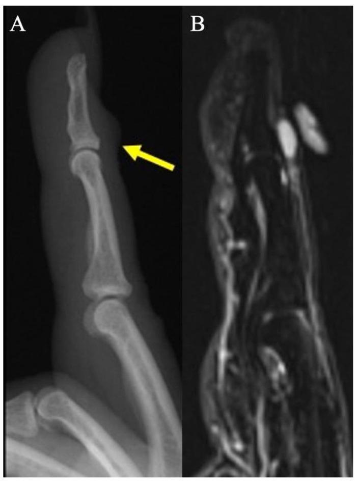
This composite figure provides a diagnostic comparison for a digital mucus cyst using two imaging modalities. Image A is a lateral plain radiograph of the index finger showing focal soft-tissue swelling (indicated by a yellow arrow) on the dorsal aspect of the distal interphalangeal (DIP) joint, located just proximal to the nail fold. There is no evidence of underlying bone erosion on the radiograph. Image B is a sagittal fat-suppressed proton density (PD) weighted MRI of the same finger. It reveals a small, well-circumscribed, uniformly hyperintense lesion consistent with a ganglion-type digital mucus cyst. The MRI confirms the absence of subungual extension or osseous erosion and demonstrates the cyst's location within the superficial soft tissues dorsal to the DIP joint. This visual material is used in medical education to differentiate between digital mucoid cysts, which are often associated with osteoarthritis and Heberden’s nodes, and other finger masses such as glomus tumors or giant cell tumors of the tendon sheath.
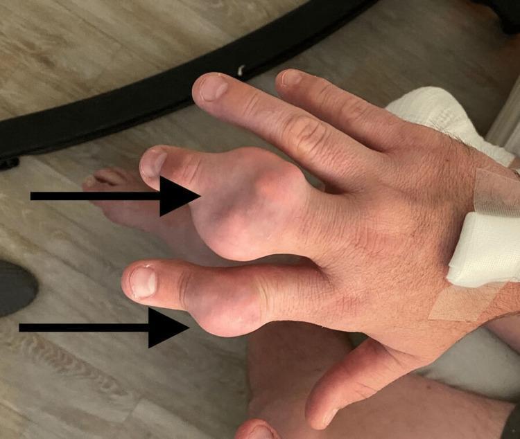
Clinical photograph of the dorsal aspect of a right hand demonstrating chronic tophaceous gout. The image prominently features two large, nodular, firm swellings (tophi) located at the distal interphalangeal (DIP) and proximal interphalangeal (PIP) joint regions of the second (index) and third (middle) digits, highlighted by black arrows. The larger mass on the index finger appears bulbous and irregular, with the overlying skin appearing thin, taut, and slightly erythematous or translucent, characteristic of underlying monosodium urate crystal deposition. Proximal to the metacarpophalangeal joints, a white medical dressing is visible on the wrist area, suggesting recent clinical intervention or intravenous access. The photograph illustrates advanced manifestations of gouty arthritis, emphasizing the typical presentation of tophi in the small joints of the hand. The clinical significance lies in identifying chronic hyperuricemia complications and distinguishing these masses from other rheumatological conditions like rheumatoid nodules or osteoarthritis-related Heberden's and Bouchard's nodes.
Confirmed: Heberden's Nodes - Osteoarthritis of DIP Joints
What Are Heberden's Nodes?
- Small, hard, bony swellings felt as a definite bony ridge across the dorsal/palmar surface of the DIP joint
- Caused by osteophyte (bone spur) formation due to cartilage degeneration at the DIP joint
- As S. Das Manual of Clinical Surgery describes: "These nodes are due to osteoarthritis... Males are rarely involved, usually due to repeated trauma to the fingers."
- Crucially: Heberden's nodes at the DIP do not mean OA is present in other joints - they can be an isolated finding
- Bradley & Daroff's Neurology: "Bony swelling of the terminal phalanges (Heberden nodes) supports the diagnosis of osteoarthritis"
Why Aceclofenac + Jointace C2 Failed
| Reason | Explanation |
|---|---|
| OA at DIP is structurally driven | Bony nodes = osteophytes - pain is from the joint lining, not just inflammation; short NSAIDs don't address the underlying degeneration |
| Aceclofenac only 1 week | Too brief for chronic OA |
| Jointace C2 needs months | Glucosamine/chondroitin/collagen takes 8-12 weeks minimum to show benefit in OA |
| Oral NSAIDs for small finger joints | Topical NSAIDs are now preferred first-line for hand OA due to equal efficacy with far less systemic risk - Rheumatology Textbook (Elsevier 2022) |
Optimized Management Plan (Hand OA with HTN on Amlodipine)
First-Line (Start Now)
- Topical diclofenac 1% gel (e.g., Voveran/Dicloren gel) - apply 2-3x daily directly over DIP joints. Same efficacy as oral NSAIDs, but minimal systemic absorption - safe in HTN
- Continue Jointace C2 for at least 3 months before judging failure
- Warm water soaks / paraffin wax therapy - 10 minutes twice daily - excellent symptomatic relief for small joint OA
If Insufficient After 4-6 Weeks
- Intra-articular corticosteroid injection into the DIP joint (done by a rheumatologist/orthopaedician) - gives 4-6 weeks of relief
- Hydroxychloroquine 200 mg OD - used off-label in hand OA with good safety profile in elderly
Important Caution re: Amlodipine & NSAIDs
- Avoid regular oral NSAIDs in this patient - NSAIDs can raise blood pressure, blunt the effect of amlodipine, and risk renal impairment in the elderly
- If oral NSAID is needed, use celecoxib (COX-2 selective) which has less BP effect than non-selective NSAIDs, always with a PPI
Lifestyle
- Avoid repetitive gripping/pinching activities that stress DIP joints
- Occupational therapy assessment; finger splints for DIP joints during activity
- Weight management if overweight (reduces systemic inflammatory load)
Investigations Still Recommended
| Test | Purpose |
|---|---|
| X-ray hands PA view | Confirm OA (joint space narrowing, osteophytes, subchondral sclerosis); rule out erosive OA or gout |
| Serum Uric Acid | Gout can co-exist and worsen joint pain |
| ESR/CRP | If elevated, suggests inflammatory component or erosive OA |
Bottom Line
Jointace used for 1 month only
glucosamine chondroitin hand osteoarthritis
Jointace C2 for 1 Month - What This Means
About the Drug Class (SYSADOAs)
- Inhibiting cartilage-degrading enzymes
- Reducing synovial inflammation
- Providing mild structural protection over time
Revised Practical Plan
Step 1 - Switch to Topical NSAID (Most Important Change)
- Topical diclofenac 1% gel (Voveran Emulgel / Dicloren gel)
- Apply directly over the DIP joints of both hands, 3x daily
- This is now the preferred first-line treatment for hand OA over oral NSAIDs - same efficacy, virtually no BP effect, no GI risk, safe with amlodipine
- Rheumatology (Elsevier 2022): "For osteoarthritis of the hand, topical NSAIDs are now preferred first-line options over oral NSAIDs due to similar efficacy with limited systemic absorption"
Step 2 - Stop Aceclofenac (oral NSAID)
- Oral NSAIDs can raise blood pressure and counteract amlodipine - not ideal for your father
- Replace entirely with topical diclofenac gel
Step 3 - Warm Soaks Daily
- Soak both hands in warm water for 10-15 minutes, twice daily
- Relieves stiffness, improves local circulation - simple and highly effective for DIP OA
Step 4 - Add Paracetamol if Needed
- Paracetamol 500 mg - 1g, TDS for breakthrough pain
- Completely safe with amlodipine and in hypertension
- Does not raise BP
Step 5 - X-ray Hands (Do This)
- X-ray bilateral hands PA view - confirms OA grade, rules out erosive changes or gout crystals
- Will guide whether an intra-articular steroid injection is needed
Timeline Summary
| Timeframe | Action |
|---|---|
| Now | Stop oral aceclofenac, start topical diclofenac gel 3x daily + warm soaks |
| Now - 2 months | Continue Jointace C2 (complete 3 month total course) |
| Within 2 weeks | Get X-ray hands PA view |
| At 3 months | Review - if still painful, rheumatology referral for intra-articular steroid injection |
One-Line Summary
These are done 2 months back. Used vit d3 for 8 weeks
python3 -c " import pdfplumber with pdfplumber.open('/tmp/workspace/attachments/a5e2dcdd-9d74-4edd-839e-1c9ce967e8d7/Gopal.pdf') as pdf: for i, page in enumerate(pdf.pages): print(f'--- PAGE {i+1} ---') print(page.extract_text()) print() "
Report Analysis: Gopal, 56M | Dated: 31 May 2026
Key Finding Relevant to Joint Pain
| Parameter | Value | Status | Significance |
|---|---|---|---|
| Vitamin D (25-OH Total) | 21.56 ng/mL | 🔴 Insufficient (20-<30) | Was insufficient BEFORE starting Vit D3 |
| Uric Acid | 6.89 mg/dL | ✅ Normal (< 7.3) | Rules out gout contributing to DIP pain |
| hs-CRP | 0.58 mg/L | ✅ Low risk (<1.0) | No systemic inflammation - strongly favours OA over inflammatory arthritis |
| Calcium | 10.15 mg/dL | ✅ Normal | Rules out hypocalcemia |
Vitamin D Status - What the Treatment Has Done
hs-CRP = 0.58 - This is Very Informative
- No active systemic inflammation
- Confirms this is osteoarthritis (non-inflammatory), NOT rheumatoid arthritis, NOT psoriatic arthritis, NOT reactive arthritis
- The Heberden's nodes + bilateral DIP involvement + normal CRP = classic primary hand OA
Other Abnormal Values - Brief Notes
| Parameter | Value | Interpretation |
|---|---|---|
| Triglycerides | 181 mg/dL (>150) | Borderline high - dietary modification needed |
| Trig/HDL ratio | 3.5 (>3.12) | Mildly elevated - insulin resistance pattern, not dangerous |
| eGFR | 82 mL/min/1.73m² | Mildly reduced - CKD Stage G2 |
| Albumin | 5.01 g/dL (>4.8) | Mildly high - likely dehydration artifact |
| Total Protein | 8.42 g/dL | Mildly high - same, dehydration |
| MCHC | 28.9 g/dL (low) | Hypochromic RBCs |
| RDW-SD | 49.5 fL (high) | Anisocytosis - mixed picture |
| Mentzer Index | 18.9 (>13) | Points toward Iron Deficiency Anemia pattern |
| HbA1c | 5.6% | Pre-diabetic range (5.7 cut-off) - monitor annually |
| TSH | 1.884 | Normal |
| Uric Acid | 6.89 | High-normal - watch dietary purines |
What This Means for Joint Pain Management
⚠️ NSAIDs are relatively contraindicated with eGFR <90 + hypertension. Oral aceclofenac was the wrong choice. This makes topical diclofenac gel even more important as the safe alternative.
Revised Action Plan (Evidence-Based)
For DIP Joint Pain (Confirmed OA):
- Topical diclofenac 1% gel - apply to DIP joints 3x daily (safe with HTN + reduced eGFR)
- Continue Jointace C2 - needs 3 months total; he's done only 1 month
- Paracetamol 500mg TDS for breakthrough pain - safe with kidneys and BP
- Warm water soaks 10-15 min, twice daily
- Recheck Vitamin D level now - after 8 weeks of supplementation; if still <30, continue maintenance dose (60,000 IU once monthly is standard maintenance)
Do NOT Use:
- ❌ Oral NSAIDs (aceclofenac, ibuprofen) - risky with eGFR 82 + HTN
- ❌ Long-term steroids
For eGFR 82 (Mild CKD):
- Monitor creatinine + eGFR every 6 months
- Stay well hydrated
- Avoid nephrotoxic drugs (NSAIDs, contrast dyes)
- Review amlodipine dose with his treating physician - actually amlodipine is safe in CKD and is a good choice here
For Triglycerides / Pre-diabetic HbA1c:
- Reduce refined carbs, sweets, fried food
- 30 minutes walking daily (also benefits joints)
- HbA1c recheck in 6 months
For Mild Hypochromic Anemia (low MCHC, high RDW):
- Consider iron-rich diet (spinach, jaggery, lean meat)
- Recheck CBC in 3 months; if persistent, add oral iron supplement
One-Paragraph Summary
Shall i add ultracet?
Ultracet (Tramadol 37.5mg + Paracetamol 325mg) - Should You Add It?
Short Answer: Yes, but with caution and only if needed
Why It Could Help
- Tramadol is a centrally-acting weak opioid (mu-receptor agonist + serotonin/norepinephrine reuptake inhibitor) - it acts on central pain pathways, which is useful in chronic OA where central sensitisation contributes to pain
- The paracetamol 325 mg component adds peripheral analgesia
- Tramadol has no effect on BP and no effect on kidneys - safe with his HTN and eGFR 82 - Katzung's Pharmacology
- No COX inhibition - does not interfere with amlodipine, no renal risk unlike NSAIDs
- Appropriate as a short-term step-up when topical NSAIDs + plain paracetamol give insufficient relief
Specific Concerns in Gopal (56M, HTN)
| Concern | Detail |
|---|---|
| Dizziness / falls | Most common side effect of tramadol is dizziness, vertigo, sedation - in a 56-year-old man, this is a real fall risk, especially at night |
| Nausea/vomiting | Very common on starting; start with half a tablet (1/2 tab) for the first 3 days |
| Serotonin syndrome | Rare but possible - check if he is on any antidepressant (SSRIs/SNRIs) - avoid combination |
| Seizure threshold | Tramadol lowers seizure threshold - relevant if he has any history of seizures |
| Constipation | Tramadol causes constipation; ensure adequate water and fibre intake |
| Dependence | Lower risk than full opioids, but avoid using daily for more than 2-3 weeks without review |
How to Use It Safely (If Added)
- Start: 1 tablet (Ultracet) at night only for the first week - this minimises dizziness and helps with sleep disruption from pain
- If tolerated, can add a daytime dose: 1 tablet BD (morning + night) with food
- Maximum: 2 tablets per day (tramadol 75mg total) - do NOT go to TDS without specialist review
- Duration: 2-4 weeks as a step-up; reassess
- Do NOT combine with plain paracetamol (Crocin/Dolo) on the same day - the Ultracet already contains 325 mg paracetamol; doubling up risks exceeding safe daily limit
Better Alternative to Consider First
Topical diclofenac gel → if insufficient → add plain paracetamol TDS → if still insufficient → add Ultracet 1 tab nocte/BD for 2-4 weeks
Bottom Line
Shall i do ESR , RA FACTOR, ANTI CCP?
ESR + RA Factor + Anti-CCP - Should You Order Them?
Short Answer:
| Test | Order It? | Reason |
|---|---|---|
| ESR | ✅ Yes - useful | Cheap, gives inflammatory baseline, complements hs-CRP |
| RA Factor | ⚠️ Low yield, but reasonable | Will almost certainly be negative - read why below |
| Anti-CCP | ⚠️ Low yield, but reasonable | Same reason - but if positive, changes everything |
The Key Clinical Reasoning
"Rheumatoid arthritis usually spares the DIP joints and the small joints of the toes."
- ✅ Bilateral DIP involvement with Heberden's nodes (bony, hard)
- ✅ hs-CRP 0.58 (completely normal - no systemic inflammation)
- ✅ Age 56, male
- ✅ Gradual onset over 2 months
Why RA Factor Alone is Unreliable
- RF is positive in only ~75% of RA patients - so 25% of true RA is RF-negative
- But more importantly, RF can be false-positive in many conditions: OA itself, infections, liver disease, aging (up to 10-15% of elderly normals are RF-positive)
- A positive RF in this context would be misleading and may cause unnecessary anxiety
Why Anti-CCP is More Useful (if you must choose one)
"Anti-CCP antibodies have high specificity 93-98% for RA, may be present years before clinical disease, and are associated with aggressive erosive disease."
- If Anti-CCP is positive despite DIP involvement → reconsider diagnosis, refer rheumatology urgently
- If Anti-CCP is negative (very likely in this case) → RA is virtually ruled out
My Recommendation
Do ESR - cheap, quick, useful baseline. If ESR is elevated (>40 mm/hr), then add anti-CCP.
Skip RF - not worth the cost or confusion. Low specificity, high false-positive rate in elderly males.
Anti-CCP - only if ESR comes back elevated OR if you see any MCP/PIP/wrist involvement on examination that was missed.
Decision Tree
ESR result
│
├── Normal (<20 mm/hr) → Confirms OA, no further serology needed
│
└── Elevated (>40 mm/hr) → Add Anti-CCP + refer rheumatology
                           (reconsider psoriatic arthritis or early RA)
Bottom Line
Xray which views
hand osteoarthritis X-ray PA view Heberden nodes DIP joint space narrowing osteophyte
osteoarthritis hand X-ray radiograph DIP joint osteophytes
X-Ray Views for Hand OA (Heberden's Nodes)
Single View is Sufficient - EULAR Guideline
"A posteroanterior (PA) radiograph of both hands on a single film or field of view is adequate for diagnosis of hand OA."
- EULAR Propositions for Hand OA, Rheumatology (Elsevier 2022), Proposition 9
What to Ask For (Write on Request Slip)
"X-ray both hands - PA view, both on single film"
- PA (posteroanterior) = palms flat down on the plate, X-ray beam from back of hand to palm
- Both hands simultaneously on one cassette - this allows direct side-by-side comparison
- No lateral view needed routinely for finger OA diagnosis
What the Radiologist Will Look For
| Finding | Significance |
|---|---|
| Joint space narrowing at DIP | Cartilage loss - confirms OA grade |
| Osteophytes | Bony spurs = Heberden's node correlate on X-ray |
| Subchondral sclerosis | Increased bone density under joint |
| Subchondral cysts | Geodes - moderate-advanced OA |
| Subchondral erosion | If present → Erosive OA (more aggressive subtype, higher pain, mildly raised CRP) |
| Bony ankylosis | Severe/late OA |
Important Addition - Request Oblique View if Erosion Suspected
"PA + Oblique (semi-pronated) view, both hands"
Practical Instructions for the Radiology Centre
| What to say | Details |
|---|---|
| Both hands | Not just the symptomatic one |
| On one film/cassette | Comparison is essential |
| PA view | Dorsum up, palms flat on plate |
| No fist/ball views needed | Those are for scaphoid fractures |
Bottom Line
Create a treatment summary document for Gopal's DIP OA management
Hi