I have all the key content I need. Let me now compile a comprehensive, exam-focused answer on GIT Neuroendocrine Tumours for MD Pathology.
Neuroendocrine Tumours of the GIT - MD Pathology
1. Introduction & Terminology
Neuroendocrine tumours (NETs) of the gastrointestinal tract were historically called carcinoid tumours - a term coined by Oberndorfer in 1907 to denote tumours that were "carcinoma-like" but apparently benign. The term "carcinoid" is now considered problematic because it implies indolent behavior and is used inconsistently. The WHO 2010/2017 classification uses "neuroendocrine tumour/neoplasm" (NET/NEN) as the preferred terminology, reserving "neuroendocrine carcinoma" (NEC) for poorly differentiated, high-grade neoplasms.
They arise from the diffuse neuroendocrine system (DNES) - previously known as the APUD cell system (Amine Precursor Uptake and Decarboxylation). The specific cells of origin in the GIT are the Kulchitsky cells (enterochromaffin cells) located at the base of intestinal crypts of Lieberkuhn.
2. Classification by Embryologic Origin (Williams & Sandler)
| Segment | Sites | Secretory Products | Carcinoid Syndrome |
|---|
| Foregut | Stomach, proximal duodenum, lung, pancreas | Histamine, catecholamines, 5-HTP (not 5-HT) | Atypical: prolonged flushing, lacrimation, facial edema |
| Midgut | Distal duodenum, jejunum, ileum, appendix, right colon | Serotonin (5-HT) - produces in large quantity | Classic carcinoid syndrome (when liver metastases present) |
| Hindgut | Transverse/descending/sigmoid colon, rectum | Usually nonsecretory | Rarely |
Key facts:
- Midgut carcinoids produce serotonin in great quantity, but it is released into the portal circulation and cleared by the liver - carcinoid syndrome only occurs if hepatic metastases are present (serotonin escapes hepatic filtration)
- Foregut carcinoids produce 5-HTP because they lack the enzyme aromatic amino acid decarboxylase (AAAD) that converts 5-HTP to 5-HT
- Hindgut tumours are most often nonsecretory
3. WHO Grading Classification (2017)
Based on Ki-67 labeling index and mitotic count - this is the most important prognostic parameter:
| Differentiation | Grade | Ki-67 Index | Mitoses per 10 HPF |
|---|
| Well differentiated (NET) | G1 | ≤3% (some sources ≤2%) | <2 |
| Well differentiated (NET) | G2 | 3-20% | 2-20 |
| Well differentiated (NET) | G3 | >20% | >20 |
| Poorly differentiated (NEC) | G3 | >20% | >20 |
Important distinction: A G3 well-differentiated NET differs from a poorly differentiated NEC - both have Ki-67 >20% but differ in morphology, behavior, and treatment response. When Ki-67 and mitotic count give discordant grades, the higher grade is assigned (NANETS 2013 update).
Source: Yamada's Textbook of Gastroenterology, 7th ed.
4. Frequency and Location
- Most common site: Appendix (most frequent GI site - approximately 50% of all GI NETs in older series), followed by ileum and rectum
- Bailey & Love: "most commonly in the appendix, ileum and rectum in decreasing order of frequency"
- Small bowel NETs: 1/3 are multiple
- The appendiceal NET is usually benign if <2 cm; those >2 cm have significant risk of metastasis
- Gastrinomas ("triangle of gastrinoma"): 90% within the gastrinoma triangle - bounded by cystic duct, junction of 2nd and 3rd parts of duodenum, and junction of head and neck of pancreas
5. Histopathology (MORPHOLOGY - High Yield)
Gross Appearance
- Primary is usually small and firm
- May cause dense desmoplastic fibrosis in surrounding mesentery - causing bowel kinking, obstruction, and a characteristic radiological "sunburst" mesenteric appearance
- Cut surface: yellow-tan in color
- Liver metastases can be bulky even with small primaries
Microscopic Appearance (H&E)
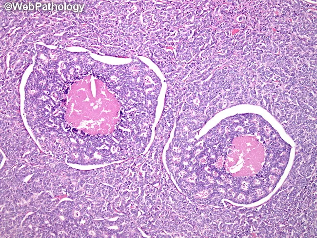
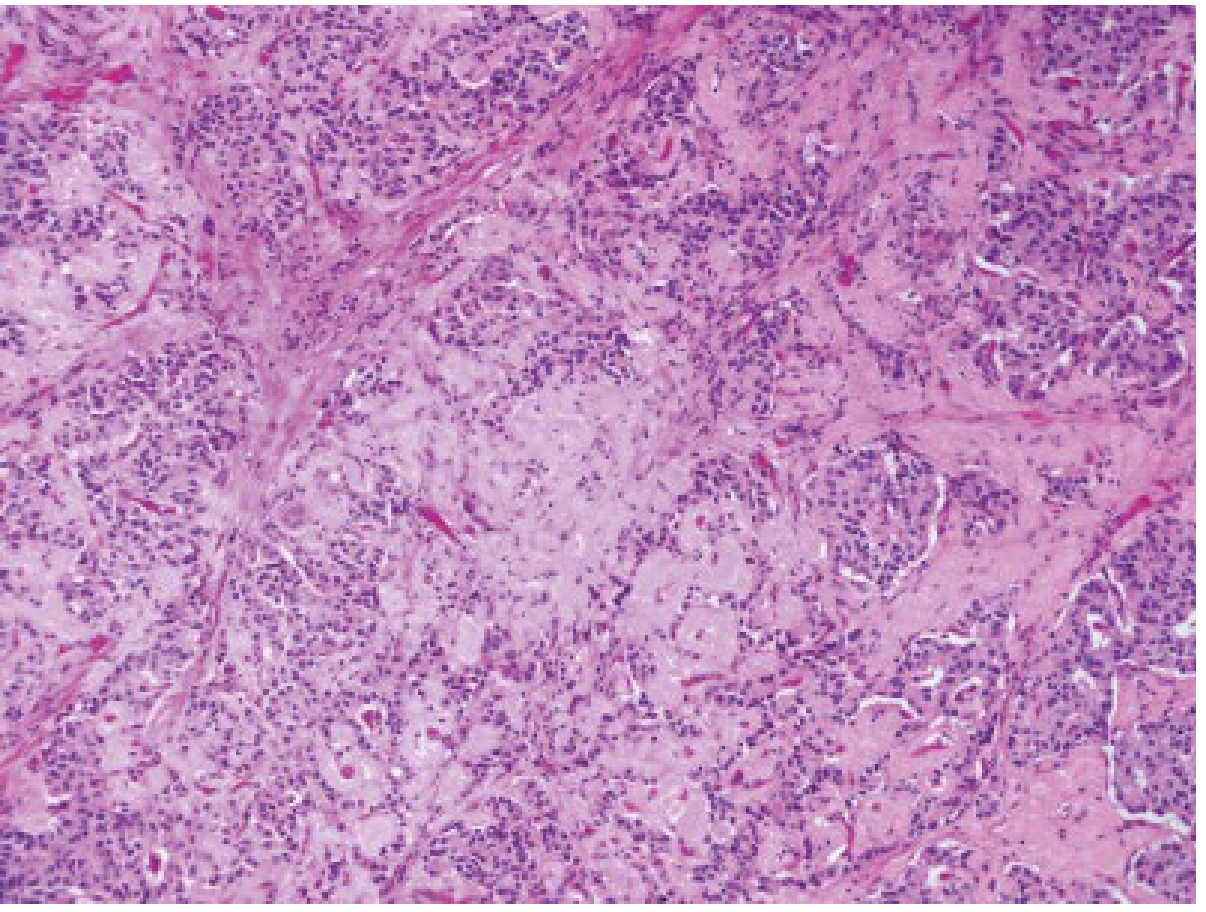
Well-differentiated NETs show a characteristic organoid architecture:
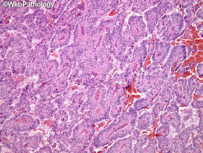
- Nesting/insular pattern (most common)
- Trabecular/ribbon-like pattern
- Glandular/tubular/acinar pattern
- Rosette (pseudorosette) formation
- Mixed patterns common
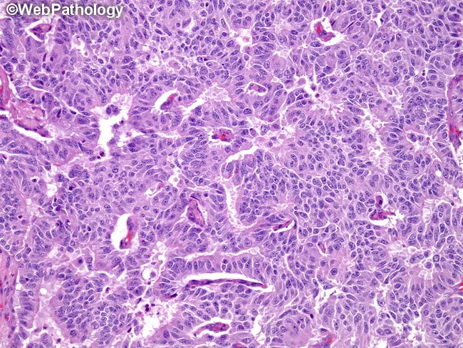
Cytology: Uniform, small-to-medium sized cells with:
- Round to oval nuclei
- "Salt and pepper" chromatin (finely stippled - the hallmark feature)
- Inconspicuous nucleoli
- Moderate eosinophilic cytoplasm
- Low mitotic activity (in G1)
- Highly vascular stroma
Poorly differentiated NEC (G3):
- Large or small cell neuroendocrine carcinoma morphology
- High nuclear:cytoplasmic ratio
- Prominent nucleoli
- Frequent mitoses and necrosis
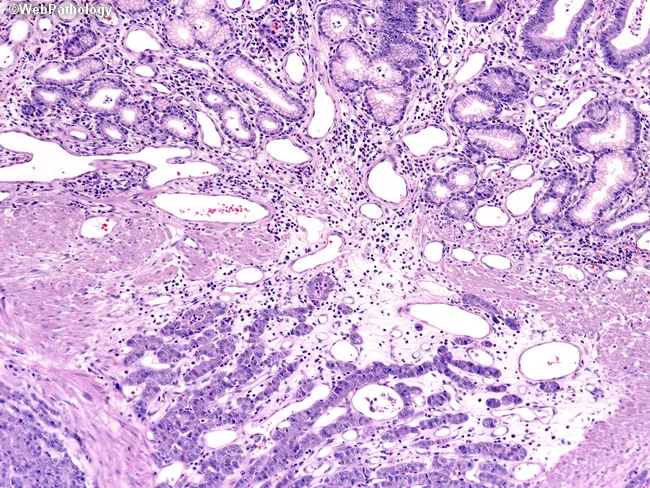
Here are the histological appearances:
H&E - Well-differentiated NET (insulinoma), showing organoid nesting pattern:
H&E - Carcinoid tumour showing classic trabecular and nesting growth patterns:
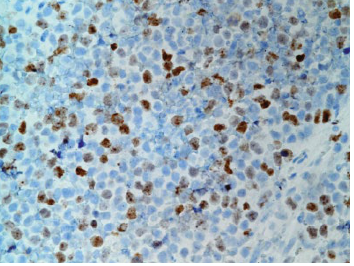
Ki-67 immunostaining of a poorly differentiated NET (>20% positivity = G3):
6. Histochemical & Immunohistochemical Staining
Special Stains
| Stain | Reaction | Basis |
|---|
| Fontana-Masson (argentaffin reaction) | Positive (black/silver) | Self-reduce silver - contains serotonin granules; midgut tumours |
| Grimelius (argyrophil reaction) | Positive | Requires external reducing agent; more sensitive, foregut + midgut |
| Periodic acid-Schiff (PAS) | Variable | Mucinous variants |
- Argentaffin +: midgut (serotonin-producing) carcinoids
- Argyrophil +: all NETs (more sensitive)
Immunohistochemistry (IHC) - Neuroendocrine Markers
| Marker | Comment |
|---|
| Synaptophysin | Most sensitive pan-neuroendocrine marker; stains all NETs |
| Chromogranin A (CgA) | Most specific; also used as serum tumour marker |
| CD56 (NCAM) | Additional neuroendocrine marker |
| NSE (Neuron-specific enolase) | Less specific |
| Ki-67 | Grading marker (MIB-1 antibody) |
Specific functional markers: Insulin, Gastrin, Glucagon, VIP, Somatostatin, Serotonin - identify the functional subtype.
7. Tumour Markers (Laboratory Diagnosis)
Serotonin Pathway
- Most 5-HT produced by midgut carcinoids is taken up by platelets in the portal circulation and stored in their dense granules
- A portion enters renal tubules → converted to 5-hydroxyindoleacetic acid (5-HIAA) by monoamine oxidase and aldehyde dehydrogenase
- 24-hour urinary 5-HIAA is the standard screening test (normal <10 mg/day)
- False positives: tryptophan-rich foods (avocado, banana, pineapple, walnuts, tomatoes), drugs (phenothiazines)
- Platelet serotonin is the most accurate marker (not affected by diet)
- 5-HIAA and 5-HT may be normal in 20-30% of patients, particularly foregut and hindgut tumours
Other Markers
| Marker | Use |
|---|
| Chromogranin A (serum) | Best overall tumour burden marker; correlates with treatment response |
| 5-HIAA (24h urine) | Serotonin-producing (midgut) NETs |
| Gastrin | Gastrinoma / ZES |
| Insulin + C-peptide | Insulinoma |
| VIP | VIPoma |
| Glucagon | Glucagonoma |
| PP (pancreatic polypeptide) | Nonfunctioning PNETs |
Source: Quick Compendium of Clinical Pathology, 5th ed.
8. Carcinoid Syndrome
Definition: Clinical syndrome resulting from systemic release of vasoactive substances, occurring almost exclusively with hepatic metastases (liver can no longer clear portal serotonin) or with bronchial/ovarian primary (drains directly to systemic circulation, bypassing portal system).
Classic Features (midgut serotonin-secreting tumours):
- Episodic flushing (reddish-blue cyanosis) - triggered by alcohol, stress, food
- Diarrhea (watery, secretory) with borborygmi
- Bronchospasm (wheezing)
- Right-sided heart disease: tricuspid regurgitation ± pulmonary stenosis (fibrous plaques - carcinoid heart disease; serotonin causes endocardial fibrosis on the right side because left side is protected by pulmonary metabolism of serotonin)
- Pellagra-like skin changes (due to diversion of tryptophan to serotonin synthesis)
Atypical carcinoid syndrome (foregut - histamine + 5-HTP secreting):
- Prolonged patchy flushing
- Lacrimation, periorbital edema
- Bronchoconstriction
- No 5-HIAA elevation
Carcinoid Crisis
- Life-threatening exacerbation of carcinoid syndrome
- Triggered by: surgical manipulation, induction of anesthesia, tumor necrosis (from chemotherapy/embolization)
- Prevention: Octreotide (somatostatin analogue) prophylaxis
9. Specific GIT NETs
A. Appendiceal NET (most common GI NET)
- Arises from subepithelial enterochromaffin cells at the base of crypts
- Most found incidentally during appendicectomy
- Usually at the tip of the appendix
- <2 cm: essentially benign, simple appendicectomy curative
- >2 cm: significant risk of metastasis, right hemicolectomy required
- Goblet cell carcinoid (adenocarcinoid): mixed neuroendocrine-mucin-secreting adenocarcinoma; more aggressive
B. Small Bowel NET (most common cause of carcinoid syndrome)
- Most common in the terminal ileum
- 1/3 are multiple (multicentric)
- Cause intense desmoplastic reaction in mesentery
- Most likely to cause carcinoid syndrome when metastatic
- IHC: serotonin positive, chromogranin A+, synaptophysin+
- Argentaffin positive
C. Gastric NET
Three types:
| Type | Association | Biology |
|---|
| Type I (75%) | Chronic atrophic gastritis, hypergastrinemia | Benign, ECL cell origin |
| Type II (5-10%) | MEN1 + ZES, hypergastrinemia | Low-grade malignant |
| Type III (15-20%) | Sporadic, normal gastrin | Most aggressive; may metastasize |
D. Gastrinoma (Zollinger-Ellison Syndrome)
- G-cell tumour secreting gastrin → massive HCl production
- ZES triad: severe peptic ulcers (often multiple, atypical sites), diarrhea, elevated gastrin
- 20-30% associated with MEN1 (look for multiple pancreatic tumours)
- Up to 5% develop ectopic ACTH secretion (Cushing syndrome)
- Gastrinoma triangle - 90% of gastrinomas occur here
- Fasting serum gastrin >1000 pg/mL highly diagnostic; secretin stimulation test
- Treated with: PPIs (for acid control), surgical resection (for cure), octreotide
E. Insulinoma
- Most common functional PNET (60% of functional pancreatic NETs)
- Whipple's triad: symptomatic hypoglycemia (symptoms with fasting/exercise) + blood glucose <2.5 mmol/L + relief with glucose
- 4-6% associated with MEN1; 5% malignant (lowest malignancy rate of all PNETs)
- Usually small (<2 cm), solitary, evenly distributed in head/body/tail
- Diagnosis: elevated fasting insulin + C-peptide, low blood glucose
F. VIPoma (WDHA syndrome / Verner-Morrison syndrome / Pancreatic cholera)
- Watery Diarrhea, Hypokalemia, Achlorhydria
- Massive secretory diarrhea (>6-8 L/day), tea-coloured, persists with fasting
- 80-90% in pancreas (often tail); 50-89% metastatic at presentation
- VIP → stimulates adenylate cyclase in intestinal epithelium → massive Cl-/water secretion
G. Glucagonoma
- Glucagon-secreting pancreatic NET; primarily >50 years
- Classic presentation: 4Ds - Diabetes (mild), Dermatitis (necrolytic migratory erythema - pathognomonic), DVT/thrombosis, Depression
- Necrolytic migratory erythema (NME): erythematous vesiculobullous rash on lower limbs, groin, perineum; due to glucagon excess and hypoaminoacidemia
H. Somatostatinoma
- Rare; Inhibitory hormone → inhibits everything
- Somatostatinoma triad: Diabetes mellitus (mild), Steatorrhea (fat malabsorption), Cholelithiasis (gallstones - reduced cholecystokinin effect)
- Most found in pancreas head or duodenum
- Duodenal somatostatinomas have association with von Recklinghausen's disease (NF1)
10. Genetics and Hereditary Associations
| Syndrome | Gene | Associated NETs |
|---|
| MEN1 | MEN1 (menin) | Gastrinoma, insulinoma, VIPoma, glucagonoma (3 Ps: Parathyroid, Pituitary, Pancreas) |
| MEN2A/2B | RET | Medullary thyroid carcinoma, pheochromocytoma |
| VHL | VHL | Pancreatic NETs (non-functional) |
| NF1 | NF1 | Duodenal somatostatinomas, carcinoids |
| TSC | TSC1/TSC2 | Pancreatic NETs |
11. Prognosis and Malignancy
- All NETs are potentially malignant - the distinction benign vs malignant cannot be made on histology alone; only metastasis confirms malignancy
- Appendiceal NETs <2 cm: very rarely malignant
- Ileal NETs: frequently malignant despite small size
- 5-year survival by grade: G1 ~95%, G2 ~75%, G3 (NEC) ~15-25%
- Hepatic metastases: most common site; patients may live many years even with metastatic disease (slow growth rate)
12. Treatment Summary
| Modality | Application |
|---|
| Surgery | First-line for resectable disease; appendicectomy for appendiceal NETs <2 cm; right hemicolectomy for >2 cm |
| Octreotide / Lanreotide | Somatostatin analogues - control carcinoid syndrome (flushing, diarrhea); antiproliferative; must cover surgery/anaesthesia to prevent carcinoid crisis |
| Octreotide scanning (somatostatin receptor scintigraphy) | Staging and detection of metastases; detects occult primaries |
| PRRT (Peptide receptor radionuclide therapy) | 177Lu-DOTATATE for somatostatin-receptor positive metastatic NETs |
| Hepatic resection / ablation | Palliative cytoreduction for liver metastases |
| Chemotherapy | Less effective for well-differentiated NETs; streptozocin + 5-FU for PNETs; platinum-based for poorly differentiated NEC |
13. High-Yield Exam Points
- Cell of origin: Kulchitsky (enterochromaffin) cells at base of crypts of Lieberkuhn
- Most common site: Appendix; most common site causing carcinoid syndrome = terminal ileum
- Argentaffin vs Argyrophil: Argentaffin (self-reduces silver) = midgut only; Argyrophil = all NETs
- Carcinoid syndrome requires liver metastases (or bronchial primary) - portal serotonin cleared by liver
- 5-HIAA in urine = screening test; false positives with tryptophan-rich foods
- Chromogranin A = best serum marker for tumour burden
- IHC: Synaptophysin (most sensitive) + Chromogranin A (most specific)
- "Salt and pepper" chromatin = hallmark nuclear feature
- Carcinoid heart disease = RIGHT-sided (tricuspid regurgitation, pulmonary stenosis); left side protected by lung degradation of serotonin
- WHO grading based on Ki-67 and mitotic count - G1/G2/G3
- Gastrinoma triangle: cystic duct + D2/D3 junction + neck/head of pancreas = 90% of gastrinomas
- Glucagonoma 4Ds: Diabetes, Dermatitis (NME - pathognomonic), DVT, Depression
- VIPoma = WDHA (Watery Diarrhea, Hypokalemia, Achlorhydria)
- Somatostatinoma triad: Diabetes + Steatorrhea + Cholelithiasis
- MEN1: gastrinoma, insulinoma - 20-30% of gastrinomas are MEN1-associated
- Malignancy cannot be determined histologically - only by presence of metastasis
- Appendiceal NETs: <2 cm = benign behavior; >2 cm = need right hemicolectomy
Primary sources: Quick Compendium of Clinical Pathology 5th ed.; Yamada's Textbook of Gastroenterology 7th ed.; Bailey and Love's Short Practice of Surgery 28th ed.; Robbins Pathologic Basis of Disease; Sleisenger and Fordtran's GI & Liver Disease