
Vision brief explanation in ophthalmology
Reading File
Reading File
Finding Sources
Searching PubMed
vision AND visual acuity
Searching the Web
American Academy Ophthalmology visual acuity vision definition ophthalmology
Reading File
Finding Sources
Vision is the ability to perceive and interpret light entering the eye. Light is focused by the cornea and lens onto the retina, where photoreceptors convert it into nerve signals. These signals travel through the optic nerve, optic chiasm, optic tracts, lateral geniculate body, and optic radiations to the visual cortex of the occipital lobe, where an image is perceived.
Visual acuity
Visual acuity is the sharpness or spatial resolution of central vision. It is tested separately in each eye, ideally with the person's best optical correction, using a Snellen chart or LogMAR chart.
- 20/20 (6/6): the person sees at 20 feet (6 m) what a normal eye sees at that distance.
- 20/60 (6/18): the person sees at 20 feet what a normal eye can see at 60 feet.
- A smaller denominator indicates better acuity, such as 20/15.
- A larger denominator indicates reduced acuity, such as 20/200.
Visual acuity reflects central macular and optic-nerve function, but it does not fully assess vision. Good visual function also needs visual fields, contrast sensitivity, colour vision, binocular vision, ocular movements, and depth perception. Bradley and Daroff's Neurology in Clinical Practice, p. 165. The AAO overview of visual acuity similarly notes that an eye chart measures only one component of vision.
Explain the visual pathway from retina to visual cortex
Reading File
Finding Sources
Finding Sources
Searching Images
labeled diagram visual pathway retina optic nerve optic chiasm optic tract lateral geniculate nucleus optic radiations visual cortex
Searching PubMed
visual pathway AND anatomy
Searching the Web
visual pathway optic chiasm lateral geniculate nucleus optic radiations primary visual cortex anatomy
Reading File
Reading File
Loading Image
Visual pathway: retina to visual cortex
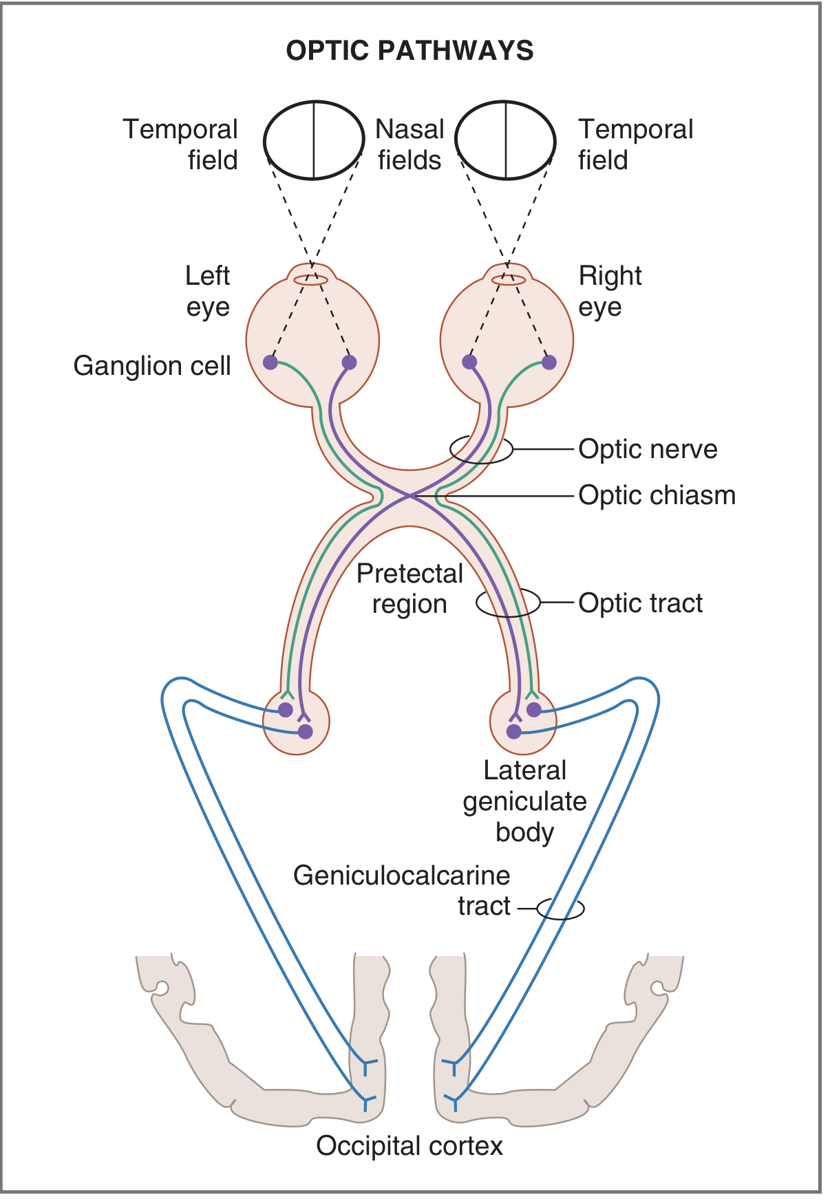
Route of visual impulses
-
Retina
- Light is detected by rods and cones.
- The signal passes through bipolar cells to retinal ganglion cells.
- Axons of ganglion cells form the optic nerve (cranial nerve II).
-
Optic nerve
- Each optic nerve carries information from one eye only.
- It travels posteriorly from the orbit through the optic canal to the optic chiasm.
-
Optic chiasm
- Fibres from the nasal half of each retina cross to the opposite side.
- Fibres from the temporal half of each retina remain uncrossed.
- This partial crossing ensures that each cerebral hemisphere processes the opposite visual hemifield:
- Right visual field → left visual cortex
- Left visual field → right visual cortex
-
Optic tract
- Each optic tract contains fibres from:
- ipsilateral temporal retina, and
- contralateral nasal retina.
- For example, the left optic tract conveys the right visual field from both eyes.
- Each optic tract contains fibres from:
-
Lateral geniculate nucleus (LGN)
- Most optic-tract fibres synapse in the lateral geniculate nucleus of the thalamus.
- The LGN maintains a precise map of the retina, called retinotopic organization.
-
Optic radiations, or geniculocalcarine tract
- Fibres from the LGN pass posteriorly to the occipital lobe.
- They divide into two major bundles:
- Inferior fibres pass through the temporal lobe as Meyer's loop. They carry information from the superior visual field.
- Superior fibres pass through the parietal lobe. They carry information from the inferior visual field.
-
Primary visual cortex
- Optic radiations terminate in the primary visual cortex (V1, Brodmann area 17) around the calcarine fissure of the occipital lobe.
- The upper bank of the calcarine fissure represents the lower visual field.
- The lower bank represents the upper visual field.
- The central macular area, responsible for detailed vision, has a large cortical representation near the occipital pole.
Important visual-field principle
The image projected on the retina is reversed and inverted:
- Temporal visual field → nasal retina
- Nasal visual field → temporal retina
- Superior visual field → inferior retina
- Inferior visual field → superior retina
Clinical correlations
- Optic nerve lesion: blindness in the affected eye.
- Optic chiasm lesion: bitemporal hemianopia, classically from a pituitary mass compressing crossing nasal retinal fibres.
- Optic tract lesion: contralateral homonymous hemianopia.
- Temporal-lobe lesion involving Meyer's loop: contralateral superior quadrantanopia, “pie in the sky.”
- Parietal optic-radiation lesion: contralateral inferior quadrantanopia, “pie on the floor.”
- Occipital cortex lesion: contralateral homonymous hemianopia, often with macular sparing in posterior cerebral artery infarction.
Costanzo Physiology, 7th ed., p. 96. A concise NCBI anatomy review also outlines the retina → optic nerve → chiasm → tract → LGN → optic radiation → occipital cortex sequence.
This is a shared conversation. Sign in to Orris to start your own chat.