
hsil morphology, biomarkers,clinical impication
"high-grade squamous intraepithelial lesion" AND biomarkers
"HSIL" AND "p16" AND cervical
HSIL: Morphology, Biomarkers, and Clinical Implications
Definition and Classification
Pathogenesis
- E6 oncoprotein: binds and mediates destruction of p53, plus upregulates telomerase
- E7 oncoprotein: binds and inactivates RB protein, displacing E2F transcription factors and driving cell cycle progression
- High-risk HPV types integrate into the host genome, disrupting the viral gene that normally suppresses E6/E7 expression - this integration also increases genomic instability
- HPV-16, -18, -31, -33, -35, -39, -45, -51, -52, -56, -58, -59, and -68 collectively account for ~90% of high-grade lesions and cancer
Morphology
Histology (Tissue Sections)
| Feature | CIN II (HSIL) | CIN III (HSIL) |
|---|---|---|
| Dysplastic cell extent | Lower 2/3 of epithelium | Full thickness |
| Koilocytic change | Usually absent | Absent |
| Mitoses | Present above basal layer, some atypical | Abnormal mitoses, diffuse |
| Nuclear features | Variation in size/chromatin | Greater variation, disorderly orientation |
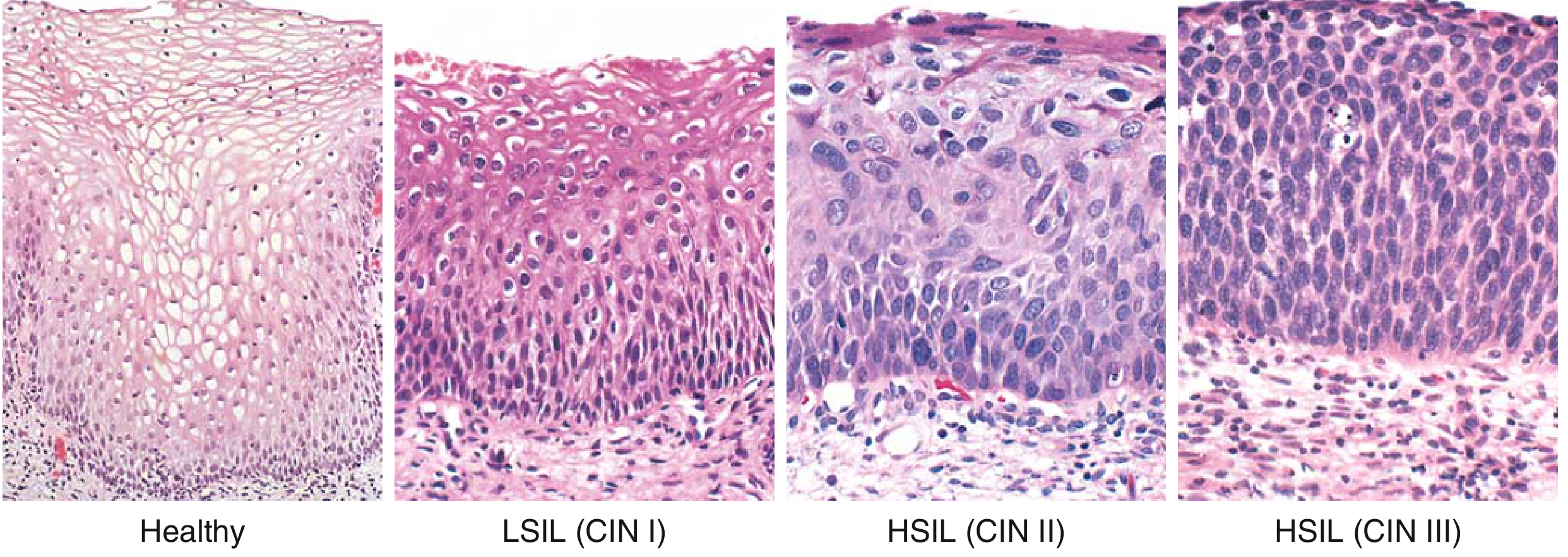
- LSIL (CIN I) shows dysplasia only in the lower 1/3 with prominent koilocytes (HPV cytopathic effect) in the upper layers
- HSIL shows no significant koilocytic change, reflecting suppressed viral replication with high proliferative activity instead
Cytology (Pap Test)
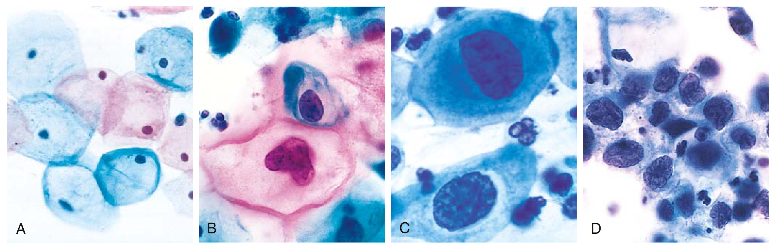
- Markedly reduced cytoplasm relative to nucleus
- High nuclear-to-cytoplasmic (N:C) ratio - the most characteristic feature
- Hyperchromatic, coarsely granular nuclei
- Nuclear membrane irregularity
- Cells smaller than LSIL cells, appearing in sheets or singly
Biomarkers
1. p16^INK4a (CDKN2A) - Most Important Biomarker
- Mechanism: Normally, p16 is a cell-cycle inhibitor that suppresses CDK4/6. In HPV-driven dysplasia, E7 inactivates RB, which in turn removes negative feedback on p16 - leading to strong diffuse p16 overexpression ("block positivity")
- Pattern in HSIL: Strong, diffuse nuclear and cytoplasmic staining throughout the full or near-full thickness of the epithelium
- Pattern in LSIL/reactive changes: Patchy, weak, or negative
- Clinical use:
- Resolves interobserver disagreement in ambiguous CIN 2 diagnoses (CIN 2 is the least reproducible grade)
- p16 IHC improves reproducibility of cervical histology interpretation (PMID 38527169, 2024)
- Guides management of CIN 2 in young women - p16-negative CIN 2 may be managed conservatively; p16-positive CIN 2 is treated as HSIL
- Per Berek & Novak's Gynecology: p16INK4a staining is useful for managing histological HSIL, and p16 IHC correlates with CIN grade
2. Ki-67 (MKI67)
- A nuclear marker of cellular proliferation
- In HSIL, Ki-67 positive cells extend into the upper epithelial layers (above the lower 1/3), reflecting loss of differentiation
- In normal or reactive epithelium, Ki-67 is confined to the basal/parabasal layers
- CINtec PLUS test: Dual staining with p16 + Ki-67 on the same cell is used in cytology triage - a cell that is simultaneously p16(+)/Ki-67(+) is an abnormal cell in an unphysiological state, highly specific for CIN 2+
- A 2025 prospective study confirmed that p16, Ki-67, and MCM2 immunocytochemistry all improve detection of cervical lesions (PMID 40530144)
3. MCM2 (Mini-Chromosome Maintenance Protein 2)
- A DNA replication licensing factor; overexpressed when HPV drives cell cycle re-entry
- Extends further up the epithelium in HSIL compared to LSIL
- Proposed as a complementary proliferation marker alongside Ki-67
4. HPV Testing
- High-risk HPV DNA/mRNA testing is a central triage tool, not a histologic biomarker per se
- HPV-16/18 genotyping has the highest individual-type risk for CIN 3+
- HPV E6/E7 mRNA testing reflects active viral oncogene expression and is more specific than DNA testing for identifying transforming infections
- Sensitivity of HPV testing for HSIL is higher than Pap cytology alone; specificity is lower - Symptom to Diagnosis, p. 1657
5. Other Emerging Markers
| Marker | Role |
|---|---|
| ProEx C (MCM2 + TOP2A) | Dual marker of aberrant S-phase induction |
| L1 capsid protein | Loss of L1 correlates with increased risk of progression |
| Methylation markers (FAM19A4, miR-124) | Epigenetic markers used in triage of HPV-positive women |
| Telomerase (hTERT) | Upregulated via E6; potential progression marker |
Natural History
| Lesion | Regression | Persistence | Progression to carcinoma |
|---|---|---|---|
| LSIL (CIN I) | 60% | 30% | 10% (to HSIL) |
| HSIL (CIN II, III) | 30% | 60% | 10% (to carcinoma) |
- ~20% of HSIL cases arise de novo (without preceding LSIL)
- HSIL demonstrates low levels of viral replication (unlike LSIL which has abundant viral shedding) - this reflects HPV integration and oncogenic transformation
- Risk factors for progression from HSIL to invasive carcinoma: cigarette smoking, HIV/immunocompromise
Clinical Implications
Symptoms
Screening and Detection
- Pap test sensitivity for HSIL: ~56% (individual test); specificity ~97%
- HPV testing is more sensitive but less specific
- Co-testing (Pap + HPV) every 5 years (ages 30-65) or Pap alone every 3 years are preferred screening strategies
- Women with HSIL cytology must undergo colposcopy and directed biopsy - Berek & Novak's Gynecology, Key Point 9
Colposcopy Findings
Management
| Situation | Management |
|---|---|
| HSIL cytology | Immediate colposcopy + directed biopsy |
| Histologic CIN 2/3 (age ≥25) | Destruction or excision of transformation zone |
| Preferred treatment | LEEP (loop electrosurgical excision procedure) |
| Alternative excisional | Cone biopsy (cold knife conization) - indicated for CIN 3 or suspicion of AIS |
| Ablative therapies | Only if invasive cancer excluded on colposcopy, ECC, and biopsy |
| Hysterectomy | Last resort for recurrent high-grade CIN |
| CIN 2 in adolescents/young women | Conservative observation acceptable (many regress), especially if p16-negative |
| Pregnant women | Defer treatment until postpartum (colposcopy in 6-12 weeks; repeat each trimester) |
Post-treatment Surveillance
Summary Table
| Domain | Key Feature |
|---|---|
| Histology | Dysplastic cells extending >2/3 thickness (CIN 2) or full thickness (CIN 3); atypical mitoses; no koilocytes |
| Cytology | High N:C ratio; hyperchromatic irregular nuclei; scant cytoplasm |
| Key biomarker | p16^INK4a - strong, diffuse, block positivity |
| Proliferation marker | Ki-67 extending into upper epithelial layers |
| Molecular driver | HPV E6 (destroys p53) + E7 (inactivates RB) |
| Natural history | 60% persist, 10% progress to carcinoma (2-10 years) |
| Clinical action | Colposcopy + biopsy mandatory; LEEP preferred treatment |
make it short
HSIL - Quick Reference
Morphology
- Dysplastic immature squamous cells extending >2/3 of epithelial thickness (CIN 2) or full thickness (CIN 3)
- Atypical mitoses above basal layer
- No koilocytic change (unlike LSIL)
- Disorderly cell orientation, nuclear pleomorphism
- High nuclear-to-cytoplasm (N:C) ratio - hallmark feature
- Hyperchromatic, coarsely irregular nuclei
- Scant cytoplasm; cells smaller than LSIL
Biomarkers
| Marker | Finding in HSIL | Use |
|---|---|---|
| p16^INK4a | Strong, diffuse "block" positivity | Confirms CIN 2/3; resolves borderline cases |
| Ki-67 | Extends into upper epithelial layers | Reflects aberrant proliferation |
| p16 + Ki-67 dual stain | Same cell positive for both | High specificity for CIN 2+ on cytology |
| HPV 16/18 | Most common high-risk types | Risk stratification |
| MCM2/ProEx C | Overexpressed | Emerging adjuncts |
Natural History
| Regress | Persist | Progress to cancer | |
|---|---|---|---|
| LSIL | 60% | 30% | 10% → HSIL |
| HSIL | 30% | 60% | 10% → carcinoma |
Clinical Implications
- Asymptomatic - detected only by screening
- HSIL cytology → mandatory colposcopy + directed biopsy
- CIN 2/3 confirmed → LEEP (preferred); cold knife cone for CIN 3/AIS suspicion
- Ablation only if invasion excluded
- Young women with p16-negative CIN 2 → conservative surveillance acceptable
- Post-treatment: remains at risk for cervical/vulvar/vaginal HPV-related cancers; long-term follow-up required
Role of IHC in differentiating primary ovarian carcinoma from metastatic carcinoma.
IHC markers primary ovarian carcinoma vs metastatic carcinoma PAX8 WT1 CK7 CK20 CDX2 pathology
"ovarian carcinoma" AND "immunohistochemistry" AND "metastatic"
IHC in Differentiating Primary Ovarian Carcinoma from Metastatic Carcinoma
Why IHC is Needed
Step 1 - Cytokeratin Profile (First-Line Screen)
| Profile | Favors |
|---|---|
| CK7+ / CK20− | Primary ovarian (serous, endometrioid, clear cell) |
| CK7− / CK20+ | Colorectal metastasis |
| CK7+ / CK20+ | Pancreaticobiliary, upper GI, or primary ovarian mucinous |
| CK7+ / CK20− | Breast, lung, endometrial |
CK7 is the single most discriminatory marker separating primary ovarian from lower GI metastases. Primary ovarian carcinomas are nearly always CK7-positive, while colorectal metastases are typically CK7-negative.
Step 2 - Site-Specific Markers (Second-Line Panel)
Markers Favoring PRIMARY Ovarian Carcinoma
| Marker | Sensitivity | Notes |
|---|---|---|
| PAX8 | 79-99% | Nuclear; Müllerian (ovarian, endometrial, cervical) + renal + thyroid. Best single positive marker for ovarian origin |
| WT1 | ~90% serous | Strong nuclear positivity in high-grade serous carcinoma; negative in mucinous, colorectal, breast, and most metastases |
| CA-125 | ~80% | Broad; more useful in combination |
| Mesothelin | ~70% | Serous and endometrioid; also positive in mesothelioma |
| ER / PR | ~60% serous, ~80% endometrioid | Positive in primary; helps exclude colorectal and gastric metastases |
Markers Favoring METASTATIC Carcinoma
| Marker | Primary Site | Notes |
|---|---|---|
| CDX2 | Colorectal / appendix | 100% sensitive for colonic metastasis to ovary; negative in serous and endometrioid; ~40% primary mucinous ovarian (less specific there) |
| CK20 | Colorectal / GI | Less specific than CDX2; ~83% primary mucinous ovarian also positive |
| GCDFP-15 / Mammaglobin | Breast | Highly specific for breast origin |
| GATA3 | Breast / urothelial | Strong nuclear staining in breast carcinoma |
| DPC4 (SMAD4) | Pancreaticobiliary | Loss of DPC4 favors pancreatic/bile duct origin (~55% of pancreatic Ca show DPC4 loss) |
| CDH17 / MUC2 | GI / colorectal | Intestinal-type mucinous adenocarcinoma |
| PSA / NKX3.1 | Prostate | Rare ovarian metastasis from prostate |
| TTF-1 | Lung | Pulmonary adenocarcinoma metastasis |
| β-catenin (nuclear) | Colorectal / endometrioid | Nuclear positivity in colorectal; also endometrioid (lower utility) |
| CA 19-9 | Pancreaticobiliary / gastric | Upper GI origin |
Differential by Clinical Scenario
Primary Ovarian (Serous) vs. Peritoneal Metastasis / Breast
- Use: PAX8 + WT1 + ER
- HGSC: PAX8(+), WT1(+), ER(+/-), CK7(+), CK20(−)
- Breast metastasis: GCDFP-15(+), mammaglobin(+), GATA3(+), PAX8(−), WT1(−)
Primary Ovarian (Mucinous) vs. Colorectal / Appendiceal Metastasis
- Most difficult differential
- Use: CK7 + CDX2 + CK20
- Primary ovarian mucinous: CK7(+), CDX2 negative or focal, CK20 variable
- Colorectal: CK7(−), CDX2(+), CK20(+) - classic "colorectal profile"
- Appendiceal: similar to colorectal; may also express CDX2
Primary Ovarian (Clear Cell) vs. Metastatic Renal Cell Carcinoma
- Use: CK7 + CA-125 + CD10 + PAX8 + RCC marker + vimentin
- Primary ovarian clear cell: CK7(+), CA-125(+/-), CD10(−), PAX8(+), vimentin(−)
- Renal clear cell: CD10(+), RCC(+), CK7(−), vimentin(+), PAX8(+)
- Note: PAX8 is positive in both - it cannot discriminate here; use CD10 + CK7
Primary Ovarian (Endometrioid) vs. Metastatic Colorectal
- Use: CK7 + CDX2 + ER + β-catenin (nuclear)
- Ovarian endometrioid: CK7(+), ER(+), CDX2(−), nuclear β-catenin(+/−)
- Colorectal: CK7(−), CDX2(+), ER(−), nuclear β-catenin(+)
Summary Panel Algorithm
Adenocarcinoma involving ovary
         │
    CK7 / CK20
         │
  ┌──────┴──────────┐
CK7+ / CK20−     CK7− / CK20+
  │                  │
Add PAX8, WT1,     CDX2 → Colorectal/
ER, GCDFP-15,      appendiceal metastasis
GATA3, TTF-1
  │
PAX8+ / WT1+ → Primary serous
PAX8+ / WT1− → Endometrioid / clear cell / renal (use CD10)
GCDFP-15+ / GATA3+ → Breast
TTF-1+ → Lung
Key Caveats
- No single marker is absolute - IHC is always interpreted as a panel in context of morphology, clinical history, and imaging
- PAX8 is the best single positive marker for ovarian/Müllerian origin but is also expressed in renal and thyroid tumors
- WT1 is highly specific for HGSC but negative in mucinous, clear cell, and most non-serous primaries
- CDX2 is superior to CK20 for detecting colorectal metastasis to ovary, but ~40% of primary ovarian mucinous tumors are CDX2-positive - limiting specificity
- DPC4 loss is highly useful when pancreatic/biliary origin is suspected (Fishman's Pulmonary Diseases textbook)
- Bilateral ovarian involvement, small tumors with surface implants, and signet-ring morphology (Krukenberg tumor) strongly suggest metastatic origin even before IHC
make it short
IHC: Primary Ovarian vs. Metastatic Carcinoma
Step 1 - CK7/CK20 Profile
| Profile | Favors |
|---|---|
| CK7+ / CK20− | Primary ovarian (serous, endometrioid, clear cell) |
| CK7− / CK20+ | Colorectal metastasis |
| CK7+ / CK20+ | Pancreaticobiliary / upper GI / primary mucinous |
Step 2 - Site-Specific Markers
Primary Ovarian Markers
| Marker | Use |
|---|---|
| PAX8 | Best single marker; positive in ~80-99% of ovarian carcinomas |
| WT1 | Highly specific for high-grade serous; negative in metastases |
| CA-125 + Mesothelin | Supports ovarian/Müllerian origin |
| ER/PR | Positive in serous & endometrioid; excludes GI metastases |
Metastatic Markers
| Marker | Origin |
|---|---|
| CDX2 | Colorectal/appendix (100% sensitive; better than CK20) |
| GCDFP-15 / Mammaglobin / GATA3 | Breast |
| TTF-1 | Lung |
| DPC4 loss | Pancreatic/biliary |
| CD10 + vimentin | Renal clear cell |
| CA 19-9 | Gastric / pancreaticobiliary |
Key Differentials at a Glance
| Scenario | Key Panel |
|---|---|
| Primary serous vs. breast | PAX8(+), WT1(+) vs. GCDFP-15(+), GATA3(+) |
| Primary mucinous vs. colorectal | CK7(+), CDX2(−) vs. CK7(−), CDX2(+) |
| Primary clear cell vs. renal | CK7(+), CD10(−) vs. CD10(+), vimentin(+) |
| Primary endometrioid vs. colorectal | ER(+), CDX2(−) vs. ER(−), CDX2(+) |
Caveats
- Always use a panel, never a single marker
- PAX8 is also positive in renal and thyroid carcinomas
- ~40% of primary mucinous ovarian tumors are CDX2-positive - reduces specificity
- Bilateral small ovarian tumors with surface implants = suspect metastasis regardless of IHC
pathology of gestational triphoblastic disease and role of beta hcg in diagnosis short notes
Gestational Trophoblastic Disease (GTD) - Short Notes
Definition
Classification & Pathology
1. Hydatidiform Mole (Benign)
| Feature | Complete Mole | Partial Mole |
|---|---|---|
| Karyotype | Diploid (46,XX) - all paternal | Triploid (69,XXY) |
| Villous edema | All villi | Some villi |
| Trophoblast proliferation | Diffuse, circumferential | Focal, slight |
| Fetal parts | Absent | Present |
| hCG | Very elevated (>100,000 IU/L) | Less elevated |
| Risk → choriocarcinoma | 2.5% | Rare |
2. Invasive Mole
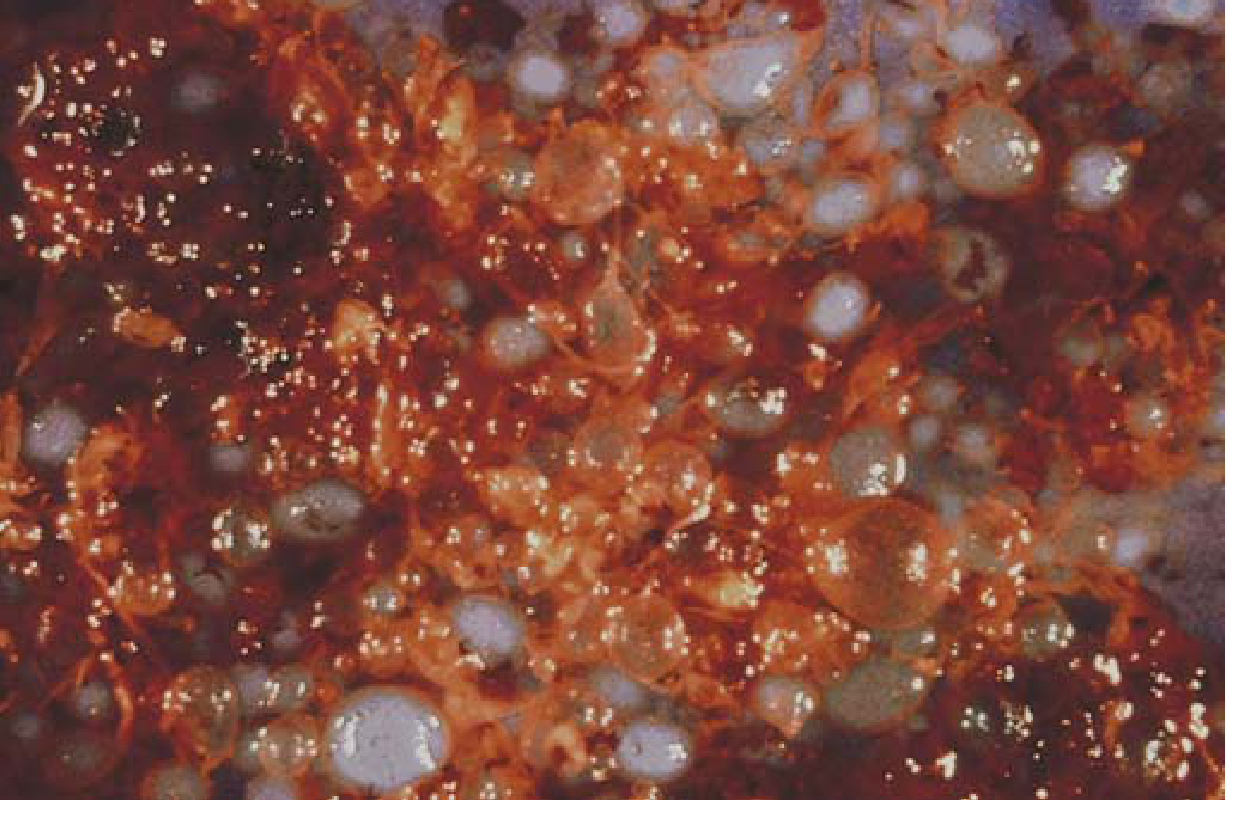
- Complete mole that penetrates deep into / perforates the myometrium
- Retains hydropic villi (unlike choriocarcinoma)
- Metastasizes to lung in ~15% of cases
- Develops in ~20% of complete moles after evacuation
- Not histologically malignant but clinically aggressive; responds to chemotherapy
3. Choriocarcinoma (Malignant)
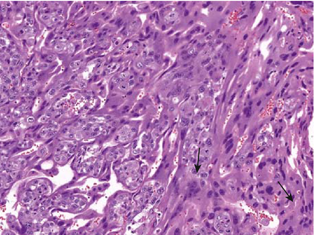
- No chorionic villi (key distinguishing feature from mole)
- Sheets of anaplastic cytotrophoblasts + syncytiotrophoblasts
- Abundant, often abnormal mitoses
- Invades myometrium and blood vessels
4. Placental Site Trophoblastic Tumor (PSTT)
- Rare (<2% of GTN); from intermediate (extravillous) trophoblasts
- Secretes hPL (human placental lactogen) >> hCG - so hCG is only mildly elevated
- Resistant to chemotherapy - treatment is surgical (hysterectomy)
- Occurs after normal pregnancy or abortion
Role of Beta-hCG
| Role | Details |
|---|---|
| Diagnosis | hCG >2× median for gestational age → suggestive of molar pregnancy; >100,000 IU/L strongly suggests complete mole |
| Staging/Prognosis | Pre-treatment hCG is one of 8 FIGO prognostic scoring factors; score ≥7 = high risk → multiagent chemo |
| Treatment monitoring | Serial hCG after evacuation; persistent rise = GTN requiring treatment |
| Remission criteria | 3 consecutive normal hCG values |
| Surveillance | hCG monitored for 6 months (if normalizes within 56 days) or 6 months from normalization (if takes longer) |
| Recurrence detection | Lifelong surveillance in high-risk women; hCG checked 6-8 weeks after any subsequent pregnancy |
| Exception - PSTT | hCG only mildly elevated; hPL is the dominant marker |
Measurement tip: Assay must detect both intact hCG and free beta-subunit for optimal sensitivity in GTD monitoring.
Summary
| Entity | Villi | Malignant | hCG | Treatment |
|---|---|---|---|---|
| Complete mole | Hydropic, all | No | Very high | Suction curettage |
| Partial mole | Hydropic, some | No | Mildly elevated | Suction curettage |
| Invasive mole | Present | Locally | Elevated | Chemotherapy |
| Choriocarcinoma | Absent | Yes | Very high | Chemotherapy (MTX) |
| PSTT | Absent | Yes | Mildly elevated | Surgery |
Create a diagnostic algorithm chart for GTD classification
A clean, professional medical diagnostic algorithm flowchart for Gestational Trophoblastic Disease (GTD) classification. The chart flows top to bottom with the following structure: TOP: "Suspected GTD" box (patient with abnormal uterine bleeding, elevated hCG, history of pregnancy) FIRST BRANCH: "Serum β-hCG + Pelvic Ultrasound" SECOND LEVEL splits into two paths: LEFT: "Molar Pregnancy (Hydropic villi on USS)" RIGHT: "No molar features / Post-pregnancy hCG elevation" Under LEFT "Molar Pregnancy": Splits into: - "Complete Mole" box: Diploid 46XX, All villi hydropic, hCG very high >100,000, No fetal parts, Diffuse trophoblast proliferation - "Partial Mole" box: Triploid 69XXY, Some villi hydropic, hCG less elevated, Fetal parts may be present, Focal trophoblast proliferation Both mole boxes lead down to: "Uterine Evacuation + Histology" Then arrow to: "Post-evacuation hCG monitoring (serial weekly)" This splits into TWO outcomes: - LEFT green box: "hCG normalizes within 56 days → BENIGN MOLE → Surveillance 6 months" - RIGHT red box: "hCG plateau or rise → GTN (Gestational Trophoblastic Neoplasia)" Under GTN red box, splits into THREE: 1. "Invasive Mole" - Hydropic villi present, invades myometrium, hCG elevated → Chemotherapy (MTX) 2. "Choriocarcinoma" - NO villi, hemorrhagic necrotic mass, anaplastic cyto+syncytiotrophoblasts, high hCG, vascular spread → Chemotherapy (MTX/ActD) 3. "PSTT / ETT" - Intermediate trophoblasts, LOW hCG, hPL dominant → Surgery (hysterectomy) Under RIGHT path "No molar features": Box: "FIGO Staging + Prognostic Scoring (includes pre-treatment hCG level)" Splits: - "Score <7 = Low Risk → Single-agent chemo (MTX)" - "Score ≥7 = High Risk → Multi-agent chemo (EMA-CO)" Bottom footer bar: "β-hCG: ~99% sensitivity & specificity | Used for: Diagnosis → Staging → Treatment monitoring → Remission → Recurrence surveillance" Color scheme: blue header boxes, yellow for mole types, red for malignant GTN, green for benign/surveillance, orange for FIGO scoring. Clean white background, clear arrows, medical textbook style.
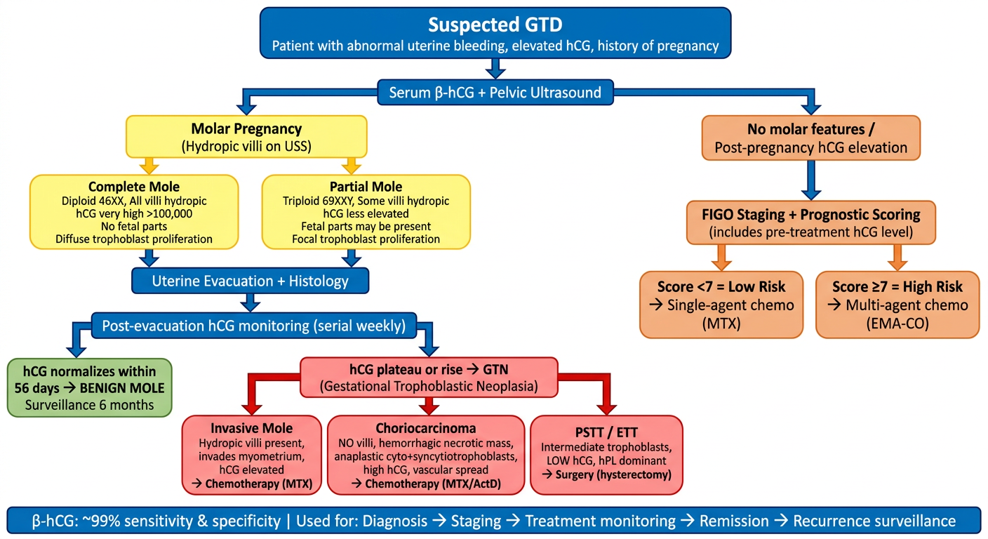
- Entry point - suspected GTD with elevated hCG + ultrasound
- Molar vs. non-molar split at first branch
- Complete vs. Partial mole differentiation by karyotype, villi extent, and hCG level
- Post-evacuation hCG monitoring - the pivotal decision point
- GTN branch - separates Invasive Mole, Choriocarcinoma, and PSTT/ETT with their distinct treatments
- FIGO scoring for risk stratification driving single-agent vs. multi-agent chemotherapy
- Footer summarizing the role of β-hCG across all stages